By Dr. Ara Deukmedjian
Board Certified Neurosurgeon
Reviewed on July 30, 2026
Disclaimer: This information is for educational purposes only and is not intended to replace the advice of a physician. Results may vary from person to person. Consult your doctor regarding your particular situation.
Key Points
✓ Disc bulge prevalence in asymptomatic individuals increases from 30% at age 20 to 84% at age 80. Having a bulge on MRI does not mean you need treatment. ¹ ²
✓ Bulging discs and herniated discs exist on a continuum of disc pathology. Both originate from annular tears and produce symptoms through the same inflammatory mechanism. ³
✓ Chemical inflammation not mechanical compression alone is the primary driver of disc-related pain. TNF-alpha, IL-6, and prostaglandins sensitize nerve endings even without significant nerve compression. ⁴ ⁵
✓ L4-L5 and L5-S1 are the most commonly affected lumbar levels. Cervical bulges most often occur at C5-C6 and C6-C7. ¹
✓ A 2025 meta-analysis confirms that exercise therapy significantly improves pain, disability, and quality of life in patients with lumbar disc problems. ⁷
✓ Physical examination findings must correlate with MRI to confirm the pain source. An MRI alone cannot diagnose the cause of your pain. ¹ ²
✓ Conservative management will work for most people when done thoroughly, which includes physical therapy, anti-inflammatories, changing activity level, and time. ⁸
✓ Progressive muscle weakness, cauda equina syndrome, or pain that continues after 6-12 weeks of conservative management is a reason for surgical review.
✓ Spinal fusion carries a documented risk of adjacent segment disease and permanently alters spinal biomechanics.
✓ Deuk Laser Disc Repair®: 99.6% success rate, 0.01% complication rate, 2,700+ procedures. Motion-preserving, outpatient, same-day recovery.
What Is a Bulging Disc?
A bulging disc refers to the condition where the whole disc protrudes out of the confines of the vertebral body. The disc itself consists of two parts. These are the nucleus pulposus, which is a jelly-like inner portion containing about 80% of water for healthy individuals. And the annulus fibrosus, a strong outer covering made up of 15–25 concentric layers of collagen fibers.
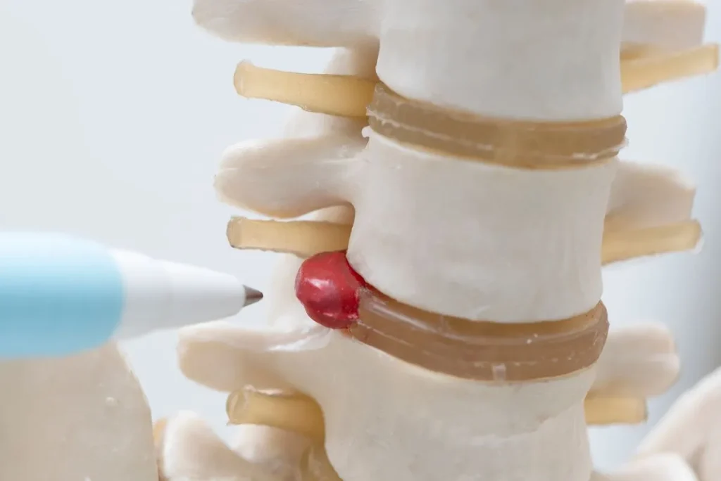
In a bulging disc, only the annulus fibrosus stretches out and bulges outwards and involves more than 25% of the perimeter of the disc. ³ This helps differentiate it from a protruding disc. Which is a more focused bulge and an extrusion or herniation. Where there is a complete tear in the annulus and the nucleus pulposus leaks out.
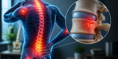
From a clinical standpoint, and based on treating thousands of patients at Deuk Spine Institute, bulging and herniated discs lie on a continuum of disc pathology. While medical literature differentiates them technically, both arise from initial posterior annular damage that is traumatic in nature, and both can cause similar symptoms through the same mechanism: inflammation triggered by damaged disc material irritating nearby nerve structures. ³ ⁴
How Common Are Bulging Discs?
Bulging discs are far more common than most patients realize. A landmark systematic review by Brinjikji et al. analyzed 33 studies involving 3,110 asymptomatic individuals and found that disc bulge prevalence increases steadily with age ¹:
- Age 20: 30% of asymptomatic individuals have disc bulges on MRI
- Age 40: approximately 50% show disc bulges
- Age 60: approximately 69% have disc bulges
- Age 80: 84% demonstrate disc bulges on imaging
A subsequent meta-analysis by the same group confirmed that while disc bulges are more prevalent in symptomatic individuals (OR 7.54; 95% CI, 1.28–44.56), the high background rate in pain-free people means that imaging findings alone cannot establish causation. ²
A vital clinical consideration is that the presence of a disc bulge on an MRI scan does not imply that you are experiencing pain or require treatment. The vital aspect is the correlation of your disc bulge with your symptoms through physical examination.
Bulging vs. Herniated Disc: Understanding the Continuum
Despite these technical differences, from a treatment perspective both conditions are fundamentally similar. Both involve trauma to the disc that creates an annular tear. Both can cause inflammation. Both respond to similar treatments when properly diagnosed. ³ ⁴
If you are confused by the different terms used to describe your disc condition, our guide on herniated disc, bulging disc, and slipped disc terminology explains all 10 names doctors and radiologists use for the same condition.
Key Facts About Bulging Discs
Degenerative discs do not heal themselves. Once the degeneration process takes place with the protrusion beyond the normal boundaries and weakening of the annulus, the degenerative process will be irreversible, although the symptoms can be alleviated by conservative therapy or by spontaneous resorption. A gradual removal of inflammatory tissue by the immune system. ⁸
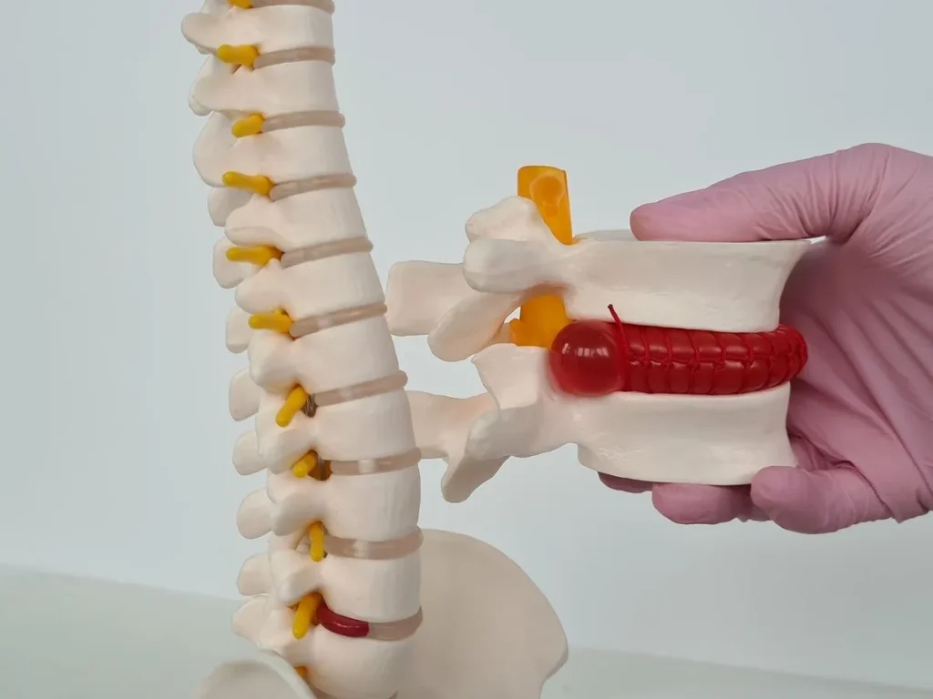
Bulging discs do not necessarily result in any symptoms. Quite often, individuals experience their entire life span with bulging discs and no symptoms whatsoever. The crucial factor is whether a person experiences inflammation and compression of nearby nerves rather than the extent of disc bulge visualized on imaging studies. ¹ ²
Bulging discs can progress. Without proper care, a bulging disc can worsen over time as the annulus continues to weaken. The bulge may eventually convert into a full herniation if the annulus tears completely. ³
Location determines symptoms. A cervical bulge at C5-C6 causes arm pain and biceps weakness. The bulging of the disc at L5-S1 causes sciatica and weak calf muscles. The same disorder located in different regions results in very different clinical pictures.
Bulging Disc Symptoms by Spinal Region
Symptoms and problems associated with bulging discs are very different depending on their location in the spine.
Cervical Bulging Discs
Cervical vertebrae are seven in number, namely C1-C7. Most bulging discs happen between C5-C6. And C6-C7 because they experience tremendous stress due to the movement of the head and weight-bearing activities.
Symptoms include neck pain and stiffness, headaches at the occipital area, shoulder pain extending to the thoracic region, arm pain associated with numbness/tingling sensations. And in serious situations, difficulty in performing fine motor tasks like fastening buttons and writing.
Nerve root patterns by level:
- C5 nerve (C4-C5 disc): Shoulder and upper arm pain, weakness lifting the arm
- C6 nerve (C5-C6 disc): Pain to the thumb, biceps weakness, thumb and index finger numbness
- C7 nerve (C6-C7 disc): Pain to the middle fingers, triceps weakness, middle finger numbness
- C8 nerve (C7-T1 disc): Ring and pinky finger pain and numbness, hand grip weakness
Thoracic Bulging Discs (Mid-Back Region)
The occurrence of thoracic disc disease is much rarer than other forms of disc herniation. Since the rib cage adds extra stability to the vertebral column. Thus restricting movement and reducing mechanical stress. ¹⁰ With the exception of lumbar disc herniation, thoracic disc herniation usually occurs more often at the lower thoracic levels (T8-T12).
Common symptoms include mid-back pain that radiates around the rib cage. Pain that is exacerbated by twisting and rotation, and chest pain. Which may be attributed to heart or lung disorders. In extreme cases, where the spinal cord is compressed, leg weakness or loss of bowel and bladder control may occur.
Since the symptoms of thoracic disc disease are similar to those of cardiac, pulmonary, and gastrointestinal disorders. Proper diagnosis is especially essential for thoracic disc diseases.
Lumbar Bulging Discs
The lumbar spine consists of five vertebrae (L1-L5) plus the sacral segments below. This region experiences the greatest mechanical stress during daily activities and is the most common site of symptomatic disc bulges. The L4-L5 and L5-S1 levels are most frequently affected.
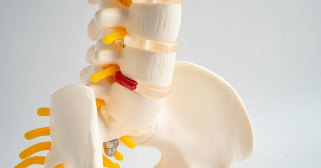
Common symptoms include lower back pain (sharp, dull, or aching), morning stiffness and limited flexibility, pain that worsens with prolonged sitting or forward bending, leg pain with numbness or tingling (sciatica or lumbar radiculopathy), and in severe cases, bowel or bladder dysfunction, which constitutes a medical emergency.
Nerve root patterns by level:
- L3 nerve (L2-L3 disc): Front-of-thigh pain, knee extension weakness
- L4 nerve (L3-L4 disc): Inner thigh and shin pain, knee-straightening weakness
- L5 nerve (L4-L5 disc): Outer leg and top-of-foot pain, foot drop (difficulty lifting the foot), numbness between the first and second toes
- S1 nerve (L5-S1 disc): Back-of-leg and foot pain, weakness pushing onto tiptoes, numbness on the bottom and outer foot
For detailed information about sciatica and its various causes, see our comprehensive guide Pain Radiating Below the Knee: When Sciatica Demands Your Attention.
Why Bulging Discs Don’t Always Cause Pain: The Inflammation Connection
For decades, the medical community believed that disc bulges caused pain primarily through mechanical compression of nerve roots. While nerve compression can certainly produce symptoms, research has fundamentally changed our understanding of disc pain: chemical inflammation, not compression alone, is the primary pain driver. ⁴ ⁵
The Chemical Inflammation Process
When a disc degenerates, develops an annular tear, or bulges significantly, inflammatory chemical mediators are released into the surrounding tissues. These include ⁴:
- Prostaglandins — lipid compounds that promote inflammation and lower pain thresholds
- Cytokines — particularly TNF-alpha (tumor necrosis factor-alpha) and IL-6 (interleukin-6), which trigger inflammatory cascades
- Proteolytic enzymes — molecules that break down the extracellular matrix and contribute to tissue damage
- Substance P — a neuropeptide involved in pain signal transmission
This is because the chemicals cause sensitization of the nerves to produce pain without any form of mechanical compression. This paradoxically occurs in many clinical scenarios where there are patients with large disc bulges who experience little pain compared to those with smaller disc bulges. The level of inflammation, not the size of the bulge, is the determining factor. ⁴ ⁵
A 2025 systematic review examining conservative treatments for lumbar disc herniation confirmed that addressing inflammation, rather than mechanical decompression alone, is crucial for successful treatment outcomes. ⁵
Clinical Implications
These insights have implications for therapeutic intervention strategies. Treatment methods such as conservative ones involving anti-inflammatory drugs, epidural corticosteroid injections, and physical therapy to reduce inflammatory stress can be very effective. Operations involving precise removal of the damaged, inflammatory disc tissue (Deuk Laser Disc Repair® surgery) eliminate the source of the pain without performing a complete disc replacement or disc fusion. In addition, the immune system can sometimes help eliminate the inflammatory disc tissues through natural absorption over time. ⁵ ⁸
Common Causes and Risk Factors
Understanding what causes disc bulges can inform prevention strategies and treatment decisions. Risk factors fall into three categories: age-related degeneration, mechanical stress and trauma, and lifestyle and genetic factors.
Age-Related Degeneration
The most common cause of bulging discs is degenerative disc disease, a set of natural changes that occur with aging. Healthy discs are approximately 80% water in youth; as we age, discs lose hydration, becoming less flexible and more prone to injury. ¹ The annulus weakens as collagen fibers deteriorate, and the disc’s already-limited blood supply becomes less efficient, impairing its ability to repair minor damage.
Mechanical Stress and Trauma
Heavy lifting with improper technique, especially with twisting, creates enormous intradiscal pressure. Lifting a 50-pound object with poor form can place over 500 pounds of pressure on lumbar discs. ¹² Prolonged sitting increases intradiscal pressure by approximately 40% compared to standing. ¹² Acute trauma from car accidents, falls, or sports injuries can cause immediate disc damage that may not become symptomatic for weeks or months. A 2025 study in Frontiers in Neurology confirmed that occupations involving repetitive lifting and bending are associated with significantly higher rates of disc problems. ¹³
Lifestyle and Genetic Factors
Excess weight exerts greater mechanical stress on intervertebral discs, especially the lumbar ones. Smoking causes narrowing of the vessels and thus worsens the insufficient blood supply to the discs, leading to degeneration of the discs and poor results from treatment. ¹⁴ Sedentary lifestyle results in weakness of the core muscles, which cannot adequately provide necessary support for the spine. Twin studies have shown that 34-74% of disc degeneration is genetically predetermined. ¹⁵
Diagnosis: The Foundation of Successful Treatment
A correct diagnosis is necessary for effective treatment. Many people undergo months or even years of failed treatments because of the wrong assessment made in the beginning.
Physical Examination: Why It Matters as Much as MRI
While MRI is invaluable, it cannot diagnose the source of your pain on its own. As I frequently tell patients: MRI scans do not tell you where pain comes from; patients do. ²
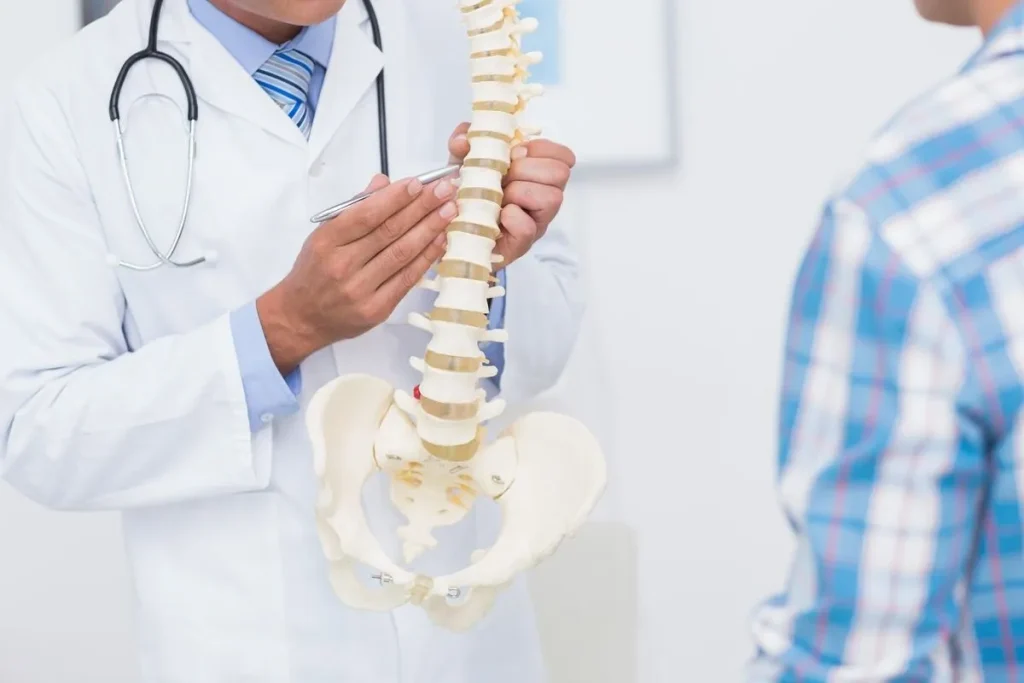
A comprehensive physical examination should include a detailed medical history, range of motion testing, neurological examination (strength, sensation, reflexes), provocative tests such as the straight leg raise for lumbar pathology and Spurling’s test for cervical pathology, and careful palpation to identify areas of tenderness and muscle spasm.
Physical examination findings must correlate with imaging studies for accurate diagnosis. A disc bulge seen on MRI that does not correspond to your symptoms may be an incidental finding unrelated to your pain. ¹ ²
Imaging Studies
MRI is the gold standard for visualizing bulging discs and their relationship to surrounding structures. ¹⁶ It demonstrates the exact location, size, and type of disc abnormalities; whether nerve roots are being compressed; the degree of disc degeneration; and the presence of co-existing conditions such as spinal stenosis or facet joint arthritis. However, MRI findings must always be interpreted in the clinical context. Studies consistently demonstrate that many people without pain have disc bulges on MRI. ¹ ²
X-rays cannot visualize discs directly but can show vertebral alignment, disc space height, bone spurs, and signs of instability.
CT scans offer superior bony detail and may be useful when MRI is contraindicated or when evaluating bony sources of nerve compression.
EMG/NCS (electrodiagnostic studies) measure electrical activity in muscles and nerves, confirming the presence and severity of nerve root compression and distinguishing radiculopathy from peripheral nerve problems.
The Value of a Second Opinion
Given the complexity of diagnosing disc-related pain and the wide variation in treatment recommendations among spine specialists, seeking a second opinion is often one of the smartest decisions a patient can make. This is especially true if surgery has been recommended, if conservative treatments have failed, if the diagnosis seems unclear, or if the recommended surgery involves fusion.
The challenge in spine surgery is that most surgeons recommend what they know how to do, rather than necessarily what is best for the patient. A surgeon who primarily performs fusion will likely recommend fusion. A pain management specialist who does not perform surgery will recommend injections. This is not dishonesty; it is human nature. Seeking opinions from specialists with different skill sets helps patients make truly informed decisions.
Upload your MRI for a free second opinion: At Deuk Spine Institute, we offer a free virtual consultation and MRI review. This service has helped countless patients discover treatment options they did not know existed.
Conservative Treatment: First-Line Management
For most patients with bulging discs, an initial trial of conservative treatment is appropriate and often successful. Research indicates that patients with disc problems can improve with comprehensive conservative care. ⁸ The keyword is “comprehensive”. Not all conservative treatments are equally effective, and the quality of implementation matters significantly.
Medications
NSAIDs (ibuprofen, naproxen, celecoxib) reduce inflammation and pain. They work best when used regularly during flare-ups rather than only when pain peaks. Long-term use carries risks including gastrointestinal bleeding, kidney problems, and cardiovascular concerns. ⁸
Neuropathic pain medications (gabapentin, pregabalin) specifically target nerve pain pathways and can be particularly effective for burning, shooting, or electric-shock sensations. Side effects include drowsiness and cognitive slowing.
Muscle relaxants (cyclobenzaprine, methocarbamol) can reduce the muscle spasms that often accompany disc problems, improving mobility and comfort. These are typically used short-term.
Oral corticosteroids (short course) may be prescribed for severe acute inflammation to rapidly reduce swelling and pain.
Important note: Opioid medications are not recommended for long-term management of chronic disc pain due to risks of dependence, tolerance, and limited long-term effectiveness. ⁸
Physical Therapy and Exercise
A well-designed physical therapy program is one of the most effective conservative treatments for bulging discs. A 2025 meta-analysis published in Frontiers in Medicine found that exercise therapy significantly improves pain, disability, range of motion, and quality of life in patients with lumbar disc problems. ⁷
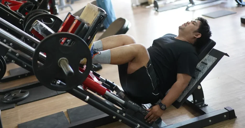
An evidence-based program typically includes core strengthening (planks, bird-dogs, dead bugs, bridges), flexibility training targeting hip flexors and hamstrings, nerve gliding exercises to help nerves move more freely through surrounding tissues, postural training for proper body mechanics, and hands-on manual therapy.
The effectiveness of physical therapy depends heavily on the quality of the program and patient compliance. Working with a therapist who specializes in spinal conditions is essential.
Epidural Steroid Injections (ESIs)
When oral medications and physical therapy provide insufficient relief, epidural steroid injections deliver corticosteroid medication and local anesthetic into the epidural space around inflamed nerve roots. ESIs can reduce inflammation and provide pain relief lasting weeks to months in some patients. ⁸
Limitations include temporary relief (3–6 months on average), the potential for weakened bone and soft tissues with repeated injections, and the fact that injections do not address the underlying disc pathology.
Activity Modification and Lifestyle Changes
Maintaining a healthy weight (even 5–10% body weight loss can significantly reduce spinal load), quitting smoking (which improves blood flow to discs and enhances healing capacity), ergonomic modifications at workstations, and staying active with low-impact exercises (walking, swimming, cycling) all support disc health and recovery. ¹⁴
When Conservative Treatment Isn’t Enough
While conservative care succeeds for most patients, recognizing when it is failing is crucial for preventing permanent damage and unnecessary prolonged disability.
Indications for Surgical Evaluation
Progressive motor weakness. Increasing weakness in a leg or foot despite conservative treatment may indicate progressing nerve damage. Prolonged compression can lead to permanent muscle atrophy and functional loss that will not fully recover even after successful decompression. Warning signs include foot drop, weakness climbing stairs, difficulty rising from a chair, and progressive loss of grip strength. ⁸
Severe, intractable pain. Pain that prevents sleep, interferes with work, or limits basic daily activities despite appropriate conservative care is a legitimate indication for definitive treatment.
Failed conservative treatment. If 6–12 weeks of comprehensive conservative care (not just one or two approaches, but a genuine multidisciplinary program) have not produced meaningful improvement, continuing the same ineffective treatments indefinitely makes little sense. ⁸
Cauda equina syndrome (MEDICAL EMERGENCY). This rare but serious condition occurs when a massive disc problem compresses multiple nerve roots, causing bowel or bladder dysfunction, saddle anesthesia (numbness in the area that would contact a saddle), bilateral leg weakness, and sexual dysfunction. This requires urgent surgical decompression to prevent permanent neurological damage.
Surgical Treatment Options
When surgery becomes necessary, understanding all available options is essential. Not all procedures are equal in invasiveness, recovery time, long-term outcomes, and impact on spinal function.
Traditional Surgical Approaches
Microdiscectomy has been the standard surgical treatment for decades. It involves accessing the spine through a 1–2 inch incision, removing a portion of the lamina (laminotomy) to access the disc, and removing the bulging or herniated material. Recovery typically requires several days in the hospital, 4–6 weeks for light activities, and 3–6 months for full recovery. ⁸
Laminectomy involves removing a larger portion of the lamina to decompress the spinal canal, typically reserved for cases with significant spinal stenosis. Recovery is generally longer than microdiscectomy.
Spinal fusion permanently joins two or more vertebrae using bone grafts and hardware (screws and rods). Fusion eliminates motion at the fused segment, permanently alters spinal biomechanics, increases stress on adjacent levels, requires 6–12 months of recovery, and carries higher complication rates than motion-preserving procedures. Research consistently shows that fusion carries an increased risk of adjacent segment disease, where levels above or below the fusion develop problems requiring additional surgery.
Deuk Laser Disc Repair®: The Motion-Preserving Alternative
After witnessing the limitations and complications of traditional spine surgery throughout my training and career, I became convinced that a better approach was needed. This led to the development of Deuk Laser Disc Repair® (DLDR), a minimally invasive, motion-preserving procedure that addresses the pathological disc material while leaving healthy structures intact.
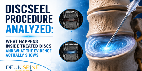
How DLDR® works: Through a 4–7 mm incision (small enough to cover with a band-aid), a specialized endoscope provides high-definition visualization. A side-firing holmium laser targets only the damaged, inflammatory disc material inside the annular tear typically just 5–10% of the total disc. No bone is cut. No hardware is implanted. The disc, lamina, facets, and ligaments are preserved.
Recovery timeline:
- Day of surgery: Most patients notice significant pain relief immediately. They leave recovery within an hour and go home the same day.
- First week: The incision heals within days. Most patients require only over-the-counter pain medication, if any. Many return to desk work within 3–5 days.
- 2–4 weeks: Complete healing occurs. Patients resume most normal activities.
- Long-term: No fusion means no adjacent segment disease. Full range of motion is preserved permanently.
Results: 99.6% patient-reported success rate in eliminating disc-related pain. 0.01% complication rate across 2,700+ procedures. Over 15 years of clinical experience. Published in peer-reviewed medical literature. Covered by most major insurance plans.
Deuk Plasma Rhizotomy®: For Facet-Related Pain
When bulging discs occur alongside facet joint arthritis, Deuk Plasma Rhizotomy® offers a solution for the facet component. This 10-minute outpatient procedure permanently disconnects pain nerves to the affected joint through a 4 mm incision, providing long-term pain-free movement without hardware or implants.
Treatment Comparison: Traditional vs. Minimally Invasive
Real Patient Story: From Daily Struggle to Total Relief
John had lived with constant lower back pain for years. Most days, his discomfort sat between a 7 and 8 on the pain scale, making simple activities feel impossible; walking the beach, climbing stairs, even enjoying retirement.
After trying conservative care without lasting results, John chose Deuk Laser Disc Repair to treat his herniated L3-4 and L4-5 discs. With tiny incisions and no fusion hardware, the procedure targeted the source of his disc pain directly.
The change was immediate. Just one day after surgery, he could climb stairs without holding on, the pain was gone, and he did not even need pain medication. Within a day, he was moving freely again and quickly returned to activities he once loved.
John’s experience highlights several key points about this treatment: long-term relief is achievable even without traditional pain management, multiple affected levels can be treated with one minimally invasive approach, recovery can occur within days rather than months, and chronic pain does not have to define your later years.
Activities to Avoid With a Bulging Disc
Certain activities can significantly aggravate bulging discs and should be avoided or modified, particularly during acute phases.
High-Risk Activities
Heavy lifting places enormous compressive forces on spinal discs. Anything over 10–15 pounds should be avoided during acute pain. ¹²
Deadlifts and squats create maximum compressive and shear forces on lumbar discs, particularly at L4-L5 and L5-S1. Avoid entirely during acute phases.
Running creates repetitive impact forces that compress discs with each foot strike, aggravating inflamed tissue and nerve roots.
Traditional sit-ups and crunches involve repeated forward flexion, the exact movement that increases pressure on posterior disc bulges. Use planks, bird-dogs, and dead bugs instead.
Golf combines rotational torque, asymmetric loading, forward flexion, and explosive force — a particularly challenging combination for people with disc problems.
Contact sports (football, hockey, rugby, martial arts) involve high-velocity collisions, twisting, and unpredictable impacts that can severely aggravate or worsen a bulging disc.
Generally Safe Activities
Walking (excellent low-impact exercise), gentle stretching within a pain-free range, water aerobics, stationary cycling with upright posture, light resistance training with proper form, and core stabilization exercises (planks, bird-dogs, dead bugs) are generally well-tolerated.
The key principle is understanding biomechanics: avoid activities involving heavy compression, forward flexion, rotation, or explosive movements. If an activity increases your pain, stop immediately.
Remove the inflamed disc tissue without fusion or hardware.
Chemical inflammation — not compression alone — is the primary driver of disc pain, which is why patients with small bulges can be in agony and patients with large bulges can be pain-free. Once you know that, the right treatment isn’t to fuse or replace the disc — it’s to remove the damaged, inflammatory tissue at the source. Send your MRI for a free review by Dr. Deukmedjian and learn whether Deuk Laser Disc Repair® can treat the exact pain-generating tissue through a 7 mm incision, with the disc, bone, ligaments, and natural motion preserved.
- 99.6%
- Success rate
- 0.01%
- Complication rate
- 2,700+
- Procedures performed
FAQs
What’s the difference between a bulging disc and a herniated disc?
While medical literature differentiates these conditions technically, they are fundamentally similar from a clinical and treatment perspective. A bulging disc extends beyond its normal boundaries in a broad, circumferential pattern with the annulus fibrosus remaining intact but stretched. A herniated disc involves a complete tear of the annulus, allowing the nucleus pulposus to leak into the spinal canal. Both conditions arise from disc trauma creating an annular tear, both can cause inflammation and nerve compression, and both respond to similar treatments when properly diagnosed. ³ ⁴
Can a bulging disc heal on its own without surgery?
The structural changes of a bulging disc are permanent. Once the disc extends beyond its normal boundaries, it does not spontaneously return to its original position. However, symptoms can improve significantly without surgical intervention for most patients. ⁸ Acute inflammation subsides over time, the immune system can gradually resorb some extruded disc material, the nervous system adapts, and strengthening surrounding muscles provides better spinal support. If symptoms persist beyond 6–12 weeks of comprehensive conservative treatment, or if progressive weakness develops, surgical evaluation is warranted.
How do I know if my bulging disc needs surgery?
Surgery should be considered when conservative treatment has failed after 6–12 weeks of comprehensive care, when progressive motor weakness is developing (foot drop, hand weakness, difficulty climbing stairs), when pain severely impacts quality of life despite appropriate conservative treatment, or in the case of cauda equina syndrome (bowel/bladder dysfunction, bilateral leg weakness — a medical emergency). ⁸ Even when surgery is needed, patients have options. Modern minimally invasive techniques like Deuk Laser Disc Repair® address the problem while preserving natural spinal motion and biomechanics.
What activities should I avoid with a bulging disc?
Avoid heavy lifting (over 10–15 pounds during acute phases), deadlifts, squats, and Romanian deadlifts, high-impact running or jumping, contact sports, golf, exercises involving repeated forward bending (sit-ups, toe touches), and prolonged sitting without breaks. ¹² Generally safe activities include walking, gentle stretching, water aerobics, stationary cycling with upright posture, and core stabilization exercises like planks and bird-dogs. As symptoms improve, activities can be reintroduced gradually under the guidance of a physical therapist.
How long does recovery take after bulging disc surgery?
Recovery time varies dramatically by procedure. Traditional microdiscectomy requires a 1–2 day hospital stay, 4–6 weeks for light activities, and 3–6 months for full recovery. Spinal fusion requires a 2–4 day hospital stay, 3–6 months for bone fusion to solidify, and up to one year for full recovery with permanent loss of motion at the fused segment. Deuk Laser Disc Repair® is a same-day outpatient procedure with most patients returning to desk work within 3–5 days and achieving complete healing in 2–4 weeks, with full range of motion preserved permanently.
Sources
View Sources
- Brinjikji W, Luetmer PH, Comstock B, et al. Systematic literature review of imaging features of spinal degeneration in asymptomatic populations. AJNR Am J Neuroradiol. 2015;36(4):811-816.
- Brinjikji W, Diehn FE, Jarvik JG, et al. MRI findings of disc degeneration are more prevalent in adults with low back pain than in asymptomatic controls: A systematic review and meta-analysis. AJNR Am J Neuroradiol. 2015;36(12):2394-2399.
- Fardon DF, Williams AL, Dohring EJ, et al. Lumbar disc nomenclature: version 2.0. Spine J. 2014;14(11):2525-2545.
- Risbud MV, Shapiro IM. Role of cytokines in intervertebral disc degeneration: pain and disc content. Nat Rev Rheumatol. 2014;10(1):44-56.
- Conservative treatments for lumbar disc herniation: systematic review. PMC. 2025.
- Exercise therapy for lumbar disc problems: meta-analysis. Front Med. 2025.
- Conservative management of lumbar disc herniation. Neurospine. 2023.
- Quint U, et al. Thoracic disc herniation: Surgical treatment. Neurosurgery. 2002.
- Nachemson AL. The lumbar spine: an orthopaedic challenge. Spine. 1976;1(1):59-71.
- Occupational risk factors for lumbar disc herniation. Front Neurol. 2025.
- Alkherayf F, Agbi C. Cigarette smoking and chronic low back pain in the adult population. Clin Invest Med. 2009;32(5):E360-E367.
- Battié MC, Videman T, Kaprio J, et al. The Twin Spine Study: contributions to a changing view of disc degeneration. Spine J. 2009;9(1):47-59.