By Dr. Ara Deukmedjian, MD
Board Certified Neurosurgeon
Medically reviewed on July 21, 2026
Medical Disclaimer: This content is for educational purposes only and does not constitute medical advice. Individual results may vary. Always consult with your healthcare provider about your specific condition and treatment options.
Key Points
✓ A disc protrusion is a contained disc herniation in which the inner nucleus pushes outward against an intact outer annulus fibrosus. ¹
✓ Paracentral protrusions are the most common subtype and typically compress one nerve root, producing sciatica or arm radiculopathy. ¹ ²
✓ Disc abnormalities are extremely common in pain-free people: 37% of asymptomatic 20-year-olds and more than 90% of asymptomatic 60-year-olds have disc bulges on MRI. ³
✓ Approximately 75–90% of patients improve within 6–12 weeks of appropriate non-operative care. ⁴ ⁵
✓ Herniated disc material can shrink or fully resorb on its own — a well-documented biological process driven by macrophage-mediated inflammation. ⁶
✓ Laminectomy and spinal fusion are almost never the correct first surgical option for an isolated disc protrusion. ⁷
✓ A landmark study documented adjacent-segment disease in 25.6% of fusion patients within 10 years, at a rate of ~2.9% per year. ⁸
✓ Full-endoscopic decompression treats a focal disc problem through a 7 mm incision, preserves motion, and reports 85–95% clinical success in the peer-reviewed literature. ⁹ ¹⁰
✓ Deuk Laser Disc Repair®: 99% pain relief for treated pain sources, 0.01% complication rate, motion-preserving, outpatient.
What Is a Disc Protrusion?
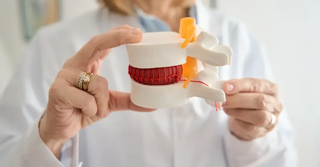
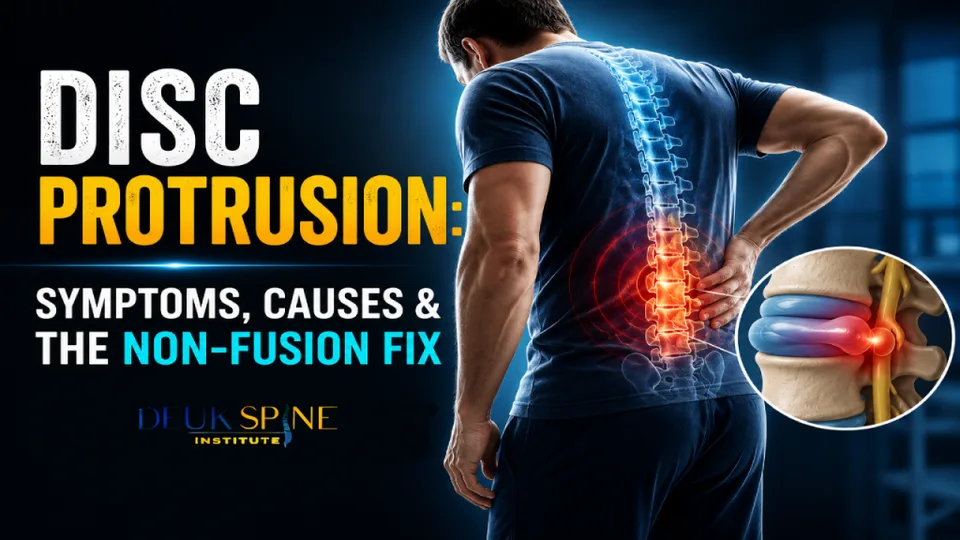
Disc protrusion is one kind of contained disc herniation where the gel-like nucleus pulposus presses on the outer part of the disc (annulus fibrosis), but does not rupture it. ¹
The intervertebral disc is composed of two different parts. The annulus fibrosus is a tough fibrous structure of the disc while the nucleus pulposus is a soft, hydrated inner structure acting as a shock absorber.
In case the annulus fibrosus is weakened but still remains intact, and its content pushes itself beyond the natural boundary of the disc, the condition is known as a protrusion. If the annulus ruptures completely and its content gets out, the condition is known as an extrusion. ¹
Radiologically, a protrusion has a base that is wider than the protruding portion. This distinguishes it from an extrusion. ¹ The distinction matters. It determines the appropriate surgical approach. It also determines whether surgery is needed at all.m an extrusion. ¹ This distinction matters because it determines the appropriate surgical approach and whether surgery is needed at all.
Types of Disc Protrusion by Location
Protrusions are classified by where they push into the spinal canal. Location determines symptoms and, ultimately, treatment planning.
1. Central Disc Protrusion
A central protrusion pushes directly into the middle of the spinal canal. It can compress the spinal cord (in the cervical or thoracic spine) or the cauda equina (in the lumbar spine). Central protrusions carry the highest risk of serious neurological consequences. They can cause bilateral symptoms.
2. Paracentral Disc Protrusion
The paracentral protrusion is the most common subtype. It occurs in the space between the central canal and the neural foramen. It typically compresses a single nerve root. This produces one sided sciatica in the lumbar spine or arm radiculopathy in the cervical spine. ²
3. Foraminal Disc Protrusion
A foraminal protrusion advances into the neural foramen. That is the opening through which a nerve root exits the spinal canal. It compresses the exiting nerve root and produces sharp, radiating pain, numbness, or weakness along that nerve’s dermatome.
Symptoms of a Disc Protrusion
Symptoms depend on the level of the spine affected. They also depend on which structures are compressed. Presentations fall into three categories.
1. Axial pain (discogenic pain)
Localized ache in the neck region or low back. May radiate into the shoulders, buttocks, or thigh region. It is related to irritation of the nerve endings of pain sensation in the outer annulus. This does not indicate any nerve entrapment. ¹ Discogenic pain increases on prolonged sitting, flexion, coughing, or sneezing. Pain decreases when lying flat.
2. Radiculopathy (nerve root symptoms)
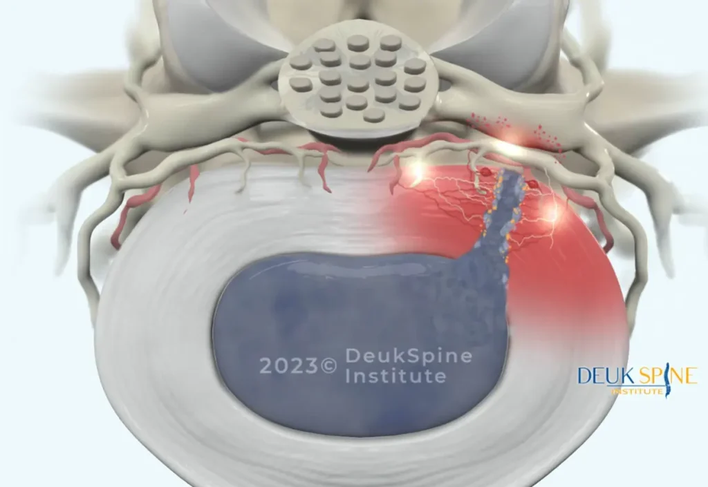
If the protrusion puts pressure on the nerve roots. The patient’s symptoms will occur in line with those nerve roots.
- Bulge in lumbar region (e.g., L4-L5, L5-S1): pain, numbness, or tingling sensation from lower back to buttocks, thigh, calf, and foot known as sciatica
- Bulge in cervical region (e.g., C5-C6, C6-C7): pain, tingling, and weakness extending from the neck to the shoulder, arm, and hand
- Weakness in motor function: foot drop, difficulty in raising feet up from ground, weak grip, or arm
- Reflex changes: diminished patellar, Achilles, biceps, or triceps reflexes depending on the level
3. Myelopathy or cauda equina (surgical red flags)
Larger central bulges may cause compression of the spinal cord or cauda equina. These represent neurosurgical emergencies that need urgent assessment. These are: ¹¹
- Progressive bilateral weakness of legs/arms
- Decreased fine motor coordination or gait disturbance
- Saddle anesthesia (anesthesia in the groin or inner thigh areas)
- Bladder/bowel dysfunction
- Increased reflexes and/or positive Hoffmann’s or Babinski sign
Any of these symptoms warrants urgent imaging and specialist referral.
Important: Not all disc protrusions cause pain. Large population studies confirm that disc bulges and protrusions are common incidental findings on MRI in people with no symptoms. ³ An MRI abnormality alone is not a diagnosis.
Disc Protrusion vs. Herniation vs. Sequestration
The terms are frequently confused and are often written as if they mean the same thing. They do not. Each describes a distinct stage of structural failure, with distinct implications on MRI and in symptoms.
Superscript numbers refer to citations in the source bibliography. ¹
All three respond to the same initial non-operative care, and all three are candidates for endoscopic decompression when surgery is genuinely required.
What Causes a Disc Protrusion?
Degeneration related to aging is the most common cause in individuals over 35 years old. ⁴ With dehydration of the disc and the development of micro tears in the annulus, there is greater chance of the nucleus pushing out through the weakened wall.
Contributing risk factors include:
- Cumulative mechanical load: repetitive bending, twisting, prolonged sitting, and forward-head posture concentrate stress on the lumbar and lower cervical discs.
- Genetics: twin studies show heritability accounts for 34–74% of disc degeneration. ¹²
- Smoking: nicotine impairs disc nutrition and accelerates dehydration.
- Obesity: every additional pound multiplies compressive force on the lumbar spine.
- Occupational exposure: heavy lifting, vibration exposure, and repetitive overhead work.
- Acute trauma: motor vehicle collisions, falls, and sports impacts can unmask an already degenerated disc, but true traumatic protrusions in a healthy disc are uncommon. ⁴
How Is a Disc Protrusion Diagnosed?
Accurate diagnosis requires three data points that agree with each other: history, physical examination, and imaging. If any one of them contradicts the other two, the diagnosis is not yet confirmed and surgery should not be on the table.
1. History and physical exam
A specialist of the spine will be able to map out the pain distribution, perform sensory tests in dermatomes and strength tests in myotomes, and do provocative tests like the SLR test for the lower back or the Spurling test for the neck region.
2. MRI
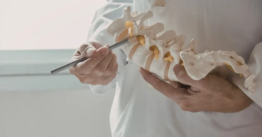
Magnetic resonance imaging is considered the imaging method of choice to evaluate disc protrusion. MRI clearly shows the details of the intervertebral disc, spinal nerves, spinal cord, and ligaments and is the only imaging modality capable of differentiating disc protrusion from other types of disc herniation. ³
3. CT scan and X-ray
X-rays evaluate bone alignment, disc space height, and instability. CT scans provide finer bony detail and are useful when MRI cannot be performed or is inconclusive.
4. EMG / nerve conduction studies
Electrodiagnostic testing pinpoints which nerve root is inflamed and rules out peripheral entrapment syndromes such as carpal or tarsal tunnel syndrome when imaging and symptoms don’t match.
The MRI only tells us so much
This is the single most important concept in modern spine care. A systematic review of imaging in asymptomatic adults found that disc bulges were present in 30% of 20-year-olds and 84% of 80-year-olds. None of whom had back pain. ³
An MRI abnormality is not a diagnosis by itself. Symptoms, physical exam, and imaging must all point to the same spinal level and the same nerve root before any treatment plan. Especially a surgical one is credible. A recommendation for surgery based on the MRI alone is a red flag.
Non-Surgical Treatment of a Disc Protrusion
Most disc protrusions do not require surgery. Current systematic reviews consistently confirm that conservative care should be the first-line approach for disc herniation and protrusion in the absence of red-flag findings. ⁷
Approximately 75–90% of patients improve within 6–12 weeks of appropriate non-operative management. ⁴ ⁵
Activity modification
A short period of relative rest during the acute inflammatory phase is appropriate. Prolonged bed rest is not. It worsens deconditioning and slows recovery. Gradual return to gentle movement is encouraged as soon as tolerable.
Physical therapy and exercise
Structured physical therapy is one of the most effective conservative interventions. A 2025 meta-analysis of eight randomized controlled trials (611 patients) found that exercise therapy significantly improved pain, disability, range of motion, and quality of life in patients with lumbar disc herniation. ¹³
The therapeutic focus should be on:
- Core stabilization: activation of the transversus abdominis and multifidus
- Directional preference exercises: McKenzie-based extension protocols where appropriate
- Hip and thoracic mobility work: offloading the lumbar spine
- Postural retraining: correcting forward-head posture and anterior pelvic tilt
Passive modalities without an active exercise component are not a substitute for supervised rehabilitation.
Medications
- NSAIDs are first-line for reducing disc-related inflammation
- Muscle relaxants can help with reflex guarding during acute flares
- Short courses of oral corticosteroids (e.g., a Medrol dose pack) are appropriate for acute severe radiculopathy
- Opioids are not appropriate long-term treatment for disc protrusion and do not address the underlying compression

Epidural steroid injections
Image-Guided Epidural/Transforaminal Steroid Injections
If treatment and medication do not alleviate the patient’s symptoms, then an image-guided epidural/transforaminal steroid injection would be helpful in delivering the drug to the inflamed nerve root. The 2024 meta-analysis and systematic review showed that epidural steroid injections have significant short-term effects for symptoms reduction in sciatica caused by lumbar disc herniation. ¹⁴ Additionally, such injections are essential to establish the cause of pain prior to performing any surgical procedure.
Spontaneous resorption: the body’s own healing process
One of the most underrated factors related to disc protrusion is that the herniated material will regress on its own. A review published in Frontiers in Medicine in 2025 highlighted how the inflammation and activation of macrophages contribute to the resorption of disc material. ⁶ It is the biological factor that explains why most cases of disc protrusion get better over time without any surgical intervention.
When Surgery Becomes Appropriate
Non-operative care is considered to have failed when:
- Symptoms persist beyond 6–12 weeks of appropriate treatment
- Motor weakness is present or progressing
- Pain is severe enough to disable normal function
- Cauda equina syndrome or myelopathy develops (surgical emergency)
At that point, decompression should be discussed. But the type of decompression matters more than the fact of it.
Surgical Treatment: The Case Against Defaulting to Fusion or Laminectomy
For decades, the default surgical options for a symptomatic disc protrusion have been:
- Laminectomy: removal of the back of the vertebra to access and decompress the nerve
- Open microdiscectomy: removal of the herniated tissue through a larger open incision with muscle stripping
- Spinal fusion (PLIF/TLIF/ACDF): removal of the entire disc and permanent fusion of adjacent vertebrae with cages, plates, and screws
Each of these procedures treats a focal, contained disc problem with a whole-segment solution and each carries durable costs:
- Permanent loss of motion at the treated level (fusion).
- Adjacent-segment disease. The landmark Hilibrand study documented symptomatic adjacent-segment degeneration in approximately 2.9% of patients per year and 25.6% within 10 years after cervical fusion. ⁸ Mechanical load a fused level cannot absorb is transferred above and below.
- Paraspinal muscle damage. Laminectomy and open discectomy strip and denervate the deep spinal stabilizers. A driver of “failed back surgery syndrome.”
- Recovery burden. Fusion typically requires an inpatient stay, weeks of restricted activity, and months of bone-healing.
A 2025 systematic review of the lumbar herniation literature confirmed that patients with longer symptom duration before surgery had worse outcomes but also emphasized that the least destructive intervention capable of solving the problem should be chosen first. ⁷ Starting with a highly destructive procedure such as fusion permanently limits future treatment options.
The critical question is not whether some surgery is warranted. It’s whether your specific disc protrusion actually requires the trades that laminectomy or fusion impose. In the absence of documented instability, cord compression with myelopathy, or multi-level severe pathology, the answer for most patients is no.
The Endoscopic Alternative: Full-Endoscopic Decompression
Full-endoscopic spinal decompression is the modern, ultra-minimally-invasive treatment for a symptomatic disc protrusion in appropriately selected patients. ⁹ ¹⁰
How it works
- A 7 mm skin incision is made under fluoroscopic guidance.
- A tubular dilator is passed between the paraspinal muscles: muscles are spread, not cut.
- An HD endoscope with a working channel is advanced to the disc under continuous saline irrigation.
- Under high-magnification live visualization, the surgeon uses micro-instruments and a side-firing holmium laser to remove the precise segment of protruding disc tissue.
- The lamina, facet joints, ligaments, and healthy disc are preserved. No implants are used.
- The wound is closed with a single suture or skin adhesive.
Because no bone is cut and no structural element is removed, the procedure is motion-preserving: no fusion, no hardware, no adjacent-segment biomechanical liability.
What the evidence shows
- Randomized and prospective studies of full-endoscopic decompression report 85–95% clinical success, with outcomes equivalent or superior to open surgery and significantly less blood loss, hospital stay, and recovery time. ⁹ ¹⁰
- Major complication rates are on the order of 1–3%, comparable to or lower than open procedures. ¹⁰
- Most patients are discharged the same day and back to desk work within a week.
Deuk Laser Disc Repair® for disc protrusion
Deuk Laser Disc Repair® is our proprietary endoscopic-laser decompression, refined by Dr. Deukmedjian over more than 15 years of dedicated endoscopic practice. For a symptomatic disc protrusion — lumbar, cervical, or thoracic — DLDR® uses a side-firing holmium laser through a 7 mm working channel to ablate the offending disc tissue and decompress the pinched nerve. The procedure is performed under local anesthesia, on an outpatient basis, in less than one hour.
Across more than 2,700 procedures, DLDR® has documented a 99% pain relief for treated pain sources and a 0.01% complication rate. The vast majority of patients return to normal activity within 72 hours.
Who Is a Candidate for Endoscopic Repair?
Best-suited candidates:
- Symptomatic disc protrusion (paracentral, foraminal, or focal central) with matching radiculopathy
- Failure of 6–12 weeks of appropriate conservative care
- MRI-confirmed disc pathology that correlates with clinical symptoms
- No radiographic instability, cauda equina, or severe myelopathy
Less-appropriate candidates:
- Severe cord compression with progressive myelopathy
- Documented segmental instability or spondylolisthesis with slippage
- Multi-level, severe degenerative disease
- Fracture, tumor, or infection
Patients in the second group may still require traditional decompression, artificial disc replacement, or fusion. An honest surgeon will tell you which category you fall into.
How to Choose the Right Spine Surgeon for a Disc Protrusion
Endoscopic and laser spine surgery is strongly surgeon-dependent, with a defined learning curve. Before consenting to any procedure endoscopic or open ask:
- Are you board-certified and fellowship-trained in neurological surgery, orthopedic spine surgery, or through the American Board of Spine Surgery?
- How many endoscopic disc decompressions have you personally performed in the last 12 months? Recent volume matters more than lifetime totals.
- What is your published success rate and complication rate? A high-volume endoscopic surgeon can quote their own outcome data.
- Why do you recommend this procedure over the alternatives? The plan should be proportional to the pathology.
- What will you not do? A surgeon who defaults to laminectomy or fusion for every protrusion is not the right surgeon for a focal disc problem.
- Have you sought a second opinion? If fusion has been recommended, get one. A phone call can prevent an irreversible surgical decision.
Preventing Disc Protrusion
While disc degeneration is a normal part of aging, several evidence-based strategies reduce the risk of a symptomatic protrusion or recurrence after treatment:
- Build core strength before symptoms arise. Planks, bridges, dead bugs, bird-dogs, and Pilates-based work offload compressive pressure from the discs.
- Practice safe lifting mechanics. Keep the load close, hinge at the hips and knees, and never twist while bearing weight. Lifting a 50-pound object with poor form can generate 500+ pounds of lumbar disc pressure.
- Maintain a healthy weight. Every pound of excess mass multiplies compressive force on the lumbar spine.
- Prioritize ergonomics. Support the natural lumbar curve, position monitors at eye level, and take standing/movement breaks every 30–45 minutes.
- Quit smoking. Nicotine reduces blood flow to the disc and accelerates dehydration.
- Stay active consistently. Walking, swimming, and cycling maintain disc hydration and spinal musculature. Long-distance runners actually show better disc hydration than sedentary controls, evidence that appropriate loading is anabolic for disc tissue.
End the injection cycle without fusion or hardware.
A facet joint injection is a diagnostic tool, not a permanent treatment. If your relief keeps wearing off every 3–6 months, send your MRI for a free review by Dr. Deukmedjian and learn whether Deuk Plasma Rhizotomy® can deactivate the medial branch nerve driving your facet pain — outpatient, motion-preserving, and without the escalation to fusion.
- Outpatient
- Same-day discharge
- No fusion
- Motion preserved
- 72hrs
- Back to normal activity
Frequently Asked Questions
Is a disc protrusion the same as a herniated disc?
A disc protrusion is a type of disc herniation. Specifically, a contained herniation in which the outer annulus remains intact. A true extrusion involves rupture of the outer wall with escape of nucleus material. A sequestration is a fragment that has detached from the parent disc. ¹
How long does a disc protrusion take to heal?
Most disc protrusions improve significantly within 6–12 weeks of appropriate conservative care, with 75–90% of patients reporting meaningful relief in that window. ⁴ ⁵ Full resolution of imaging findings. Including spontaneous resorption of protruded tissue can take several months to over a year. ⁶
Can a disc protrusion heal on its own?
Yes. The herniated disc material can shrink or fully disappear through a well-documented biological process involving macrophage-mediated inflammation and resorption. ⁶ This is why time and conservative care are appropriate for the majority of patients.
Do I need surgery for a disc protrusion?
Most patients do not. Surgery becomes appropriate when conservative care has failed after 6–12 weeks, when motor weakness is progressing, when pain disables normal function, or when myelopathy or cauda equina is present. ⁷ Even then, surgery does not have to mean laminectomy or fusion.
Is a laminectomy or spinal fusion necessary for a disc protrusion?
Almost never for an isolated protrusion, unless there is documented instability, severe multi-level disease, or cord compression that cannot be addressed endoscopically. A focal disc problem calls for a focal decompression, not permanent hardware. Fusion is followed by symptomatic adjacent-segment degeneration in ~2.9% of patients per year. ⁸ If fusion or laminectomy has been recommended for a single-level disc protrusion, get a second opinion.
What’s the difference between a disc protrusion and a disc bulge?
A disc bulge is a diffuse, symmetric extension of disc tissue greater than 25% of the disc’s circumference. A protrusion is a focal extension involving less than 25% of the circumference. ¹ Bulges are extremely common incidental findings; protrusions are more likely to cause symptoms.
How successful is endoscopic surgery for a disc protrusion?
Published outcomes for full-endoscopic decompression report meaningful improvement in 85–95% of appropriately selected patients. Statistically equivalent to or better than open surgery, with less blood loss, shorter stays, and faster return to work. ⁹ ¹⁰ Deuk Laser Disc Repair® reports a 99% pain relief for treated pain sources across more than 2,700 procedures.
How long is recovery after endoscopic disc decompression?
Most patients are discharged the same day, walking within hours, and back to desk work within 3–7 days. Return to full activity typically takes 4–6 weeks. ⁹
Can a disc protrusion come back after treatment?
Yes, recurrence is possible; particularly when underlying risk factors (weak core, poor ergonomics, continued heavy lifting, smoking) are not addressed. Patients who complete structured rehabilitation and adopt spine-protective habits significantly reduce recurrence risk. Motion-preserving procedures like DLDR® maintain healthy disc tissue, which better positions the segment biomechanically compared with fusion.
Does insurance cover endoscopic disc decompression?
Most major U.S. insurance plans, Medicare, and workers’ compensation cover medically necessary endoscopic spine procedures, though coverage for specific advanced techniques varies by carrier. Deuk Spine Institute verifies benefits during a free MRI review.
When should I go to the emergency room for a disc protrusion?
Seek emergency care immediately for sudden loss of bladder or bowel control, saddle anesthesia (numbness in the groin or inner thighs), or rapidly progressive bilateral leg weakness. These are signs of cauda equina syndrome; a surgical emergency. ¹¹
Sources
View Sources
- Fardon DF, Williams AL, Dohring EJ, et al. Lumbar disc nomenclature: version 2.0. Spine J. 2014;14(11):2525–2545. PubMed
- Iyer S, Kim HJ. Cervical radiculopathy. Curr Rev Musculoskelet Med. 2016;9(3):272–280. PMC
- Brinjikji W, Luetmer PH, Comstock B, et al. Systematic literature review of imaging features of spinal degeneration in asymptomatic populations. AJNR Am J Neuroradiol. 2015;36(4):811–816. PubMed
- Wong JJ, Côté P, Quesnele JJ, Stern PJ, Mior SA. The course and prognostic factors of symptomatic cervical disc herniation with radiculopathy: a systematic review of the literature. Spine J. 2014;14(8):1781–1789. PubMed
- Kreiner DS, Hwang SW, Easa JE, et al. An evidence-based clinical guideline for the diagnosis and treatment of lumbar disc herniation with radiculopathy. Spine J. 2014;14(1):180–191. PubMed
- Lumbar disc herniation reabsorption: a review of clinical manifestations, mechanisms, and conservative treatments. Front Med (Lausanne). 2025;12:1633762. Frontiers
- Jin H, Lopez AM, Garza Romero F, et al. A systematic review of treatment guidelines for lumbar disc herniation. Neurospine. 2025;22(2):389–402. Neurospine
- Hilibrand AS, Carlson GD, Palumbo MA, Jones PK, Bohlman HH. Radiculopathy and myelopathy at segments adjacent to the site of a previous anterior cervical arthrodesis. J Bone Joint Surg Am. 1999;81(4):519–528. PubMed
- Ruetten S, Komp M, Merk H, Godolias G. Full-endoscopic interlaminar and transforaminal lumbar discectomy versus conventional microsurgical technique: a prospective, randomized, controlled study. Spine (Phila Pa 1976). 2008;33(9):931–939. PubMed
- Ahn Y. Endoscopic spine discectomy: indications and outcomes. Int Orthop. 2019;43(4):909–916. PubMed
- Todd NV, Dickson RA. Standards of care in cauda equina syndrome. Br J Neurosurg. 2016;30(5):518–522. PubMed
- Battié MC, Videman T, Kaprio J, et al. The Twin Spine Study: contributions to a changing view of disc degeneration. Spine J. 2009;9(1):47–59. PubMed
- Du S, Cui Z, Peng S, et al. Clinical efficacy of exercise therapy for lumbar disc herniation: a systematic review and meta-analysis of randomized controlled trials. Front Med (Lausanne). 2025;12:1531637. Frontiers
- Efficacy of epidural steroid injection in the treatment of sciatica secondary to lumbar disc herniation: a systematic review and meta-analysis. Front Neurol. 2024;15:1406504. Frontiers