Dr. Ara J. Deukmedjian, MD
Board Certified Neurosurgeon
Updated November 5, 2026
Key Points
✓ The L5-S1 segment takes more mechanical pressure than any other part of the lower back. It is also where about 95% of back disc herniations happen.¹
✓ Common signs are pain in the lower back and sciatica that goes into the buttock back of the thigh, calf and foot. Sometimes there is also a feeling of numbness, tingling or weakness in the foot.²
✓ Pain in the stomach is not usually linked to a herniation at L5-S1. However it can happen in three ways.³,⁴
✓ Feeling numb in the saddle area changes, in bowel or bladder control or quickly getting weaker in the legs are signs of a serious condition called cauda equina syndrome. This needs treatment usually within 24 to 48 hours.⁵
✓ MRI is the best way to find out if there is a problem. CT scans, X-rays and EMG can help in some cases when checking for breaks, tumors or nerve issues.⁶
✓ Deuk Laser Disc Repair® is a treatment that does not involve fusing parts. It is done in an outpatient setting and only fixes the part of the disc that is herniated. It keeps the movement of the spine.⁸
Understanding L5-S1 Disc Herniation and the Abdominal Pain Connection
An L5-S1 disc herniation happens when the soft center of the disc called the nucleus pulposus pushes out through a tear in the layer the annulus fibrosus. This happens where the fifth lumbar vertebra meets the sacrum.
This area is the common spot for disc herniation in the lower back. It is also a leading cause of sciatica missed work and spine surgery, in the United States.¹,²

Most people think they will feel pain or leg pain.. Many are surprised when they instead have pressure discomfort in the groin or unclear digestive issues. These symptoms don’t seem like problems at all.
For this reason, patients may first be tested for appendicitis, kidney stones, ovarian cysts, hernias, or irritable bowel syndrome before undergoing any investigation into their lumbar MRI.³,⁴
This is a bit more complicated than how the two sides portray it. Saying “all the problem is, in your back” or “the spine cannot cause belly pain” does not give the picture. An L5-S1 disc herniation can cause symptoms but only via particular anatomical and physical routes that most patients and many primary‑care doctors have not learned. This article describes those routes, what the evidence actually indicates, when abdominal symptoms call for an emergency check and why the usual surgical option of laminectomy and fusion is seldom the first choice when a motion‑preserving alternative is available.
Anatomy: Why the L5-S1 Disc Fails Before the Rest of Your Spine
The L5-S1 disc sits at the lumbosacral junction, where the mobile lumbar spine meets the fixed pelvis. Three features make it uniquely vulnerable:
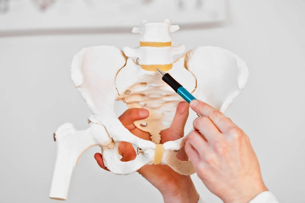
- Load. It carries the weight of the upper body and makes every bend, lift and twist feel stronger. Studies done inside the body show that pressure inside the disc, at L5-S1 can go above 1.5 to 2 times a person’s body weight when standing normally. When bending forward while carrying a load the pressure gets higher.¹
- Geometry. The disc has a wedge shape. It tilts forward at an angle and rests on the sloped top surface of the sacrum. This shape pushes shear forces to the part of the disc’s outer ring. That’s where most disc herniations happen.²
- Transition. The lumbosacral junction is a transition zone between a mobile segment above and a rigid one below, which exaggerates torsional strain on the annulus over decades.
The disc itself works like a cushion. A gel‑like core called the nucleus pulposus sits inside layers of collagen called the annulus fibrosus. When the outer fibers tear, either from a heavy lift or from many small injuries over time the core can push out. It may bulge, protrude, extrude or even separate from the disc. Where it pushes determines which nerves feel pain and what symptoms appear. At this level two nerve roots are in danger. The L5 nerve root leaves the spine above the L5‑S1 disc. Supply the outer part of the leg, the top of the foot and the big toe. If the L5 root is weak the patient drops the foot. Shows a positive Trendelenburg sign. The S1 nerve root leaves below the L5‑S1 disc. Supply the back calf and the outer foot and the sole. When the S1 root is affected the patient has trouble pushing off the toes. Loses the ankle reflex. Which root is hurting depends on whether the herniation’s central, paracentral, foraminal or extraforaminal. These details are very important when planning treatment.
The Classic Symptoms: Back, Leg, and Beyond
Symptoms of an L5-S1 herniation fall into three groups: axial pain, radicular pain and neurologic deficit.²
Axial back pain is often the symptom that people notice. People talk about an ache that sits across the belt line and gets worse when they sit, bend forward, lift something heavy or ride in a car. The pain usually eases when the person lies down. This kind of pain comes from pain fibers in the ring of the disc. It also comes from chemicals that are released when disc material touches the tissues, around it a situation called chemical radiculitis. Because of this a severe pain can happen even if MRI shows a herniation. ²
Radicular pain, also known as sciatica is a sign that a nerve root is compressed or irritated. Radicular pain feels sharp, shooting, burning or electric. Radicular pain follows a dermatomal map:
- L5 radiculopathy: pain radiating down the lateral thigh and calf into the top of the foot and great toe; weakness of ankle and great-toe dorsiflexion; a slapping gait or foot drop in severe cases.
- S1 radiculopathy: pain radiating down the posterior thigh and calf into the lateral foot and small toes; weakness of plantarflexion and difficulty rising on the toes; diminished or absent ankle reflex.²
Neurologic deficit is numbness, weakness or loss of reflexes. Shows that the nerve root is not just inflamed but is actually losing function. When the deficit keeps getting worse or is very bad it changes how we look at treatment. It can turn a patient who might have waited for -surgical care into someone who now needs quick decompression surgery.
How an L5-S1 Herniation Causes Abdominal Pain
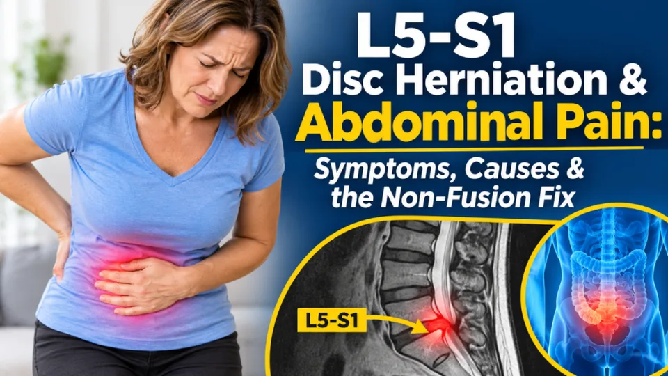
A disc herniation at L5-S1 rarely causes abdominal pain the way, say, appendicitis does. But it is not accurate to say the spine cannot produce abdominal symptoms. Three mechanisms are recognized in the peer-reviewed literature.³,⁴
1. Ventral disc herniation is a type of lumbar disc herniation. Most lumbar herniations push backward either posterior or posterolateral into the spinal canal. That is why leg pain is the symptom that patients notice. Only a small number of cases than 1 percent, in most imaging studies push forward. The forward migrating lumbar disc herniation moves into this area that is present in front of the vertebral body. In this area, the nucleus pulposus that comes out from its usual place may cause either mechanical compression or chemical irritation of the superior hypogastric plexus and sympathetic chain to give rise to deep pelvic pain, cramps in the lower abdomen, abdominal bloating, and in some cases, urgency of micturition, constipation, or sexual dysfunction. This was stated in a case series in 2023 as a type of visceral complex regional pain syndrome.
2. Visceral‑type pain happens when sensory fibers from the spine and from organs meet in the area of the spinal cord. When there is inflammation at the L5‑S1 nerve root the brain may misinterpret the signal. Visceral‑type pain can feel like pain, in the abdomen, groin or front of the thigh. This is known as convergence. The pain can be sharp or burning and real even though imaging of the belly shows nothing. Antispasmodic medications don’t help. Many patients visit gastroenterology, urology and gynecology departments repeatedly before anyone thinks to order a lumbar MRI. ⁴
3. Secondary and Indirect Effects. This is the most frequent type and the one least likely to be talked about. The pain that is chronic at the level of L5-S1 affects the life of the individual – they sit protectively, stand unevenly, breathe shallowly, exercise less, have poor sleep, and usually use increasingly larger doses of opioids, NSAIDs, and muscle relaxers. Each of these has secondary effects on the GI tract – opioid-induced constipation, NSAID induced dyspepsia, visceral hypersensitivity from chronic stress, and decreased motility from lack of physical activity.⁷ A patient suffering from a spinal disorder for months may develop a very real abdominal disorder as a secondary effect of the disease.
Red Flags: When Abdominal and Back Symptoms Become an Emergency
A big herniation at the L5-S1 area can press on the cauda equina, which’s the group of nerves below the spinal cord in the lower back and it can lead to cauda equina syndrome (CES). CES needs surgery. Any of the following symptoms together requires quick visit, to the emergency room:⁵
- Saddle anesthesia: numbness in the inner thighs, perineum, or genital region.
- New bowel or bladder dysfunction: urinary retention, overflow incontinence, loss of the urge to void, or fecal incontinence.
- Bilateral leg weakness or numbness, especially if progressing.
- Sudden sexual dysfunction paired with any of the above.
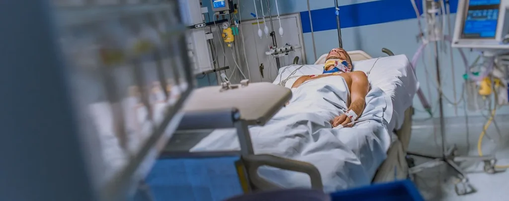
Surgical decompression performed within twenty‑four to forty‑eight hours is linked to recovery of bladder and bowel function than surgical intervention performed later. If any of these signs appear, do not wait for an outpatient MRI. Go to the emergency room.
Red flag abdominal patterns. Sudden severe abdominal pain accompanied by fever, rigidity, syncope, blood in stool or a pulsatile abdominal mass is not related to spinal problems. These red flag abdominal patterns point to vascular emergencies such as appendicitis, perforation or aortic aneurysm. Immediate medical evaluation is required for these red flag patterns, independent of any disc disease.
Getting the Right Diagnosis
A sound L5-S1 diagnosis rests on four pillars: history, physical examination, imaging, and, when abdominal symptoms are present, purposeful exclusion of visceral disease.⁶
History. A board-certified spine specialist should document the location, quality and radiation of pain. They should also note the inciting event if there was one. It is important to record what positions make the pain worse. What positions make it better. Bladder and bowel function must be checked. Any red-flag symptoms need to be identified. Timing matters a lot. Pain that gets worse, with sitting and bending points to a source. Night pain, fever and weight loss are concerning. These symptoms suggest infection or tumor. They change the workup.
Physical examination. I find that a straight‑leg‑raise test that reproduces sciatica below the knee is sensitive, for L5 or S1 radiculopathy. I also perform motor testing of ankle and great‑toe dorsiflexion (L5) and plantarflexion (S1) testing in the dermatomes and reflex testing of the Achilles tendon (S1) and patellar tendon (L3‑L4) to localise the problem.²
Imaging
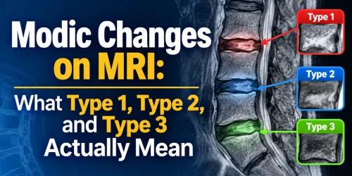
- MRI is the standard. MRI shows the disc, the degree and direction of herniation the nerve-root compression and any associated stenosis or facet disease.⁶
- I use CT when MRI is contraindicated or when bony anatomy must be clarified.
- X-ray adds alignment and stability information. X-ray does not visualize the disc.
- EMG and nerve-conduction studies are reserved for cases in which a competing peripheral nerve lesion, such as diabetic neuropathy or piriformis syndrome must be ruled out.
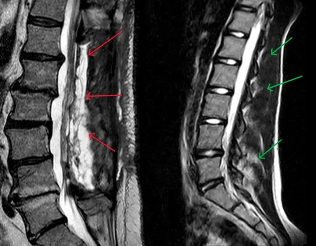
No causes. In case there are abdominal signs, the investigation process shouldn’t end with the MRI scan. The lab tests like CBC, inflammatory parameters, urinalysis, lipase abdominal and pelvic physical exam and if necessary, an ultrasound or CT will prove that the abdominal complaints are caused by referred pain and not by any problems in the organs themselves. Diagnostic discipline is the way to save the patient from abdominal surgery and from its opposite, when L5-S1 herniation is treated instead of abdominal condition.
What Conservative Care Can and Cannot Do
Most patients with an L5-S1 herniation get better without surgery. In studies that look at how the condition progresses about 85% to 90% of lumbar disc herniations improve a lot, within six to twelve weeks when treated with conservative care. A good first-line approach includes:
- Modification of short-term activities with avoidance of bedrest is essential.
- Specific physiotherapy for core stability, hip range of movement and neural mobilization is what we recommend for lumbar flexion exercises.
- Pharmacotherapy includes NSAIDs as anti‑inflammatories where appropriate. A short course of steroids is used for acute radiculopathy. Neuropathics such as gabapentinoids are used when severe neuropathic pain is present. Chronic use of opioids is an amber alert, not a management tool.
- Patients who try conservative treatment, for pain and when pain does not get better after four to six weeks I consider an epidural steroid injection. Epidural steroid injection can help. It only gives short‑term relief and it does not heal the disc.
Conservative care has limits. Conservative care cannot shrink a disc fragment that has moved out of place. Conservative care cannot repair the fibers around the disc. For patients whose pain in the back or legs is caused by pressure and chemical irritation, injections and physical therapy can give relief for a while. Sometimes, for a long time but they do not repair the underlying damage. Patients who stay disabled after a conservative trial, who get weaker over time or who keep a quality of life even with the best non-surgical care deserve a surgical opinion from a specialist who offers a motion-preserving option, not just fusion.
Do injections wear off? Ask about an option.
Repeated epidural steroid injections are a signal, not a solution.
Patients who need a fourth injection, in a year have a structural problem that deserves a surgical evaluation.
Why Laminectomy and Fusion Are Almost Never the Correct First Surgery
The two common treatments available for an L5-S1 herniation in the United States are laminectomy (taking away part of the bone and the ligament to take pressure off the nerve) and spinal fusion (joining the bones of L5 to S1 together permanently often using screws and a cage placed between the bones). Both of these options have a place. Both of these options are almost never the right first choice, for a single L5-S1 disc herniation.
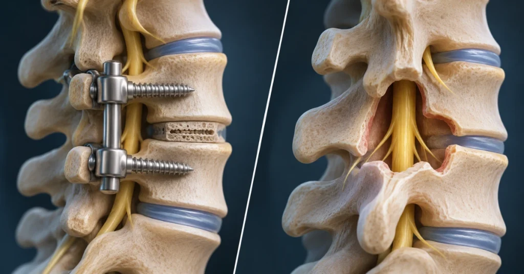
- Laminectomy removes stabilizing posterior structures to reach a disc problem. In many patients, the result is relief of leg pain followed by months or years of mechanical back pain from the altered biomechanics. A recurrence rate of 10% to 25% at the index level has been reported in long-term series.⁸
- Fusion eliminates motion at L5-S1 by design. Motion is not optional. It is how the lumbar spine distributes load. Eliminating motion at one level transfers that load to the levels above and below, which explains why adjacent-segment disease is one of the best-documented complications of fusion, with reported rates of 2% to 4% per year in long-term follow-up. Patients often come back five to ten years later needing another operation at L4-L5.⁹
Fusion is the appropriate operation for true segmental instability, including spondylolisthesis with motion on flexion-extension films, high-grade deformity, trauma, infection, or tumor. It is not the appropriate first operation for a contained or small extruded L5-S1 herniation in a neurologically stable patient. A patient told “you need a fusion” for an isolated L5-S1 herniation should obtain an independent second opinion.
Deuk Laser Disc Repair® (DLDR): A Non-Fusion Alternative
Deuk Laser Disc Repair® (DLDR) is a minimally invasive, non-fusion endoscopic procedure developed at Deuk Spine Institute. It is designed to treat the herniated or torn portion of the disc without removing stabilizing bone, without fusing vertebrae, and without implants.
How it works. Under light sedation and local anesthesia, a narrow working channel is placed through the lateral foramen under live imaging. An endoscope with a side-firing Holmium:YAG laser is introduced to the annular tear. The laser precisely ablates only the extruded or torn disc tissue and seals the annular defect. The healthy nucleus pulposus is preserved. Patients are typically discharged the same day and resume ordinary activity within days, not months.⁸
Who qualifies. DLDR is appropriate for patients whose primary problem is a contained or small extruded L5-S1 disc herniation with radicular pain, or a symptomatic annular tear, who have failed six or more weeks of conservative care. It is not for patients with gross segmental instability, high-grade spondylolisthesis, tumor, or infection. A careful MRI review and a conversation with a surgeon who performs the procedure routinely are the only way to know whether a given disc is a candidate.
Why it matters for patients with abdominal symptoms. Because DLDR is performed from a lateral approach and targets only the pathologic tissue, it is particularly attractive when the mechanism of pain is a ventral or paracentral herniation, which are the same configurations most likely to produce referred abdominal and pelvic symptoms. Preserving motion also avoids the gait and postural changes that drive many of the indirect gut problems described earlier.
Not sure if your disc qualifies? A board-certified spine specialist can review your MRI at no cost and tell you, in plain English, whether you are a DLDR candidate or whether another option is better for your anatomy.
A Patient’s Perspective: Why He Flew to Florida Instead of Accepting Fusion
A recent Deuk Spine Institute patient from the Kansas City, Missouri area traveled out of state this week for a combination of procedures addressing both cervical and lumbar pain that had persisted for four years following a motor vehicle accident. He underwent Deuk Laser Disc Repair® on the lumbar spine on Tuesday, followed the next day by Deuk Laser Disc Repair® on the cervical spine and a Deuk Laser Foraminotomy to decompress the affected facet joints in the lower back.
As an active-duty service member, he had lived with chronic neck and back pain for four years, with the discomfort limiting his ability to perform his military duties and spend quality time with his family. Like many patients who research alternatives before committing to invasive spinal surgery, he sought out a treatment option that could resolve multiple pain generators without the extended recovery, hardware, and adjacent-segment complications associated with traditional fusion.
Less than 24 hours after the second procedure, he reported that the pain and discomfort he had been experiencing had disappeared overnight, even while the surgical sites were still in the earliest phase of healing. He required no fusion hardware, no rods, and no pins across any of the treated levels. Each procedure was performed on an outpatient basis through a small incision using the precision laser technique developed by Dr. Deuk.
His own message to others weighing their options was direct: if you are dealing with back pain, neck pain, or related radicular symptoms, come see Dr. Deuk. He described the experience as life-changing and said he is looking forward to returning to his family and continuing his military service pain-free. Luis Galarza, who has worked at Deuk Spine Institute for the past 12 years, noted that outcomes like this one are consistent with what he has observed across more than a decade of patients choosing endoscopic laser repair over fusion.
Questions to Ask Before You Let Anyone Operate on Your Spine
Before you consent to any operation on your lumbar spine, ask your surgeon the following questions in writing. The answers tell you more than any marketing page.
- Is fusion really necessary for me, or is motion preservation possible? If the answer is “fusion is the only option,” get a second opinion from a surgeon who routinely performs non-fusion procedures.
- What percentage of my disc tissue will be removed? A procedure that removes most of the nucleus destabilizes the segment; one that targets only the herniated fragment preserves function.
- What is your personal complication rate and reoperation rate for this procedure? National averages do not matter. Your surgeon’s numbers do.
- How many of these procedures have you performed in the last 12 months? Volume predicts outcome; a procedure performed once a month is not the same as one performed daily.
- What happens to the levels above and below after fusion? A surgeon who dismisses adjacent-segment disease is not a surgeon to trust with your spine.
- Can I return to work and normal activity in days, weeks, or months? The honest answer tells you the real invasiveness of what is being proposed.
Prevention: Protecting the L5-S1 Disc for Life
Prevention is unglamorous and effective. The L5-S1 disc is remarkably durable when it is loaded well and remarkably fragile when it is not.
- Keep the core strong. Deep abdominal and gluteal strength offloads the discs. Prioritize planks, bird-dogs, dead bugs, and hip bridges over sit-ups.
- Protect the hinge, not the back. Lift from the hips with a neutral lumbar spine; the back is a transmission, not a crane.
- Fix the sitting problem. Prolonged seated flexion is the single most repeatable disc stressor in modern life. Stand or walk at least five minutes every half hour, and set the lumbar support on your chair to maintain a gentle inward curve.
- Keep weight proportional. Every additional kilogram of trunk mass multiplies L5-S1 load through the day. Modest, sustained weight loss unloads the disc more than any supplement.
- Stop smoking. Nicotine reduces disc nutrition and is an independent risk factor for herniation and for poor surgical outcome.
- Sleep well, hydrate, move often. The disc rehydrates overnight; chronic sleep deprivation and dehydration worsen its mechanical properties.
- Address pain early. A new episode of radicular pain that lasts more than two weeks deserves a professional evaluation, not a drawer of ibuprofen.
~95% of lumbar disc herniations happen at L5-S1 — the segment that bears 1.5–2× your body weight with every step
L5-S1 herniations can mimic abdominal disease.
Fusion locks the joint. DLDR preserves motion.
The L5-S1 disc sits at the base of your spine and absorbs more load than any other segment. When it herniates, nerve compression can radiate pain into the groin, pelvis, and lower abdomen—sending patients through rounds of GI and urological testing before anyone checks the spine. Laminectomy and fusion respond by removing bone and locking the segment, with recurrence rates of 10–25% and adjacent-segment disease that compounds over time. DLDR treats only the herniated fragment through a 4 mm incision, preserves full motion, and sends you home the same day.
occur at L5-S1
rate over time
2,800+ procedures
- Bone removed to access the disc
- Segment permanently locked with hardware
- 10–25% recurrence rate
- 2–4% adjacent-segment disease per year
- 3–6 month recovery, prolonged opioid use
- Only herniated fragment treated; healthy disc preserved
- Full L5-S1 motion maintained
- 95%+ success rate, 2,800+ procedures
- 0.01% complication rate
- 4 mm incision, same-day discharge
Board-certified neurosurgeon review · No insurance approval required · 100,000+ procedures performed
FAQ
Can an L5-S1 disc herniation really cause abdominal pain, or is my doctor just missing something in my belly?
Answer
Yes, it can, but only through specific mechanisms, and visceral disease still needs to be ruled out. Ventral herniation, referred pain via dermatomal convergence, and indirect effects of chronic pain all contribute. In reported ventral-herniation series, up to 1% of symptomatic lumbar herniations produce pelvic or lower-abdominal symptoms through sympathetic plexus irritation.³ A proper workup is parallel: a lumbar MRI and an abdominal evaluation, not one or the other.
How do I tell spine-referred abdominal pain from true organ pain?
Answer
Referred spinal pain tends to shift with spinal position. It is worse with sitting or forward bending and better lying flat, and it is not accompanied by fever, rebound tenderness, or abnormal lab values. True visceral pain is usually constant, position-independent, and often paired with GI, GU, or systemic signs. Position-dependent pain that respects a dermatome is rarely intra-abdominal.⁴
Should I wait to seek treatment if my leg pain is severe but my back pain is mild?
Answer
No. Severity of leg symptoms, not back symptoms, is the better predictor of nerve-root compromise. Progressive weakness, foot drop, or saddle numbness is a reason to be seen today, not next month.⁵
How long does recovery take after Deuk Laser Disc Repair® for an L5-S1 disc?
Answer
Most patients walk the same day and return to desk work within three to seven days, with gradual return to full activity over four to six weeks. Because the procedure preserves bone and motion, recovery is faster and less variable than after laminectomy or fusion.⁸
If I have had multiple epidural steroid injections, is it too late to consider a motion-preserving option?
Answer
No. Prior injections do not disqualify a patient from DLDR. In fact, a pattern of injections that work briefly and then wear off is one of the clearer indications that there is a persistent mechanical lesion worth addressing directly.
Sources
View Sources
- Amin RM, Andrade NS, Neuman BJ. Lumbar Disc Herniation. Current Reviews in Musculoskeletal Medicine. 2017;10(4):507-516. https://pmc.ncbi.nlm.nih.gov/articles/PMC5685963/
- Dydyk AM, Massa RN, Mesfin FB. Disc Herniation. StatPearls. NCBI Bookshelf. Updated 2024. https://www.ncbi.nlm.nih.gov/books/NBK441822/
- Aydin SM, Brunetti G, Everest A, et al. Ventral Lumbar Disc Herniation and Visceral Complex Regional Pain Syndrome: A Case Series. Journal of Pain Research. 2023;16:1233-1240. https://pmc.ncbi.nlm.nih.gov/articles/PMC10105593/
- Jin X, Guo W, Zhang J, et al. Mechanisms of Referred Visceral Pain: Convergence of Spinal and Visceral Afferents. Frontiers in Neuroscience. 2022;16:825623. https://pmc.ncbi.nlm.nih.gov/articles/PMC8984263/
- Gardner A, Gardner E, Morley T. Cauda Equina Syndrome: A Review of the Current Clinical and Medico-Legal Position. European Spine Journal. 2011;20(5):690-697. https://pmc.ncbi.nlm.nih.gov/articles/PMC3082683/
- Modic MT, Ross JS. Lumbar Degenerative Disk Disease. Radiology. 2007;245(1):43-61. https://pubs.rsna.org/doi/10.1148/radiol.2451051706
- Benoist M. The Natural History of Lumbar Disc Herniation and Radiculopathy. Joint Bone Spine. 2002;69(2):155-160. https://pubmed.ncbi.nlm.nih.gov/12027305/
- Deukmedjian AJ, Cutright J, Cianciabella A, Deukmedjian A. Deuk Laser Disc Repair: An Alternative to Conservative Therapy for Treatment of Herniated Disc. Cureus. 2017;9(6):e1388. https://pmc.ncbi.nlm.nih.gov/articles/PMC5557670/
- Hilibrand AS, Robbins M. Adjacent Segment Degeneration and Adjacent Segment Disease: The Consequences of Spinal Fusion? The Spine Journal. 2004;4(6):S190-S194. https://pubmed.ncbi.nlm.nih.gov/15541666/