
By Dr. Ara J. Deukmedjian, MD
Board Certified Neurosurgeon
Updated: November 30, 2026.
Disclaimer: This article is for educational purposes only and is not a substitute for a formal medical evaluation. Please consult a professional, for personal advice.
Quick Answer: Modic changes are changes in the bone marrow of a vertebra right next to a spinal disc, seen on MRI. Radiologists sort them into three types: Type 1 (inflamed marrow), Type 2 (fatty marrow), and Type 3 (hardened bone). They are common, describe how the bone looks, and are neither a disease name nor an emergency.
A report line about Type 2 endplate changes can read like a warning on a first pass. The name comes from Dr. Michael Modic, the radiologist who classified these patterns. This guide covers what each type means, how it reads on MRI, whether it causes pain, and what helps.
What Are Modic Changes?
The term describes signal changes in the bone marrow of a vertebra, just behind the endplate beside a disc. The endplate is a thin layer of bone between the disc and the vertebral body. Behind it sits the marrow, which is the soft inner tissue of the bone. Modic endplate changes appear on an MRI as shifts in how that marrow looks.
The finding sits in the bone rather than the disc. Many readers assume the opposite, yet every later point in this guide rests on this distinction.
The answer to what is modic is simple: it is the surname of the radiologist who described these marrow patterns with colleagues in 1988. Radiologists still use his three-type system. The glossary of other findings on your MRI report explains the related terms in plain language. Schmorl’s nodes are a separate endplate finding that people frequently confuse with this one.
How Modic Changes Show Up on an MRI
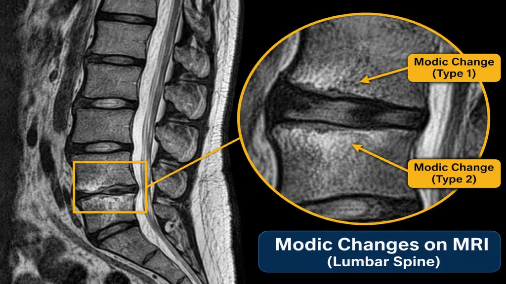
An MRI is the one scan that reveals these marrow changes, because it reads the water and fat content inside bone. X-ray and CT scans lack that ability, so the marrow signal stays invisible on those images.
MRI uses two common image settings, called T1 and T2, to show tissue in different shades. Inflamed, swollen marrow looks one way across those images. Fatty marrow looks another way, while dense scarred bone looks a third way. Those three patterns are the entire basis of the three types.
Radiologists usually describe Modic changes on MRI in short technical phrases, so the report can feel hard to read. Common examples appear below.
- “Modic type 2 changes at L4-L5” points to fatty marrow beside the disc between the fourth and fifth lumbar vertebrae
- “Endplate marrow signal change” means the marrow behind an endplate looks different from normal bone on the scan
- “Degenerative endplate changes” describes a marrow shift tied to wear in the neighboring disc
Reports frequently mention disc findings on the same line. A separate guide covers how a disc bulge or herniation is described.
The Three Types of Modic Changes
Modic types describe how the marrow looks rather than the pain level. The marrow may appear inflamed, fatty, or sclerotic (hardened). Modic type changes are the MRI changes seen in the bone marrow next to a vertebral endplate. A Modic type can change over time, but the changes do not follow a fixed Type 1 → 2 → 3 path. For example, Modic type 1 and 2 endplate changes can shift in either direction, and some stay the same across follow-up scans.
Modic Type 1: Active Inflammation
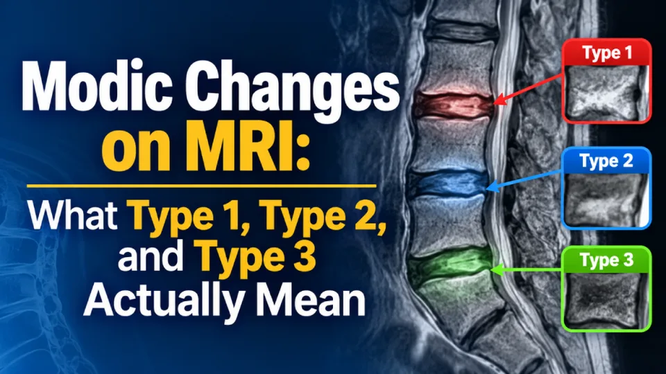
Type 1 appears dark on T1 images and bright on T2 images. This pattern describes marrow that is inflamed and swollen, with extra fluid and an increased blood supply. In plain terms, the bone behind the endplate is irritated and holds more water than usual.
Of the three types, Type 1 shows the most consistent link to back pain in the research. A review in the Journal of Clinical Orthopaedics and Trauma reports this association. Even so, the link describes a pattern across groups of patients rather than a verdict on one person.
For a reader with a Type 1 note on a report, the practical step is a review of symptoms alongside the images. Pain history, exam findings, and the scan together help show whether this marrow change matters in a given case.
Type 1 is the least stable of the three and can change over time, such as a conversion to Type 2.
Modic Type 2: Fatty Marrow Change
Type 2 appears bright on T1 images, while T2 images show it as bright or the same as nearby normal tissue. Fat has replaced the inflamed marrow in this type, and fat shows up brightly on T1 images. Under a microscope, the finding is simply fat replacement.
Modic type 2 endplate changes are the most commonly seen of the three types, and specialists generally view them as a more stable, quieter state of the marrow. Type 2 is a different pattern from Type 1 rather than a worse or more advanced stage. That stability offers a good measure of reassurance to readers who find it on a scan.
A study in BMC Musculoskeletal Disorders adds two details worth knowing. It found that Type 2 was the predominant type, seen most frequently at the L4 and L5 levels of the lower back. The authors also link the changes with body fat and its distribution, which points to a possible metabolic factor.
On its own, this finding says little about pain levels. The rest of the scan, along with symptoms and a physical exam, helps show whether the level matters clinically. Treatment options for this type appear in the treatment section further down this page.
Modic Type 3: Sclerotic Bone
Type 3 appears dark on T1 and T2 images alike. Here, dense, hardened bone has replaced the marrow, and radiologists call this change sclerosis.
Modic type 3 endplate changes are the least common of the three types. They are also the most likely to appear alongside advanced degeneration at the same spinal level. Degeneration here refers to wear in the disc over time.
A Type 3 finding still deserves a clear explanation from a specialist. That review places the dense bone in context with the disc, the nearby joints, and the symptoms at that level.
Do Modic Changes Actually Cause Back Pain?

The honest answer is sometimes, because these marrow changes can relate to back pain in some people and cause no symptoms in others. An MRI report alone leaves the cause of pain an open question. According to Physiopedia, these changes appear in 18 to 62 percent of clinical populations, a wide range across studies. Prevalence means the share of people in a group who have a finding, so the range shows how widespread the changes are.
A single report can list several findings at one spinal level:
- A Modic signal change in the marrow beside the endplate
- A degenerated disc with lost height or water content
- Facet joint wear, which falls under spinal arthritis
Which of these structures drives the pain is a clinical question. The report describes how each structure looks, while a specialist exam connects those images to real symptoms.
The disc beside the marrow change is frequently the actual pain generator. A common culprit is an annular tear, which is a crack in the tough outer ring of the disc. A careful evaluation weighs the disc and the endplate together before any conclusion about the cause.
What Modic Changes Feel Like
Pain tied to the endplate is commonly described as a deep, central ache in the low back rather than sharp or shooting pain. It may feel worse with long periods of sitting or standing still. Some people also notice it more in the morning or at night.
Nerve-driven pain follows a different path and travels down into the leg. That pattern points to another source, which the guide to other causes of chronic back pain explains further.
Some symptoms call for emergency care rather than a routine appointment. These include new weakness, numbness in the saddle area around the groin and inner thighs, loss of bladder or bowel control, or fever with back pain. Fever deserves special attention here, since infection is one possible explanation for endplate changes.
Worth a specialist conversation if:
- A deep, central ache in the low back keeps returning
- Pain feels worse with long periods of sitting or standing still
- Discomfort stands out in the morning or at night
- An MRI report mentions an endplate change beside a worn disc
What Causes Modic Changes
These changes develop alongside disc degeneration, which is the main driver. As a disc dries out and loses height, more load shifts onto the endplate. The marrow behind the endplate then reacts to that extra stress. The medical term for a dried-out disc is disc desiccation, and it frequently appears on the same report.
Physiopedia lists several factors associated with these changes, such as age, male sex, smoking, diabetes, higher body weight, and genetics. The link is an association, so research has yet to show that any of them causes the change.
Researchers have also proposed a bacterial theory for some cases, and the idea remains under debate. The treatment section explains what the antibiotic evidence shows.
Since these changes follow disc wear, they frequently appear in people with degenerative disc disease. Some reports also describe changes at more than one level of the spine, which calls for a review of each segment on its own.
Treatment: What Actually Helps
No current treatment targets the marrow signal itself. Care aims at the pain and the structure behind it. For that reason, the source of pain matters more than the Modic type on the report.
Conservative Care First
Options for Modic type 2 endplate changes treatment usually starts with conservative care, such as physical therapy and activity changes. Common options include:
- Physical therapy under the guidance of a licensed therapist
- Graded activity, which raises movement in small, planned steps
- Load management that reduces strain on the affected level
- Short-term anti-inflammatory medicine on a doctor’s advice
- Support to quit smoking or reach a healthier weight where relevant
These options aim to ease pain and restore everyday movement. Progress with this approach tends to build gradually over weeks or months rather than days. A care team can adjust the plan if improvement stalls.
When the Disc Beside the Change Is the Pain Generator
When an evaluation points to the disc at that level rather than the endplate, the conversation shifts to that disc. Many people are unaware of disc-focused options that sit between conservative care and invasive open-back surgery.
Deuk Laser Disc Repair is one such option worth a discussion with a specialist. The procedure uses an endoscopic laser, which travels through a thin camera-guided tube to the inflamed tissue inside the damaged disc. The treatment involves no bone removal or segment fusion, and the structure of the spine stays intact.
The procedure targets the disc, so the marrow change in the neighboring bone remains a separate finding. When the endplate itself drives the pain, a different care pathway applies.
What the Evidence Does Not Support
Current evidence offers no support for routine antibiotics in the care of these marrow changes. That holds true despite the bacterial theory that circulates widely online. Any decision about antibiotics belongs with a doctor who knows the full case.
Other procedures exist that target the endplate pathway specifically. A specialist evaluation decides the right route, since the source of pain shapes which option fits. This approach keeps the focus on the true pain generator rather than one procedure for every patient.
FAQ
Are Modic changes permanent, or can they change over time?
Answer
They can change over time, especially Type 1, which is the least stable type. Type 1 can convert to Type 2, while Type 2 is generally the more stable state. A follow-up MRI shows whether any shift has occurred.
Are Modic changes a form of arthritis?
Answer
Not in the usual sense, since radiologists report them as a marrow finding beside a disc, separate from arthritis. Arthritis refers to wear in joints, such as the facet joints at the back of the spine. A report can still list the two findings at the same level.
Can Modic changes be seen on an X-ray or CT scan?
Answer
MRI is the scan that reveals them, since it reads water and fat content in bone marrow. X-ray and CT images lack that view of marrow content, so these changes stay hidden on those scans.
Do Modic changes mean I need surgery?
Answer
A Modic finding alone points to no specific treatment, surgical or otherwise. Care depends on the structure behind the pain, with conservative care as the usual first step. A specialist review can explain which options fit a particular case.
Are Modic changes serious?
Answer
They are common findings that describe the bone, rather than a disease or an emergency. Their importance depends on symptoms and the other findings at that level. Fever, new weakness, or loss of bladder control with back pain calls for emergency care.
Can you exercise or stay active with Modic changes?
Answer
In general, yes, and graded activity forms part of standard conservative care. A physical therapist can shape an exercise plan around pain levels and the scan findings. A sharp rise in pain during exercise is a reason to check back with a clinician.
Conclusion
Modic changes are common MRI findings in the bone beside a disc. The type describes the state of that bone rather than how serious the condition is. The more useful question is which structure at that level generates the pain, since that answer guides treatment.
For anyone with a report in hand, a free MRI review offers a low-pressure next step. A specialist can walk through the findings and explain which options fit, so the next decision feels clearer.
Modic changes appear in up to 62% of back-pain patients — but the disc beside the marrow change is frequently the actual pain generator, not the bone itself
Your MRI says “Modic changes.” The real question is
which structure is driving your pain.
Modic changes describe the bone marrow beside a disc—not a diagnosis and not an emergency. The disc at that same level is often the true pain generator, especially when an annular tear or herniation sits on the same report line. Fusion responds by removing the disc entirely and locking the segment with hardware. DLDR targets only the damaged disc tissue through a 4 mm incision, preserves the spine’s natural motion, and sends you home the same day.
Modic changes on MRI
often at L4–L5
2,800+ procedures
- Entire disc removed & replaced with hardware
- Segment permanently locked
- Adjacent-segment disease risk compounds yearly
- Extended recovery & prolonged opioid use
- Does not address the marrow change itself
- Only damaged disc tissue treated; healthy disc preserved
- No bone removed, no vertebrae fused
- 95%+ success rate, 2,800+ procedures
- 0.01% complication rate
- 4 mm incision, same-day discharge
Board-certified neurosurgeon review · No insurance approval required · 100,000+ procedures performed