


By Dr. Ara Deukmedjian, MD
Board-Certified Neurosurgeon
Medically reviewed on Aug 6, 2026
Medical Disclaimer: This content is for educational purposes only and does not constitute medical advice. Individual results may vary. Always consult with your healthcare provider about your specific condition and treatment options.
Key Points
✓ PS involves compression of the sciatic nerve in the buttock region, resulting in sciatica-like symptoms and mimicking a lumbar disc herniation. ¹ ²
✓ Affects 5–8% of low back and sciatic pain cases; female to male ratio of 6:1. ³ ⁴ ¹ ²
✓ Often confused with lumbar radiculopathy, disc herniation, or SI joint dysfunction due to lack of a gold standard diagnostic test. ¹ ⁵ ⁶
✓ MR neurography demonstrates 93% specificity for piriformis asymmetry and hyperintense sciatic nerve. ⁸
✓ Conservative treatment (PT, NSAIDs, injections) controls symptoms, however, it is not a cure for PS as it is often impossible to completely relieve pain using conservative measures. ¹ ³ ¹⁰ ¹¹
✓ It is estimated that about 99% of piriformis injuries will not heal without treatment because of the constant mechanical loading of the muscle. ²⁴
✓ Deuk Piriformis Release® – patented minimally invasive outpatient procedure that cures piriformis syndrome with only a 4 mm incision providing 99% pain relief, 0% complications and 30 minutes of recovery time. ²⁴
What Is Piriformis Syndrome?
The clinical name for this syndrome is ‘piriformis syndrome‘. Wherein the sciatic nerve gets entrapped at the level of ischial tuberosity due to the compression and irritation of the piriformis muscle. ¹ ² The piriformis muscle is the flat and pyramidal shaped muscle that lies deeply in the buttocks and arises from the anterior surface of sacrum and inserts onto the greater trochanter of the femur. ¹ This muscle helps to rotate the hip joint externally in extended hip joint and abduction of hip joint in flexed hip joint.
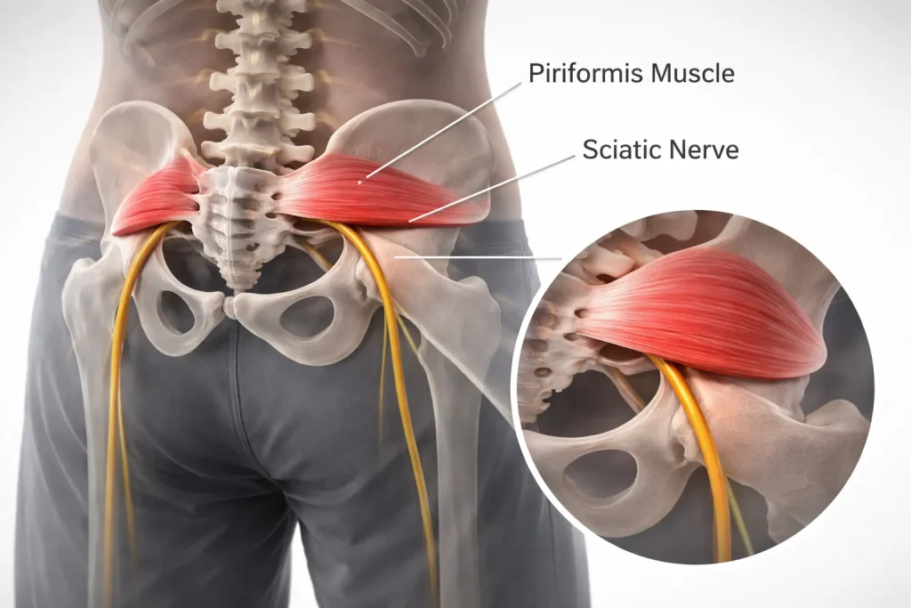
As this nerve is the largest nerve in the peripheral nervous system, it either passes close to or even inside the piriformis muscle in some people. ¹ ⁹ Due to the irritation, inflammation, enlargement, or spasm of the piriformis muscle, this sciatic nerve gets compressed, leading to a condition similar to the disc herniation and radiculopathy.
This syndrome has traditionally been known under other terms such as deep gluteal syndrome, extra-spinal sciatica, and wallet neuritis. ² Now the literature on this subject is starting to use the term of DGS for the classification of the piriformis syndrome as one of its subtypes. DGS is described as an umbrella term covering all causes of sciatic nerve entrapment in the deep gluteal space, and not only the one caused by the piriformis muscle. ¹⁵ ¹⁶
How common is piriformis syndrome?
The exact prevalence of piriformis syndrome is still being argued, mainly because of the lack of a gold standard test for its diagnosis. ¹ ³ The prevalence rates of patients with low back pain and sciatica among those who suffer from piriformis syndrome are 0.3% to 36% based on various criteria used for diagnosing this condition. ³ According to several reviews, the prevalence of PS among patients with low back and sciatic-type pain ranges from 5% to 8%. ⁴ ⁶

A meta-analysis performed in 2026 resulted in the pooled prevalence of 46.79% of patients with buttock pain and low back radiation pain; however, as the presence of rigorous criteria was absent in 96% of studies, the result should be carefully treated. ¹⁷
The condition predominantly affects middle-aged individuals and is significantly more common in women, with a reported female-to-male ratio of 6:1. ¹ ² This gender disparity may relate to biomechanical differences in the female pelvis, including a wider Q-angle that places increased stress on the piriformis during hip movement.
A 2025 systematic review of published case reports (1980–2024) collected data from 212 patients (117 females, 95 males, mean age 43.6 ± 14.8 years), confirming the characteristic demographic profile of the condition. ⁵
Causes of Piriformis Syndrome
Piriformis syndrome can arise from a variety of mechanisms. StatPearls classifies these causes into two broad categories: primary causes related to intrinsic pathology of the piriformis muscle itself, and secondary causes resulting from external factors that lead to piriformis irritation or sciatic nerve compression. ¹
1. Muscle Overuse and Repetitive Strain
Overuse injury is the most prevalent etiology behind the occurrence of piriformis syndrome among athletes. ¹ ³ Endurance athletes such as runners and cyclists are at risk due to their hip’s repeated actions of rotating and flexing and extending, resulting in the irritation of the piriformis and other surrounding structures. The piriformis is put under strain whenever there is an internal hip rotation.
2. Prolonged Sitting
Extended periods of sitting. Whether at a desk, in a vehicle, or during air travel. Place direct compressive force on the piriformis muscle and the underlying sciatic nerve against the ischial tuberosity. ¹ ² ³ This is one of the most frequently reported triggers. Patients commonly describe worsening buttock pain after sitting for more than 20 –30 minutes, which is often the first symptom that brings them to clinical attention.
3. Trauma and Injury
Direct injury to the gluteal muscles, such as a fall on the buttocks, a car accident, or an impact during sporting activity, may result in irritation, bleeding, or scar formation within the piriformis muscle. ¹ This may cause fibrosis and adhesion that fixes the sciatic nerve to the adjacent soft tissues. Post-traumatic piriformis syndrome is a known condition, and gluteal trauma is an important clue to diagnosis.
4. Anatomic Variants of the Sciatic Nerve
In approximately 85% of the population, the sciatic nerve passes beneath the piriformis muscle as a single trunk. ⁹ ¹⁸ However, anatomic variants exist in roughly 13% of individuals, with the most common variant involving the common peroneal division of the sciatic nerve piercing directly through the piriformis muscle while the tibial division passes below it. ⁹
A 2020 systematic review and meta-analysis of 44 cadaveric studies found that the typical pattern (nerve passing undivided below the muscle) had a 90% pooled prevalence, while total sciatic nerve variants had a 13% pooled prevalence. ⁹ East Asian populations demonstrated the highest prevalence of variants at 31%. ⁹ Although these variants are commonly associated with piriformis syndrome, at least one MRI study of 783 cases found no significant difference in the prevalence of piriformis syndrome between normal and variant sciatic nerve anatomy. ¹⁸
5. Piriformis Muscle Hypertrophy or Spasm
Chronic spasm, hypertrophy, or contracture of the piriformis muscle can reduce the space available for the sciatic nerve at the greater sciatic notch. ¹ ³ MRI and ultrasound studies in patients with piriformis syndrome frequently demonstrate asymmetric enlargement of the piriformis muscle on the affected side compared to the unaffected side. ⁷ ⁸
6. Other Contributing Factors
Additional factors that can contribute to piriformis syndrome include sacroiliac joint dysfunction, leg length discrepancy, abnormal gait patterns, hip surgery or arthroplasty, pelvic masses or tumors that compress the piriformis, inferior gluteal artery aneurysms, and fibrous bands within the deep gluteal space. ¹ ¹⁵ ¹⁶
Symptoms of Piriformis Syndrome
The clinical presentation of piriformis syndrome is fairly consistent across patients, though the severity and combination of symptoms can vary considerably. ¹ ² ³
1. Deep Buttock Pain
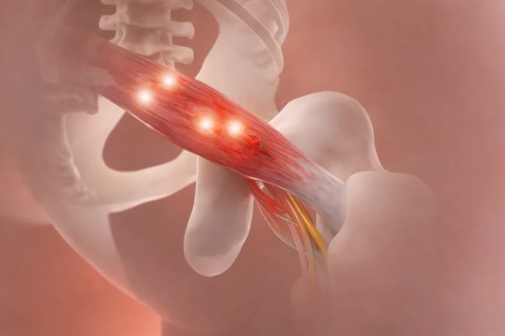
The characteristic sign of piriformis syndrome is deep and achy pain felt centrally in the gluteal area, usually unilaterally. ¹ ² ³ Patients may feel deep pain that is hard to locate exactly. This pain tends to worsen with long periods of sitting, going up stairs, squatting, or doing anything involving hip rotation. Walking may intensify the pain in acute cases, though in chronic piriformis syndrome, ambulation may actually lessen symptoms temporarily. ³
2. Sciatica-Like Radiating Pain
Pain frequently radiates from the buttock down the posterior thigh, following the distribution of the sciatic nerve. ¹ ² The pain may extend to the calf and foot in some patients, closely mimicking lumbar disc herniation or radiculopathy. The radiating component may be described as shooting, burning, or aching in quality.
3. Numbness and Tingling
Paresthesia’s including numbness, tingling, and pins-and-needles sensations can occur along the distribution of the sciatic nerve, affecting the buttock, posterior thigh, calf, and foot. ¹ ² However, true neurologic deficits (motor weakness, reflex changes) are less common in piriformis syndrome than in lumbar radiculopathy and may be completely absent.
4. Difficulty Sitting
Intolerance to prolonged sitting is one of the most consistently reported features. ¹ ² ³ Patients frequently report that they need to stand up, shift positions, or avoid sitting on the affected side altogether. This is sometimes called the “wallet sign,” as sitting on a thick wallet in the back pocket can directly compress the piriformis and exacerbate symptoms.
5. Hip and Pelvic Symptoms
Some cases involve stiffness of the hip, decreased range of motion of the hip joint, or pain when internally rotating the hip joint. ³ The patient may suffer from referred pain to the groin, perineum, and/or sacroiliac areas. Defecation pain and coccygeal pain can be found due to the anatomical relation of the piriformis muscle to the pelvis.
6. Worsening with Specific Movements
Symptoms are usually triggered by movements involving the stretching and contracting of the piriformis muscle, such as climbing stairs, sitting with crossed legs, squatting, running, and walking for long periods. ¹ ² ³ Relief from symptoms can be achieved through lying down with the legs bent and spread apart.
Why Piriformis Syndrome Is Often Misdiagnosed
Piriformis syndrome is the most frequently un-diagnosed and misdiagnosed medical condition out of all the musculoskeletal and nervous system diseases. ¹ ⁵ ⁶ There are multiple causes for this misdiagnosis and poor treatment that arise due to diagnostic delay resulting in chronic pain, hyperesthesia, paresthesia, and muscle weakness. ¹⁹

1. Symptoms mimic lumbar disc herniation
The sciatica-like pain pattern of piriformis syndrome closely resembles that of a herniated lumbar disc compressing the L5 or S1 nerve root. ¹ ⁵ Without careful clinical differentiation, many patients receive unnecessary lumbar MRIs, epidural injections, or even spinal surgery for a condition that originates in the buttock, not the spine.
2. No gold-standard diagnostic test exists
Unlike a herniated disc (which is clearly visible on MRI), piriformis syndrome lacks a single confirmatory test. ¹ ³ ⁷ Diagnosis relies on a combination of clinical history, provocative physical examination maneuvers, and the exclusion of spinal pathology. All of which require a high index of clinical suspicion.
3. The condition is not well-covered in medical training
Piriformis syndrome receives relatively little attention in standard medical school and residency curricula. ⁵ ⁶ Many physicians default to spinal explanations for sciatica-pattern pain and may not consider extra-spinal causes unless imaging of the lumbar spine is unrevealing.
4. Imaging findings can be subtle or absent
Standard MRI of the pelvis may appear entirely normal in patients with piriformis syndrome. ⁷ ⁸ Advanced imaging techniques such as MR neurography, which can identify piriformis asymmetry and sciatic nerve hyperintensity, are not routinely ordered and may not be available at all imaging centers.
5. Multiple conditions can coexist
Piriformis syndrome can occur simultaneously with lumbar disc disease, sacroiliac joint dysfunction, hip osteoarthritis, and other conditions, making it difficult to isolate the piriformis as the primary pain generator without targeted diagnostic injections.
Differential Diagnosis for Piriformis Syndrome
Buttock pain with radiating leg symptoms can arise from several conditions beyond piriformis syndrome. Here is how each condition distinguishes itself.
What Happens If Piriformis Syndrome Is Left Untreated?
Despite the fact that piriformis syndrome cannot be compared with spinal cord compression in terms of the seriousness of its consequences, the neglect of this condition can have extremely negative effects on the function of the body. ¹ ¹⁹
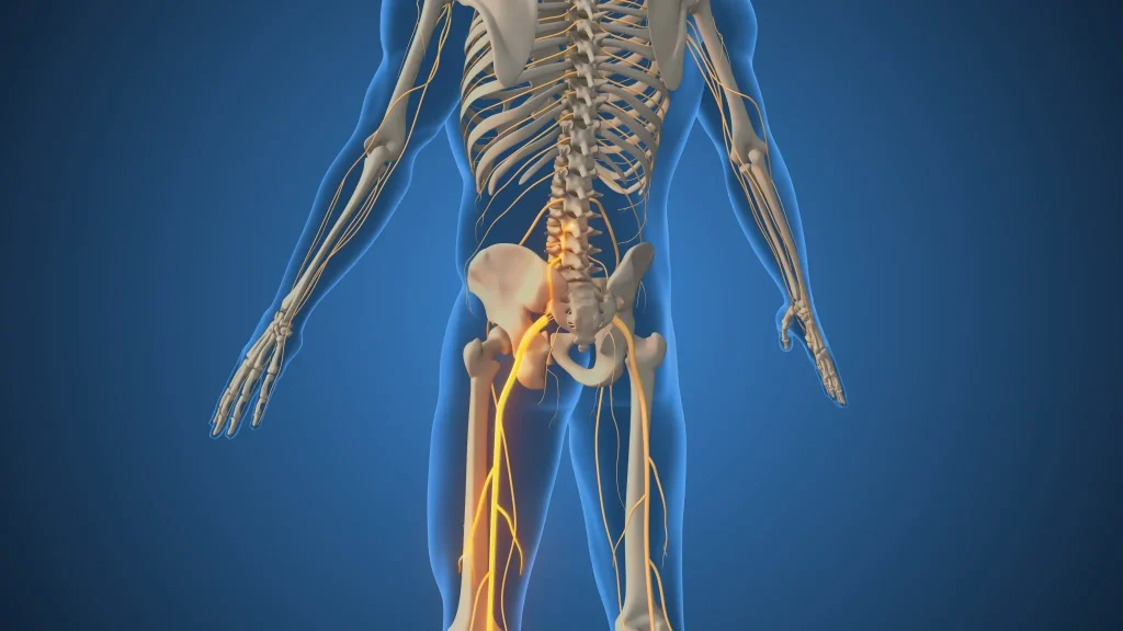
- Chronic pain syndrome. In case of prolonged irritation and compression of the sciatic nerve, they may develop neuropathic pain syndrome which is rather hard to eliminate. ¹ ¹⁹
- Muscle weakness and atrophy. Prolonged irritation and compression of the sciatic nerve may cause weakness of muscle innervated by this nerve, such as muscles of the legs and foot and even cause foot drop. ¹ ⁵
- Gait abnormalities. Chronic pain and weakness can alter gait mechanics, leading to compensatory movement patterns that place additional stress on the hip, knee, and lumbar spine. ¹
- Activity limitation and disability. Patients with untreated piriformis syndrome frequently report significant limitations in sitting tolerance, walking endurance, exercise capacity, and participation in work and recreational activities. The condition accounts for many cases of partial or total disability. ¹⁹
- Fibrosis and adhesions. Chronic inflammation around the piriformis muscle and sciatic nerve can produce fibrosis and adhesions within the deep gluteal space, making the condition progressively more difficult to treat with conservative measures and potentially necessitating surgical intervention. ¹⁵ ¹⁶
Diagnosing Piriformis Syndrome
Diagnosis of piriformis syndrome is mainly a clinical one. No lab investigation, no imaging test, no electrodiagnostic study can diagnose this condition. ¹ ³ ⁷ This diagnosis is made based on a thorough evaluation of the history, physical examination, imaging studies to rule out spinal disorders, and, in some cases, diagnostic blocks.
The diagnostic sequence
1. Comprehensive history and physical examination. The clinician evaluates the onset, location, quality, and aggravating/relieving factors of the pain. Key historical features include deep buttock pain worsened by sitting for more than 20–30 minutes, radiating pain in the sciatic distribution, and symptom reproduction with activities involving hip rotation. ¹ ² ³
Physical examination includes palpation of the piriformis (tenderness on deep palpation of the buttock is found in up to 92% of patients with piriformis syndrome), ²⁰ assessment of hip range of motion, and a series of provocative tests:
- FAIR test (Flexion, Adduction, Internal Rotation): Reproduction of sciatic-type symptoms with passive flexion, adduction, and internal rotation of the hip. This is the most widely used provocative test. ¹ ³ ⁷
- Pace sign: Pain and weakness on resisted abduction and external rotation of the hip while seated. ³
- Freiberg test: Pain on passive forced internal rotation of the extended hip, stretching the piriformis. ³
- Beatty test: Pain when the patient, in the lateral decubitus position, holds the knee of the affected side several inches off the table. ³
- Active piriformis test: One study evaluating diagnostic accuracy against endoscopically confirmed sciatic nerve entrapment found the active piriformis test to have a sensitivity of 78% and specificity of 80%. ²¹
2. MRI of the lumbar spine (to exclude spinal pathology). The main reason why MRI is done in the work-up of possible piriformis syndrome is to rule out lumbar disc protrusion, spinal stenosis, or any other spinal cause of sciatica. ¹ ⁷ Normal lumbar MRI results when there is typical piriformis syndrome can confirm the diagnosis.
3. MRI or ultrasound of the pelvis/hip. The regular pelvic MRI can reveal hypertrophy, asymmetry, and edema of the piriformis on the side where there is pain. ⁷ ⁸ In one study from 2025, piriformis muscle cross-sectional area (PMCSA) was found to be a more sensitive diagnostic criterion than the thickness of the piriformis muscle, with an AUC of 0.81, 75% sensitivity, and 75% specificity at the cutoff point of 611.67 mm². ⁷
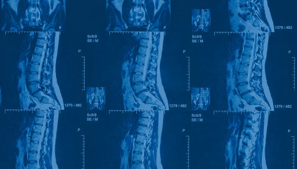
Special MRI technique called the MR neurography is known to have 93% specificity in revealing piriformis muscle asymmetry and sciatic nerve hyperintensity in the sciatic notch. ⁸ But its sensitivity is only 64%, which means that the negative result doesn’t rule out the presence of this condition.
Diagnostic musculoskeletal ultrasound examination can be considered as a helpful method in diagnosing piriformis syndrome. ²²
4. Electrodiagnostic studies. Electromyography (EMG) and nerve conduction studies (NCS) can help differentiate piriformis syndrome from lumbar radiculopathy and peripheral neuropathy. ¹ The most useful electrodiagnostic finding is prolongation of the H-reflex during the FAIR position, which indicates sciatic nerve conduction slowing at the level of the piriformis. ⁶
5. Diagnostic injection. An image-guided injection of local anesthetic (with or without corticosteroid) directly into the piriformis muscle can serve as both a diagnostic and therapeutic tool. ¹ ¹⁰ ¹¹ Significant pain relief following a targeted piriformis injection strongly supports the diagnosis.
Non-Surgical Treatment of Piriformis Syndrome
Conservative management is the first-line approach for piriformis syndrome and resolves symptoms in the majority of patients. ¹ ³ ¹⁰ A structured, multimodal approach typically produces the best outcomes.
Physical therapy and targeted stretching
The combination of physical therapy involving piriformis muscle stretching, hip strengthening, and core stabilizing exercises forms the backbone of conservative management. ¹ ³ ¹⁰ It is recommended to include deep stretching of the piriformis muscles, hip extensor strengthening exercises, hip abductor strengthening exercises, hip external rotator muscle strengthening exercises, core stabilizing exercises, and neuromobilization of the sciatic nerves.

Patients should be prescribed specific piriformis stretching exercises rather than generic low-back programs. Stretching positions that combine hip flexion with adduction and internal rotation have been shown to effectively elongate the piriformis muscle.
Medications
- Non-steroidal anti-inflammatory drugs (NSAIDs). Ibuprofen, naproxen, or celecoxib are first-line agents for reducing inflammation and pain.
- Muscle relaxants. Short-term use of cyclobenzaprine or tizanidine can help manage piriformis spasm.
- Neuropathic pain medications. Gabapentin or pregabalin may help when significant neuropathic (burning, tingling) symptoms are present.
Activity modification
Prolonged sitting should be avoided. And the patient should refrain from sitting on any hard surfaces such as a wallet in his back pocket. The patient can use a seat cushion for comfort. While sitting and should change the exercises that cause pain in the piriformis muscle.
Image-guided injections
In cases where physical therapy and medication have not been sufficient enough, the injection of corticosteroid or botulinum toxin into the piriformis muscle can help:
- Corticosteroid injections. This is the injection of corticosteroid and local anesthetic under the guidance of fluoroscopy, ultrasound, or computed tomography to reduce the inflammatory processes in the area, which allows for better participation in physical therapy. ¹ ¹⁰ ¹¹
- Botulinum toxin (Botox) injections. A systematic review suggests that injections of botulinum toxin into the piriformis muscle are effective for the reduction of pain. ¹¹ The dosage is usually from 100 to 300 units. ¹¹ A study conducted by the UT Southwestern researchers has shown that CT-guided injections of botulinum toxin resulted in more effective response than non-botulinum toxin injections. ¹²
- Pulsed radiofrequency (PRF). PRF targeting the sciatic nerve under imaging guidance is an emerging option for refractory cases. A 2025 propensity score-matched study comparing PRF and endoscopic piriformis release found that both provided meaningful pain reduction at 3 and 6 months. ²³
When conservative care is not enough
Nonsurgical treatment may be reconsidered if symptoms continue for 3 months or longer of appropriate conservative treatment, if there is severe pain that significantly affects daily activities, if there are progressive neurologic deficits such as weakness or foot drop, or if an injection has localized the source of pain to the piriformis muscle but the effect is not long-lasting.
Surgical Treatment for Piriformis Syndrome
Surgical treatment of piriformis syndrome is indicated only after the failure of a full course of conservative treatment consisting of physical therapy, medication, and injection therapy. ¹ ¹³ ¹⁴ In case of surgery, there are two main procedures that can be done, which are open piriformis release and endoscopic piriformis release with sciatic neurolysis.
Open Surgical Release
The conventional open surgery method entails full release of the piriformis tendon from its attachment to the femur at the greater trochanter. ¹ ¹⁹ The sciatic nerve neurolysis procedure, which entails freeing up the nerve from adherent scar tissues, can be done concurrently in cases of substantial fibrosis. Despite being highly efficacious, open surgery is more invasive than endoscopic surgery methods.
Endoscopic Piriformis Release and Sciatic Neurolysis
Endoscopic piriformis tendon release is a method that is favored for surgical management of resistant cases of piriformis syndrome. ¹³ ¹⁴ ¹⁵ It entails insertion of arthroscopic instruments into a number of small openings in order to explore the deep gluteal region and relieve the sciatic nerve from adhesions by releasing the piriformis tendon.
The following study on the effectiveness of endoscopic sciatic neurolysis and piriformis tendon release is expected to be published in Arthroscopy in 2025. The investigators reported favorable patient-reported outcomes, high satisfaction rates, and a high percentage of patients reaching clinically important thresholds, with a low rate of complications. ¹³
An earlier series using the Benson operative scale found that outcomes were excellent in 53.4% of patients and good in 22.2%, with 88% of patients reporting satisfaction at a mean 24-month follow-up. ¹⁴ Notably, the three patients with poor results in that series were identified as having incorrect diagnoses, underscoring the critical importance of precise preoperative diagnosis.
Key advantages of the endoscopic approach over open surgery include smaller incisions and reduced tissue disruption, direct visualization of the sciatic nerve and all structures in the deep gluteal space, the ability to identify and address additional causes of deep gluteal syndrome (fibrous bands, vascular compression, gemelli-obturator internus pathology) during the same procedure, ¹⁵ ¹⁶ faster recovery and earlier return to activity, and lower complication rates compared to open surgery.
A 2025 propensity score-matched cohort study between endoscopic piriformis release (EPR) and pulsed radiofrequency demonstrated superior and more sustained pain relief in the former at 6 months although both modalities delivered clinical improvement. ²³
Why a Second Opinion Can Change Your Outcome
By far the most critical thing one can do as a patient with presumed piriformis syndrome is to be seen by a physician that recognizes extra-spinal reasons for sciatic pain and has expertise in diagnosing and treating piriformis syndrome.
Since piriformis syndrome is a diagnosis by exclusion and presents very similar to many other much more common disorders, it is often overlooked, and many patients get unnecessary procedures and interventions on the lumbar spine while the cause of their pain is still unknown. ¹ ⁵ ⁶
If you have been advised to have surgery on your lower back due to sciatica, but your pain is centered in the buttocks, aggravated by sitting down and is not adequately explained by your spinal radiology results, then it is advisable to seek a second opinion.
Stop living with piriformis pain.
Get back to your life.
Piriformis syndrome accounts for up to 8% of all sciatica cases — and is one of the most misdiagnosed conditions in medicine. If you’ve been told you need back surgery but your pain is in the buttock, you may have the wrong diagnosis.
Patented, minimally invasive, outpatient procedure · Board-certified neurosurgeon · Thousands of patients treated
FAQs
What is the difference between piriformis syndrome and sciatica?
Sciatica is a sign of pain transmission through the distribution of the sciatic nerve, and it is not a diagnosis itself. ¹ There are numerous causes of sciatica, ranging from lumbar disc herniation to spinal stenosis and piriformis syndrome. One of such causes is piriformis syndrome, which implies entrapment of the sciatic nerve by the piriformis muscle in the buttocks. ¹ ² The essential distinction between these two syndromes lies in the fact that spinal sciatica occurs due to nerve compression in the lumbar spine, whereas piriformis syndrome develops because of nerve entrapment in the deep gluteal space.
How is piriformis syndrome diagnosed?
Yes. The great majority of patients suffering from piriformis syndrome can be treated successfully with conservative treatment methods like physical therapy and piriformis stretches, anti-inflammatory drugs, exercise modifications (especially avoiding long periods of sitting), and in some cases, guided injections of corticosteroids or botulinum toxin. ¹ ³ ¹⁰ ¹¹ Surgery can be recommended only to those who have failed conservative treatment after months.
Can piriformis syndrome be treated without surgery?
Yes. The majority of patients with piriformis syndrome improve with conservative treatment, including targeted physical therapy and piriformis stretching, anti-inflammatory medications, activity modification (particularly reducing prolonged sitting), and in refractory cases, image-guided corticosteroid or botulinum toxin injections. ¹ ³ ¹⁰ ¹¹ Surgery is reserved only for patients who fail a comprehensive conservative treatment program of at least three months.
Does piriformis syndrome show up on MRI?
Standard lumbar MRI will not show piriformis syndrome, which is a common reason the diagnosis is missed. ¹ ⁷ However, MRI of the pelvis and hip may show piriformis muscle hypertrophy, asymmetry, or edema. ⁷ MR neurography a specialized MRI technique has shown 93% specificity for identifying piriformis muscle asymmetry and sciatic nerve signal changes. ⁸ A 2025 study demonstrated that piriformis muscle cross-sectional area on hip MRI could serve as an independent diagnostic parameter for piriformis syndrome. ⁷
What is deep gluteal syndrome, and how does it differ from piriformis syndrome?
Deep gluteal syndrome is the general name used to refer to any case where the sciatic nerve gets trapped in the deep gluteal space. ¹⁵ ¹⁶ The piriformis syndrome is the most common form of DGS; however, entrapment of the sciatic nerve can occur due to the presence of fibrous bands, vascular issues such as inferior gluteal artery, gemelli-obturator internus complex, hamstrings, and ischiofemoral impingement. ¹⁵ ¹⁶ With the advancement in technology and the use of endoscopy in the study of the deep gluteal space, it has been found out that the piriformis is not the only structure that compresses the nerve.
How long does piriformis syndrome take to heal?
Recovery depends on the seriousness and longevity of the illness. If the condition is mild, then recovery could be possible within a few weeks through stretching and activity modification. ¹ ³ For more serious cases, recovery can be achieved through 6–12 weeks of physical therapy. Cases that require injection therapy may take additional weeks to reach maximum benefit. Patients who undergo endoscopic surgical release generally achieve significant improvement within weeks to months, with outcomes assessed at a minimum of two years in published studies. ¹³ ¹⁴
Can piriformis syndrome cause foot drop?
In rare and severe cases of prolonged sciatic nerve compression, piriformis syndrome can cause significant weakness in the muscles of the leg and foot, potentially including foot drop (weakness of ankle dorsiflexion). ¹ This is uncommon, as most patients seek treatment before nerve damage reaches this severity. The development of progressive motor weakness should prompt urgent specialist evaluation and consideration of surgical decompression.
Is piriformis syndrome more common in women?
Yes. The reported female-to-male ratio is approximately 6:1. ¹ ² This is thought to be related to biomechanical differences in the female pelvis, including a wider Q-angle at the hip, which may place the piriformis under greater mechanical stress during activities such as walking and running.
Sources
View Sources
- Hicks BL, Lam JC, Varacallo M. Piriformis syndrome. StatPearls [Internet]. StatPearls Publishing; Updated Aug 4, 2023.
- Physiopedia contributors. Piriformis syndrome. Physiopedia. 2024.
- Siddiq MAB. Piriformis Syndrome: Epidemiology, Clinical Features, Diagnosis, and Treatment. Springer. 2023.
- Piriformis Syndrome Is Often Overlooked as a Cause of Gluteal Pain and Sciatica: Diagnostic Challenges and the Role of Imaging — A Narrative Review. Clinical Medicine Insights: Arthritis and Musculoskeletal Disorders. 2025.
- Monteleone G, et al. Piriformis syndrome: a systematic review of case reports. BMC Surgery. 2025;25(1).
- Davis D, Taqi M, Vasudevan A. Sciatica. StatPearls [Internet]. StatPearls Publishing; Updated Jan 4, 2024.
- Lim C, Park HB, Kim YU. Diagnosis of piriformis syndrome based on the piriformis muscle cross-sectional area on hip MRI. Medicine. 2025;104(8):e41689.
- Filler AG, et al. MRI of piriformis syndrome. AJR Am J Roentgenol. 2004.
- Defined A, et al. Sciatic nerve variants and the piriformis muscle: a systematic review and meta-analysis. Cureus. 2020.
- Spine-Health editorial staff. Piriformis syndrome treatment. Spine-Health. Updated 2023.
- Al-Hashel JY, et al. Use of botulinum neurotoxin in the treatment of piriformis syndrome: a systematic review. J Musculoskelet Surg Res. 2022.
- Piriformis syndrome: pain response outcomes following CT-guided injection and incremental value of botulinum toxin injection. Skeletal Radiol. 2021.
- Quesada-Jimenez C, et al. Comprehensive management of piriformis syndrome with endoscopic release and sciatic neurolysis provides favorable outcomes and low complication rate. Arthroscopy. 2025;41(11):4596–4603.
- Ilizaliturri VM Jr, et al. Endoscopic treatment of piriformis syndrome results in a significant improvement in pain visual analog scale scores. Arthrosc Sports Med Rehabil. 2022;4(1).
- Martin HD, et al. From piriformis syndrome to deep gluteal syndrome. Practical Neurology. 2019.
- Ilizaliturri VM Jr. Editorial Commentary: Piriformis syndrome is a complex condition that requires precise diagnosis. Arthroscopy. 2025;41(11).
- Global prevalence of piriformis syndrome: a systematic review and meta-analysis. ScienceDirect. 2026.
- Pearce JMS. The deep gluteal (piriformis) syndrome. ACNR. 2025.
- Anatomy, bony pelvis and lower limb: piriformis muscle. StatPearls [Internet]. StatPearls Publishing; Updated Nov 13, 2023.
- Cholewa J, et al. Piriformis syndrome — anatomical causes, diagnosis, treatment. J Pre-Clin Clin Res. 2024.
- Martin HD, et al. Deep gluteal syndrome clinical tests. Physiotutors. Referenced 2024.
- Manske RC, et al. Use of diagnostic musculoskeletal ultrasound in the evaluation of piriformis syndrome: a review for rehabilitation providers. Int J Sports Phys Ther. 2024;19(6):768–772.
- Comparison of pulsed radiofrequency and endoscopic piriformis release for refractory piriformis syndrome: a propensity score-matched retrospective cohort study. Medicina. 2025.
- Deuk Spine Institute. Deuk Piriformis Release®. deukspine.com.