

Most people never think about spine anatomy until something starts to hurt. Or until a report lands in their hands with words like “L4-L5” and “foraminal narrowing” on it. Knowing the anatomy of the spine makes that moment a lot less confusing.
The spine does two jobs. It holds the body up and lets it move, and it protects the spinal cord running through its center. This guide covers the 5 regions of the spine, what a vertebra is made of, the discs and soft tissue, the spinal cord and nerve roots, why the spine curves, and how all of it connects to the words in an MRI report.
What Is the Spine? A Plain-English Overview of Spinal Anatomy
Put simply, the spine is a stack of bones reaching from the skull down to the pelvis. Try to name everything it does in a morning, and the list gets long fast. Sitting up in bed, tying shoes, checking a blind spot. All of that leans on the spine, and the whole time it’s also guarding the spinal cord tucked inside it.
Spinal anatomy gets easier once it’s split into four groups of parts. Bones come first, and doctors call them vertebrae. Discs are the cushions squeezed in between. A small facet joint on each side hooks one bone to the next, while ligaments, muscles, and tendons keep everything tied together.
The 5 Sections of the Spine (And What Each One Does)
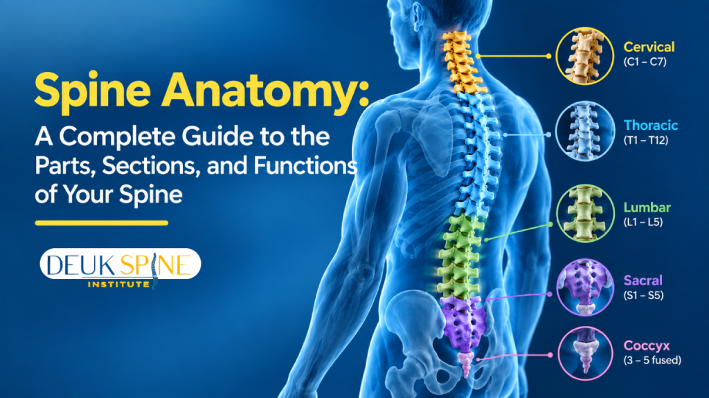
The five sections of the spine run from the neck to the tailbone, and all five spine sections have their own letter code and job.
Cervical Spine (Neck)
Seven small vertebrae make up the neck, numbered C1 through C7. Together they balance a head weighing roughly 10 to 12 pounds. No other region moves as freely, and from the side, the neck bends slightly inward in what doctors call a lordotic curve.
Thoracic Spine (Mid-Back)
Drop down to the mid-back and the count jumps to 12, labeled T1 to T12. Ribs attach to each of these bones, so this stretch anchors the rib cage. That makes it the least mobile part of the spine, with a curve that bows outward, known as kyphotic.
Lumbar Spine (Low Back)
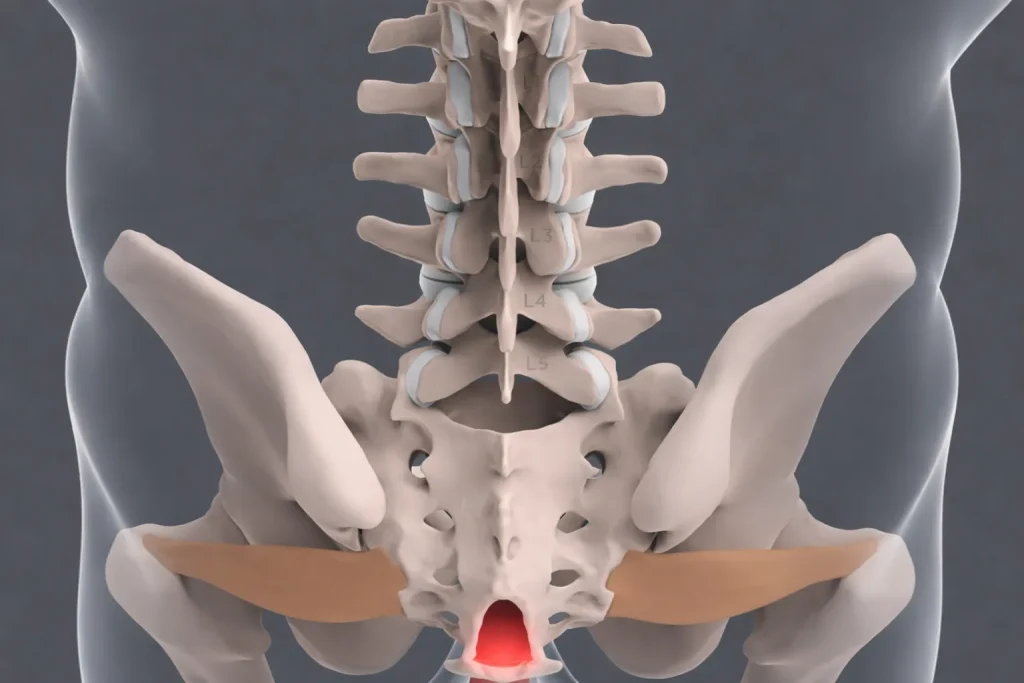
The low back runs on just 5 vertebrae, L1 to L5, but they’re the sturdiest in the stack because they carry most of the body’s weight. The curve here swings inward again, matching the neck’s lordotic shape.
Sacrum
The sacrum sits below the lumbar spine. It starts as 5 separate vertebrae, S1 to S5, which fuse during development. The sacrum connects the spine to the pelvis.
Coccyx (Tailbone)
The coccyx, or tailbone, is made of 4 small fused vertebrae. Several pelvic floor muscles and ligaments attach to it.
Added together, the running total is 7 + 12 + 5 + 5 + 4 = 33 vertebrae. This count is a basic part of the anatomy of the spine. In adults, the fused sacrum and coccyx are usually counted as one bone each, which brings the number of separate bones to 26.
The 5 Spine Sections at a Glance
The spine is divided into five regions, each with a distinct structure, a distinct job, and a distinct set of symptoms when something goes wrong.
What Are the Parts of a Vertebra? (Back Bones Anatomy)
Here’s the good news. Nearly every vertebra shares the same basic design, so once one makes sense, the rest follow. That’s really the core of back bones anatomy. And the names below? They’re the parts of the spine bones that keep popping up in doctors’ notes.
- Vertebral body: The solid front block that carries most of the weight.
- Vertebral arch: A curved ring of bone sitting behind the body. The spinal cord runs safely through its center.
- Pedicles and laminae: Pedicles are two short posts joining the arch and body. Laminae are flat plates closing the ring at the back.
- Spinous and transverse processes: The bony points sticking out backward and sideways. Muscles and ligaments grab on here.
- Facet joints: The spots where one vertebra touches the next. They guide movement and keep it from going too far.
Now stack those bones. The arches line up and create a tunnel, the spinal canal, where the cord runs. Along each side, gaps between neighboring vertebrae form small openings. Each one is called a neural foramen, and a nerve root slips out through it.
Why learn these two spaces? Because any “narrowing” on an MRI means one of them, either the canal or a foramen.
Discs, Joints, Ligaments and Muscles: The Soft Parts That Hold It Together
The spine may look like a stack of bones, but there is more to it. Between the vertebrae are soft discs that provide cushioning. Each disc has a strong outer layer, called the annulus fibrosus, and a softer center known as the nucleus pulposus.
These two parts work together whenever the spine moves. Walking, climbing stairs, or lifting a bag puts pressure on the discs. They compress slightly under that pressure and help keep the vertebrae from rubbing against each other.
Now move toward the back of the spine. On the left and right of each segment you’ll find a facet joint, and these two small joints glide against each other whenever the back turns. Twist too far and the joint surfaces butt up against one another, which keeps that segment from overrotating.
Holding everything in line are the ligaments, strong fibrous straps that run from bone to bone. Muscles handle the actual work of moving and keeping good posture, while tendons tie those muscles onto the vertebrae.
Cleveland Clinic counts this soft tissue among the five core parts of the spine. Discs and soft tissue also happen to be some of the most common sources of back pain, so these names show up constantly in conversations about sore backs.
The Spinal Cord and Spinal Nerves: What Each Level Controls
Think of the spinal cord as a two-way cable between the brain and everything below the neck. Stub a toe, and the pain signal races up the cord to the brain. Decide to pull that foot back, and the command rides the same cable down to the muscles. A few reflexes never reach the brain, since the cord handles them alone.
Oddly enough, the cord is shorter than the spine. It begins at the base of the skull and wraps up near the top of the lower back, around L1 or L2. On the way down, 31 pairs of nerve roots peel off and slip out through small gaps between the bones.
Because the cord quits early, the roots meant for the lower body have to keep traveling down the canal before they find their exit. Doctors named that trailing bunch the cauda equina, Latin for “horse’s tail,” and a drawing shows why. You’ll see the term in articles about spinal warning signs.
The roots divide up the body like this:
- Cervical roots: The shoulders, arms, and hands
- Thoracic roots: Bands circling the chest and belly
- Lumbar and sacral roots: The hips, legs, and feet
A pinched root usually makes itself felt wherever that root travels, not at the spine, so a neck problem can show up as tingling in the hand.
Why Does the Spine Curve?
Viewed from the front, a healthy spine looks straight, but a side view tells a different story. It bends into a gentle S shape made of three natural curves. The neck curves inward, which is called cervical lordosis.
The upper and middle back curve outward in what’s known as thoracic kyphosis, and the low back curves inward again to form lumbar lordosis. These curves work together a lot like a spring. Each step sends a small jolt up through the body, and the alternating bends spread that force out rather than letting it travel straight up to the skull.
They also keep the head balanced over the pelvis, a quiet but important part of the anatomy of the spine. Posture habits can gradually change the shape of these curves over the years, which is why curve changes sometimes appear on imaging reports.
How Spine Anatomy Shows Up in Your MRI Report
Most of the wording on an MRI report points back to the parts covered above. Take “L4-L5.” It names the segment between the fourth and fifth lumbar vertebrae, including the disc, joints, and foramina at that level, not a single bone.
Direction words each have a simple meaning:
- Anterior and posterior: Toward the front or the back
- Left and right: The patient’s own sides
- Central or foraminal: The middle of the canal, or the side opening
Common phrases decode the same way, as the table below shows. Remember, a report describes structures, not symptoms, and many findings appear in people with no pain at all. It’s a map of anatomy, not a diagnosis.
For a closer look at each term, see the guide to reading your MRI report. Anyone who wants their own scan explained can also request a free MRI review from some spine centers.
MRI Report Terms Decoded
Radiology reports are written for other physicians. Here is what each term actually means in plain language.
Which Part of the Spine Is Involved in Common Back Problems?
Most common back problems are named after the structure they involve. The map below simply pairs each structure with the conditions linked to it, and every condition has its own page with the full details.
- Disc: A herniation involves the intervertebral discs, specifically the gel-like center and the outer ring that surrounds it.
- Facet joints: Facet pain and facet arthritis both involve the small paired joints at the back of each spinal segment.
- Foramen or canal: A pinched nerve and spinal stenosis both involve these spaces, either the side doorway or the central tunnel.
- Nerve root: Sciatica and other radiating symptoms involve the nerve roots that branch off the spinal cord.
- Muscles and ligaments: A strain involves the soft tissue that supports and moves the spine.
An MRI describes structures, not a diagnosis. That’s what a neurosurgeon is for
You learned the anatomy.
Now find your pain generator.
MRI findings like “disc bulge,” “foraminal narrowing,” and “canal stenosis” appear in people with no pain at all. The difference is whether those structures are actually generating your symptoms. Dr. Deukmedjian personally reviews every MRI to connect what the scan shows to where your pain is coming from—with 99% accuracy.
- Lists structural findings at each level
- Cannot tell you which finding causes pain
- Many findings appear in pain-free people
- Correlates imaging with your actual symptoms
- Pinpoints the exact pain generator
- 99% diagnostic accuracy across all spine regions
Deuk Spine Institute · Board-certified neurosurgeon · No cost, no obligation · Cervical, thoracic & lumbar
FAQs: Spine Anatomy
What are the 5 sections of the spine?
The neck, mid back, low back, sacrum, and tailbone, also known medically as the cervical, thoracic, lumbar, sacral, and coccygeal regions. They are often referred to by doctors as C, T, L, S and Co respectively, hence their codes on scans.
What is the function of the spinal cord?
Mostly, it passes signals back and forth between the brain and the rest of the body. Reach for a hot pan, and the “that burns” message goes up the cord while “let go” comes back down, all in a split second. A handful of reflexes skip the brain completely.
How many bones make up the spine in total?
Thirty-three, if the fused ones are counted individually. That breaks down to 7 in the neck, 12 through the mid-back, 5 in the low back, 5 making up the sacrum, and 4 in the tailbone. Count the sacrum and tailbone as one bone each, and adults end up with 26.
What’s the difference between a vertebra and a disc?
A vertebra is one of the bones that make up the spine, while a disc is the soft cushion that sits between two vertebrae. Each disc has a tough outer ring and a gel-like center, which lets it absorb pressure and keep the bones from rubbing against each other.
What’s the difference between the spine and the backbone?
Nope, same thing. “Backbone” is what most people say at the kitchen table, while a doctor is more likely to write “spine,” “spinal column,” or even “vertebral column” in a chart.
Does the spine get shorter as people age?
A bit, for most people. Discs slowly dry out and get thinner over the decades, and those tiny losses at every level add up. The curves can change, too, and some people barely notice, while others lose a more noticeable amount.
What does “L4-L5” mean on an MRI report?
The space between the fourth and fifth bones of the low back. It covers the whole level rather than a single bone, so the disc, the joints on either side, and the nerve openings all fall under that one label. Think of it as an address that says where, not what’s wrong.
Conclusion
Spine terms look intimidating on paper. But each one points to a real structure, and none of them are hard to picture once the basics click. Regions, bones, discs, nerves, curves. Put those together and a report, or a doctor’s explanation, gets much easier to follow.
Knowing the anatomy of the spine also makes the next appointment more useful, since questions get sharper. Some spine centers offer a free MRI review, which gives people with an existing report a clear explanation of what it means.
Sources
View Sources
- https://my.clevelandclinic.org/health/body/10040-spine-structure-and-function
- https://spinehealth.org/spine-basics/spine-anatomy/
- https://www.spinenerve.com/anatomy-of-the-spine30c807e6
- https://www.ucirvinespinecenter.com/anatomy-of-spine-orthopaedic-spine-surgery-orange-costa-mesa-ca/
- https://www.umms.org/ummc/health-services/orthopedics/services/spine/patient-guides/anatomy-function