


What is an Intervertebral Disc?
The intervertebral disc is a central component of the spine, providing flexibility, support, and shock absorption. Without these discs, spinal movement would be severely limited, and the vertebrae would experience excessive stress, leading to rapid deterioration.
This article explores the structure, function, and biomechanics of intervertebral discs. We will examine their anatomical components as well as their blood supply and innervation.
We will also discuss physiologic variations, important functions, and the biomechanical properties that enable movement. Finally, we will analyze intervertebral disc degeneration, its role as a leading cause of back pain, and how it progresses over time.
Clinical Anatomy
The intervertebral disc is made up of specialized tissues that work together to maintain spinal stability. Each part of the disc plays a foundational role in ensuring both strength and flexibility.
Nucleus Pulposus
The nucleus pulposus is the central, gel-like portion of the intervertebral disc, which is composed mainly of water, proteoglycans, and collagen. It functions as the primary shock absorber within the spine, redistributing mechanical forces exerted during movement.
The high water content, which ranges from 70% to 90%, allows the nucleus pulposus to retain its cushioning properties, preventing excessive strain on surrounding spinal structures.
Proteoglycans within the nucleus attract and retain water, ensuring the disc remains hydrated and able to resist force.
Annulus Fibrosus
The annulus fibrosus is the tough outer layer that surrounds the nucleus pulposus, providing structural integrity to the intervertebral disc.
Based on our observations, it is composed of 15 to 25 concentric fibrocartilaginous sheets known as lamellae, each arranged in alternating directions.
This unique configuration enables the annulus to resist tensile forces while maintaining flexibility. The annulus primarily contains type I and type II collagen, providing strong yet elastic properties.
These layers work together to confine the nucleus pulposus and protect the disc from excessive strain.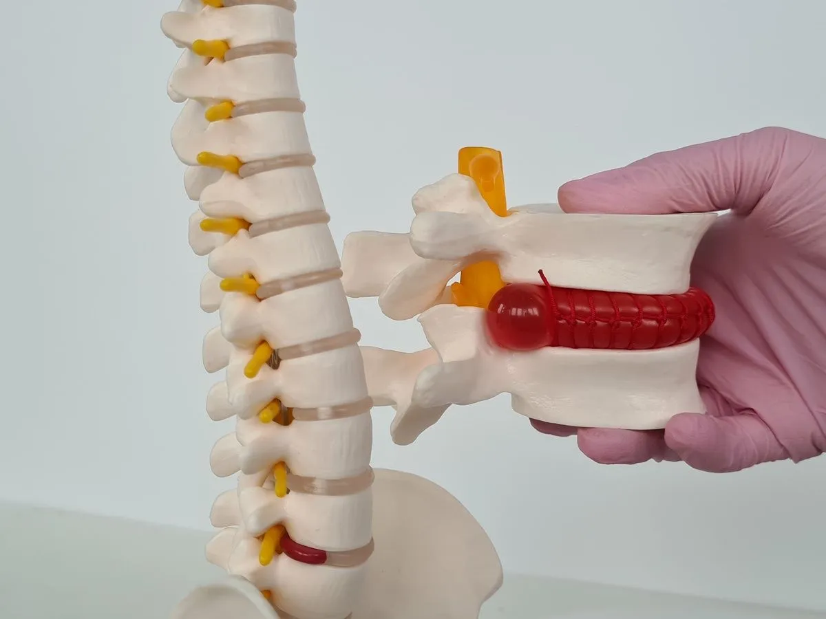
Vertebral Endplate
The vertebral endplate is a thin layer of hyaline cartilage that separates the intervertebral disc from the adjacent vertebral bodies. Our findings show that it serves two key functions: mechanical support and nutrient exchange.
The endplate acts as a semi-permeable membrane, allowing essential nutrients such as glucose and oxygen to diffuse from the vertebral bone into the largely avascular disc.
This nutrient flow is critical for maintaining the health of both the nucleus pulposus and annulus fibrosus.
Blood Supply And Lymphatics
As mentioned, intervertebral discs are largely avascular, meaning they lack a direct blood supply. Only the outermost layer of the annulus fibrosus receives blood flow, while the inner portions of the disc rely on diffusion to obtain nutrients and remove metabolic waste.
This process occurs through the blood vessels located near the disc-bone junction and those in the outer annulus.
Because intervertebral discs lack a dedicated lymphatic system, the removal of waste products is entirely dependent on passive diffusion.
When diffusion is impaired due to aging, injury, or reduced spinal mobility, the disc becomes more susceptible to inflammation, degradation, and structural failure.
Nerves
In a healthy intervertebral disc, only the outer third of the annulus fibrosus contains nerve fibers, which receive input from branches such as the sinuvertebral nerve.
This limited innervation means that the inner two-thirds of the annulus and the nucleus pulposus are devoid of nerve fibers.
Physiologic Variations
The intervertebral discs exhibit several physiologic variations based on their location within the spine. Generally, disc thickness increases near the front and tail parts of the body, with a slight decrease at the T3-T4 level.
The cervical and lumbar discs tend to have the highest ratio of disc thickness to vertebral body height, correlating with the greater range of motion observed in these regions.
Our research indicates that the intervertebral discs are thicker anteriorly in the cervical and lumbar regions, contributing to the natural curve of the spine.

Important Functions
The intervertebral discs play a crucial role in maintaining spinal integrity and facilitating movement. They serve as primary shock absorbers for the spine, reducing the impact of mechanical stress during everyday activities such as walking, running, and bending.
Without the intervertebral discs, the vertebrae would endure excessive wear and tear, leading to rapid deterioration and chronic pain.
Each component of the intervertebral disc plays a specialized role in its function, which we will touch on next.
Biomechanics
When the spine undergoes compression, the nucleus pulposus functions like a hydraulic cushion, distributing pressure in all directions. This action reduces localized stress on the vertebral endplates and annulus fibrosus.
Meanwhile, the annulus fibrosus, with its unique layered architecture, provides strength, preventing excessive bulging or rupture of the nucleus pulposus, under healthy conditions.
Together, these components ensure that the spine can withstand daily movements, high-impact activities, and heavy lifting.

Intervertebral Disc Degeneration
Intervertebral disc degeneration, such as herniated disc and bulging disc, is a progressive condition that contributes to chronic back and neck pain.
Degenerative disc disease occurs in several stages. First, a small tear occurs in the outer fibrous ring of the disc. Then this tear allows fragments of the nucleus pulposus to leak into the annular space, triggering inflammation and discogenic pain.
More strain causes further tearing. As the annular tear expands, the disc may undergo bulging, protrusion, herniation, or extrusion, where the nucleus pulposus extends beyond its normal boundaries.
When the protruding disc comes into contact with spinal nerves or the spinal cord, it can cause radiating pain, numbness, tingling, and weakness in the affected area.
In severe cases, disc degeneration can lead to spondylosis (disc narrowing and osteophyte formation).
Traditional treatments for disc degeneration include physical therapy, pain medications, and steroid injections, but these methods provide only temporary relief without addressing the underlying damage.
Similarly, surgeries like laminectomy and artificial disc replacement come with high risks: spinal fluid leaks, wound infections, hardware failure or migration, and failed back surgery syndrome,
A superior alternative is Deuk Laser Disc Repair, which precisely removes the inflamed nucleus pulposus material from within the annular tear while preserving the healthy disc structure.
Over 95% of chronic disc-related pain can be cured with Deuk Laser Disc Repair®, with a 0% complication rate, making it the safest treatment for disc degeneration today.
Conclusion
Intervertebral disc injury is the most common cause of back pain, affecting millions of people worldwide. The breakdown of these discs leads to reduced functionality and, consequently, quality of life. The anatomy of intervertebral discs is foundational to our understanding of degenerative disc disease.
To this end, we have explored their biomechanical properties, how they facilitate movement, and their role in maintaining spinal stability. We have also discussed the degenerative process, highlighting its impact on spinal function and overall health.
If you are experiencing back pain, Deuk Spine Institute offers a cure. Get your free MRI review or contact Deuk Spine Institute today to return to a pain-free life.
If you want to learn more, why not check out these articles below:


