
Dr. Ara J. Deukmedjian, MD
Board Certified Neurosurgeon
Published: August 26, 2026
Disclaimer: The contents provided in this article are for learning purposes only. It is always recommended to speak to a doctor about your personal health condition.
Key Points
✓ US Patent 12,623,035 B2 describes a minimally-invasive process that provides copper particles to injured spinal discs. 1
✓ Copper serves as a cofactor to lysyl oxidase, which links collagen and elastin in annulus fibrosis. 2 3
✓ Copper nanoparticles enhance wound healing, decrease inflammation, and stimulate angiogenesis according to peer-reviewed publications. 4 5
✓ Delivery uses a 1–12 mm shaft. Either a needle or endoscopic retractor with minimal blood loss. 1
✓ Annular tears can take 12–24 months to heal on their own due to poor blood supply. 6 1
✓ The technique integrates with Deuk Laser Disc Repair®, adding a regenerative step to proven endoscopic debridement. 7 8
What Are Copper Nanoparticles for Spinal Disc Repair?
On May 12, 2026, the United States Patent and Trademark Office issued U.S. Patent No. 12,623,035 B2 to Dr. Ara Deukmedjian and Panacea Spine, LLC, titled “Systems and Methods for Repairing Spinal Disc Injury or Treating Spinal Disc Disease Using Copper.”1
The patent describes a method for delivering copper nanoparticles directly to a damaged spinal disc using medical imaging guidance. The concept is straightforward but powerful: position a needle at the site of annular tear, inject the copper particles onto the injured tissue. The whole process is totally non-invasive, where the amount of blood lost is just a few drops.1
This is what makes the process important because it changes the treatment strategy from excision of damaged tissues to repair of the same. Current spine treatments: discectomy, laminectomy, spinal fusion.
Focus on taking something out or immobilizing segments. This patent proposes adding Copper to the disc that helps it heal from within.1
Why Copper? The Science Behind the Metal
Copper is not new to medicine. It is one of the oldest known antimicrobial metals, used in wound care since ancient Egypt. But the modern scientific interest in copper for tissue repair goes far deeper than its antibacterial properties.
The presence of copper is necessary for the activity of lysyl oxidase, which catalyzes the formation of cross-links in collagen and elastin, the two structural proteins responsible for the strength and elasticity of the connective tissues. 2 3 Lysyl oxidase cannot function without sufficient amounts of copper, and thus collagen cannot undergo cross-linking and mature into its mature form, giving tensile strength to connective tissue such as the annulus fibrosus of a spinal disc. 3
The use of copper in spinal disc repair is especially applicable due to the nature of the structure of the annulus fibrosus. The annulus fibrosus is made up mainly of collagen fibers in concentric rings, which when ruptured require repair using collagen fibers that have undergone proper cross-linking. Thus, the use of copper in this repair seems biologically plausible.
Beyond collagen cross-linking, peer-reviewed research has identified several additional mechanisms through which copper nanoparticles promote tissue healing: 4 5
- Angiogenesis. Copper stimulates the development of new blood vessels via stimulation of the production of vascular endothelial growth factor (VEGF) through the HIF-1α pathway. Oxygen and nutrient supply is carried out with the help of newly developed blood vessels. 4
- Anti-inflammatory effect. Copper nanoparticles enable the delivery of growth factors which contribute to the anti-inflammatory stage of the wound healing process, thus alleviating the inflammatory process leading to the generation of discogenic pain. 5
- Antibacterial effect. The copper particles prevent the growth of bacteria through inhibition of protein synthesis and membrane peroxidation. 4
- Biocompatibility. According to systematic reviews, copper has been found to have higher biocompatibility compared to other metallic nanoparticles, thus stimulating regeneration. 5
Why Damaged Spinal Discs Need Better Healing Solutions
Each spinal disc includes an outer tough band called annulus fibrosus around a soft interior gel core known as the nucleus pulposus. Annular tears are caused by injuries, degenerative changes or repetitive stresses and when the nucleus moves through a tear in the annulus, it results in a disc herniation, which becomes one of the most common causes of chronic back and neck pain. 1
The underlying issue with annular tears lies in a very poor blood supply of the annulus fibrosus. For any healing process to occur, there must be enough blood flow providing necessary cells and nutrients for repair processes in damaged tissues. Due to being poorly supplied with blood, annular tears take up to 12 to 24 months to heal and some never do. 6 1
While undergoing such a long healing process, a tear generates pro-inflammatory cytokines that sensitize the nerve endings in the posterior part of the annulus and result in chronic pain called discogenic pain. The inflammatory cascade may extend to nearby nerve roots and give rise to the so-called radicular pain – arm pain after cervical herniations and leg pain after lumbar herniations. 1 7
Current treatment options for damaged discs include: 1
- Management through physical therapy and pain medication that only controls symptoms without helping to heal the tear.
- Surgical management such as discectomy or spinal fusion that removes or immobilizes the involved structure without repairing the tear.
- Deuk Laser Disc Repair®, a minimally invasive, motion-preserving endoscopic procedure that debrides inflamed tissue and vaporizes herniated material while preserving the disc.7 8
What has been missing is a regenerative component. Something that actively accelerates the biological healing of the torn annulus after debridement. The copper nanoparticle approach described in this patent is designed to fill that gap.1
How the Procedure Works: Two Delivery Methods
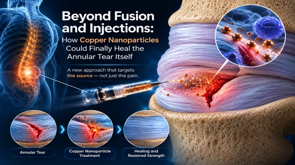
The patent describes two primary approaches for delivering copper particles to a damaged spinal disc. Both are minimally invasive and performed under medical imaging guidance such as fluoroscopy, CT, stereotactic techniques, or robotic navigation.1
1. Needle Injection
The simplest delivery method uses a syringe pre-loaded with copper particles. The physician inserts a hypodermic or spinal needle, guided by medical imaging, directly to the damaged disc. The plunger is actuated to push the copper particles through the needle and distribute them onto the damaged tissue. Then the needle is removed.1
This approach is ideal for straightforward disc injuries where debridement is not required. The copper particles can be delivered as a dry powder, in a liquid solution (such as saline or Lactated Ringer’s solution), or in a gel suspension. The entire procedure can potentially be performed as an outpatient visit.1
2. Endoscopic Delivery (Combined with Debridement)
For more advanced disc damage. Especially herniations with significant inflammatory tissue. The patent describes a comprehensive endoscopic procedure that combines tissue debridement with copper particle delivery:1
- Step 1: A guidewire is inserted under medical imaging guidance to the damaged disc.
- Step 2: One or more hollow dilators are advanced over the guidewire.
- Step 3: An endoscopic tubular retractor (typically around 4 mm in diameter) is inserted over the dilator(s) to create a working channel from the skin to the disc.
- Step 4: The guidewire and dilators are removed, leaving the retractor in place.
- Step 5: A laser (e.g., Holmium:YAG or CO2) or RF probe is used to debride inflamed tissue, remove herniated nucleus material, and clean the annular tear.
- Step 6: After debridement, copper particles are dispensed through the tubular retractor directly onto the debrided surfaces of the damaged disc.
- Step 7: An endoscopic camera confirms adequate distribution of the copper particles. The retractor is then removed.
This second approach integrates directly with the Deuk Laser Disc Repair® surgical technique, adding a regenerative step to an already proven endoscopic debridement procedure.7 8 1
Formulation and Delivery Options
One of the most notable aspects of the patent is the range of formulation options it covers for the copper particles. This flexibility allows clinicians to choose the delivery format best suited to the specific clinical scenario:1
- Dry powder—copper particles in powder form for direct application.
- Liquid solution—copper particles suspended in saline or Lactated Ringer’s solution for injection.
- Gel suspension—copper particles in a hydrogel or biocompatible polymer for sustained-release delivery and improved retention at the site.
- Aerosolized delivery—copper particles atomized using sterile compressed air or syringe air to coat the torn surfaces of the annulus.
The copper particles can be coated with hydroxyethyl cellulose, a biocompatible coating studied for its very low toxicity to human cells. A color additive dye may be included to enhance visualization of particle distribution through the endoscopic camera.1
Copper is also radiopaque. It shows up on X-ray imaging. The patent describes using a combination of smaller nanoparticles and a smaller proportion of larger microparticles allowing the surgeon to confirm particle placement under fluoroscopy in real time.1
What Conditions Can This Treat?
While the patent focuses heavily on annular tears and disc herniations, it explicitly states that the copper particle approach applies to a broad range of spinal disc conditions:1
- Herniated discs
- Bulging discs
- Ruptured discs
- Degenerated and desiccated discs
- Annular tears and fissures
- Extruded discs
- Degenerative disc disease
- Disc osteophyte complex
- Spondylosis and spondylolisthesis
- Spinal stenosis and neural foraminal narrowing
The patent covers treatment of discs in the cervical, thoracic, and lumbar regions of the spine. For cervical discs, the approach enters from the front of the neck through the disc to reach the damaged posterior region. For lumbar discs, the approach enters from the patient’s side through the neural foramen.1
How This Builds on Deuk Laser Disc Repair®
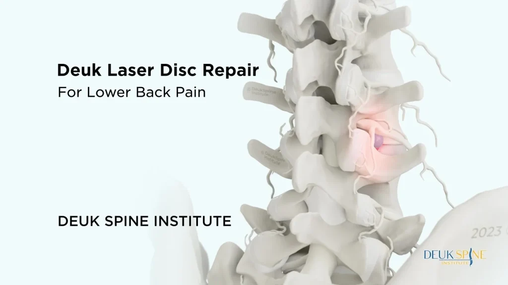
Deuk Laser Disc Repair® (DLDR) is a full-endoscopic, outpatient, motion-preserving procedure developed by Dr. Ara Deukmedjian for treating symptomatic disc disease. Peer-reviewed studies published in Surgical Neurology International have documented its safety and effectiveness, with an overall clinical success rate of 94.6% in treating symptomatic cervical disc herniations.7 8
The copper nanoparticle technique described in this patent is designed to integrate seamlessly with the DLDR procedure. After the laser debrides the inflamed tissue and removes herniated disc material, copper particles can be dispensed through the same endoscopic tubular retractor onto the freshly prepared annular surfaces. This adds a regenerative component to what is already a proven debridement and decompression procedure.1 7
The combination of endoscopic debridement and copper particle application represents a potential two-phase approach to disc repair: first remove the damaged, inflammatory tissue that is causing pain, then provide the biological stimulus needed to accelerate healing of the underlying tear.
What This Patent Does Not Tell Us
It is important to understand what a patent is and what it is not. A patent protects an invention. It does not prove clinical efficacy. The patent document does not include clinical trial data demonstrating that copper particles accelerate spinal disc healing in human patients. The language is appropriately measured: the patent states that copper particles are “believed” to speed up healing.1
The biological rationale is well supported by peer-reviewed research on copper’s role in tissue regeneration, collagen synthesis, and wound healing.2 3 4 5 The procedural foundation is also well established through published studies on Deuk Laser Disc Repair®.7 8 However, validating the specific application of copper particles to spinal disc tissue will require clinical studies, regulatory evaluation, and long-term follow-up data.
What is clear is that the approach represents a meaningful shift in how we think about spinal disc treatment. From removal and immobilization to repair and regeneration.
The disc was never meant to be removed.
It was meant to be repaired.
Annular tears can take 12–24 months to heal — if they heal at all. Current treatments remove tissue or lock vertebrae together. Dr. Deukmedjian’s patented copper nanoparticle technology introduces a regenerative step: deliver copper directly to the damaged disc to accelerate the collagen cross-linking the annulus needs to repair itself.
Board-certified neurosurgeon · Pioneer in endoscopic laser spine surgery · 2,700+ DLDR procedures since 2004
FAQ’s
What is the copper nanoparticle disc repair procedure?
It is a minimally invasive method for delivering copper particles (nanoparticles and/or microparticles) directly to a damaged spinal disc using medical imaging guidance. The copper particles are dispensed through a hollow shaft. Either a needle or an endoscopic tubular retractor to promote healing of the damaged disc tissue.1
Why are copper particles used instead of other metals?
Copper is a required cofactor for lysyl oxidase, the enzyme that cross-links collagen and elastin in connective tissue. Since the annulus fibrosus of a spinal disc is composed primarily of collagen. Copper’s role in collagen maturation makes it biologically suited for promoting disc repair. Copper nanoparticles also demonstrate anti-inflammatory, antimicrobial, and angiogenic properties that support multiple phases of tissue healing.2 3 4
Is this procedure currently available to patients?
The technique is currently protected by a U.S. patent issued on May 12, 2026. While the procedural framework builds on the established Deuk Laser Disc Repair® technique, the specific copper nanoparticle component would need to undergo further clinical validation and regulatory review before becoming widely available as a standard treatment.1
How long does an annular tear take to heal without treatment?
Limited blood supply makes it difficult for the annulus fibrosus to repair itself. An annular tear takes between 12 to 24 months to heal on its own, and some may not heal at all. The long healing period is one of the main issues that the copper nanoparticle technology seeks to solve.6 1
What forms can the copper particles be delivered in?
The patent covers multiple delivery formats: dry powder, liquid solution (such as saline or Lactated Ringer’s solution), gel suspension, and aerosolized spray. The particles can also be coated with hydroxyethyl cellulose for improved biocompatibility and may include a color additive to enhance endoscopic visualization.1
How does this relate to Deuk Laser Disc Repair®?
The copper nanoparticle technique is designed to integrate with the Deuk Laser Disc Repair® procedure. DLDR uses an endoscopic laser to debride damaged and inflamed tissue from the disc. After debridement, the copper particles can be dispensed through the same tubular retractor onto the prepared disc surfaces, combining proven debridement with a regenerative step.1 7 8
What sizes are the copper particles?
The patent covers copper nanoparticles (1 nm to 1,000 nm) and microparticles (1 µm to 1,000 µm). Some embodiments use a combination of smaller nanoparticles for biological activity and a smaller proportion of larger microparticles for enhanced visibility under X-ray imaging, since copper is radiopaque.1
What conditions can this approach treat?
The patent covers a broad range of spinal disc conditions including herniated discs, bulging discs, annular tears, degenerative disc disease, ruptured discs, desiccated discs, extruded discs, spinal stenosis, neural foraminal narrowing, spondylolisthesis, and spondylosis. It applies to discs in the cervical, thoracic, and lumbar regions.1
Is the procedure minimally invasive?
Yes. The hollow shaft used in the procedure ranges from 1 mm to 12 mm in diameter, with the endoscopic tubular retractor typically around 4 mm. Blood loss is described as minimal; a few drops. The procedure is designed to be performed on an outpatient basis under medical imaging guidance.1
Sources
View Sources
- Deukmedjian A. Systems and Methods for Repairing Spinal Disc Injury or Treating Spinal Disc Disease Using Copper. U.S. Patent No. 12,623,035 B2. May 12, 2026.
- Rucker RB, et al. Copper and the synthesis of elastin and collagen. Ciba Foundation Symposium. 1980;79:180–207.
- Wu M, et al. Biochemistry, Collagen Synthesis. StatPearls. National Library of Medicine. Updated June 2026.
- Sandoval C, et al. Role of copper nanoparticles in wound healing for chronic wounds: literature review. Burns & Trauma. 2022;10:tkab047.
- Sandoval C, et al. Effectiveness of Copper Nanoparticles in Wound Healing Process Using In Vivo and In Vitro Studies: A Systematic Review. Pharmaceutics. 2022;14(9):1838.
- Mork T. Everything You Need to Know About An Annular Tear. Updated September 2024.
- Deukmedjian AJ, et al. Cervical Deuk Laser Disc Repair® A novel, full-endoscopic surgical technique for the treatment of symptomatic cervical disc disease. Surgical Neurology International. 2012;3:142.
- Deukmedjian AJ, et al. Deuk Laser Disc Repair® is a safe and effective treatment for symptomatic cervical disc disease. Surgical Neurology International. 2013;4:68.