By Dr. Ara Deukmedjian
Board Certified Neurosurgeon
Medically reviewed on Jul 22, 2026
Medical Disclaimer: This information is intended for educational purposes only and is not to be taken as a substitute for medical advice. Results will vary from individual to individual. Consult your doctor regarding your own situation.
Key Points
✓ The L1-L2 disc is located at the thoracolumbar junction where the stiff thoracic spine ends and gives way to the flexible lumbar spine. ¹
✓ While L1-L2 disc herniations make up only 1–2% of all lumbar disc herniations, they have an especially high rate of causing nerve damage. ²
✓ The end of the spinal cord (conus medullaris) is usually at or near the L1-L2 level in adults, so even a small disc herniation at this level will press on the spinal cord itself and not just the nerves. ³
✓ Classic symptoms include groin pain, pain in the upper front part of the thigh, and hip flexor weakness, which may be thought to come from a hip condition. ⁴
✓ MRI confirms the diagnosis, but abnormal disc findings are common in pain-free adults, so imaging must match the exam. ⁵
✓ Most L1-L2 disc bulges and small herniations respond to 6–12 weeks of conservative care. ⁶
✓ Traditional surgery at L1-L2 (fusion or open discectomy) carries higher risk than at lower lumbar levels because of the conus. ⁷
✓ Full-endoscopic laser decompression removes the offending disc fragment through a 7 mm incision: no fusion, no hardware, and the spinal cord is never manipulated. ⁸ ⁹
✓ Deuk Laser Disc Repair®: 99% pain relief for treated pain sources, 0.01% complication rate, 2,700+ procedures.
What Is the L1-L2 Disc, and Why Is It Special?
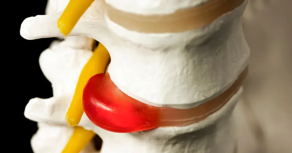
In the lumbar region, there are five vertebrae known as L1 through L5, between which lie intervertebral discs as shock absorbers. This nucleus pulposus consists of a soft gel, which is covered by a tough annulus fibrosus.
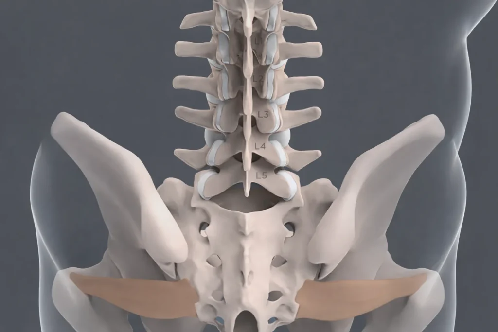
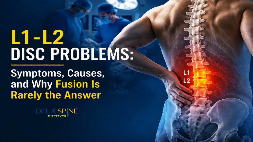
L1-L2 disc lies at the upper end of the lumbar spine and lies just below the last thoracic vertebra, T12. The Thoracolumbar junction is a biometric transitional zone from the rib supported stiff thoracic spine to the lumbar spine. ¹
Anatomically, L1-L2 is unlike any lower lumbar level for one critical reason. The spinal cord itself usually ends here. In roughly 94% of adults, the conus medullaris. The tapered terminal end of the spinal cord. Sits between the T12 and L2 vertebral bodies with the most common position being at L1. ³ Below the conus, the spinal canal contains a bundle of free-floating nerve roots called the cauda equina.
This is why an L1-L2 disc herniation is more serious than a comparable herniation at L4-L5. At L4-L5, a disc presses on a single nerve root. At L1-L2, a large central herniation can press on the spinal cord itself, producing a distinct and more urgent syndrome called conus medullaris syndrome. ³
Symptoms of an L1-L2 Disc Problem
Symptoms depend on what the disc is pressing on. The disc wall itself is an exiting nerve root (L1 or L2 radiculopathy), or the conus medullaris/cauda equina.
1. Axial back pain (discogenic pain)
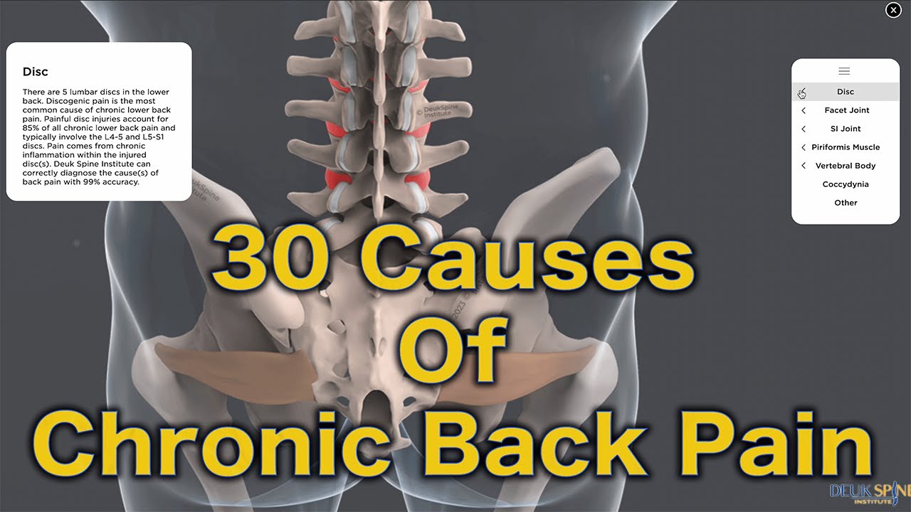
The earliest and most common symptom is a deep, aching pain across the upper lumbar region. Sometimes described as a “belt of pain” just above the waistline, occasionally wrapping into the flank. This is irritation of nerve endings in the outer disc wall, not compression of a spinal nerve. ¹ Discogenic pain at L1-L2 typically worsens with prolonged sitting, forward bending, or heavy lifting, and eases with lying flat.
2. L1 and L2 radiculopathy
When the disc bulges laterally and compresses an exiting nerve root, symptoms follow that specific nerve. Unlike lower lumbar radiculopathies, upper lumbar radiculopathies do not produce sciatica down the back of the leg. Instead, patients report: ⁴
- L1 radiculopathy: pain, burning, or numbness across the groin, the front of the hip, and the very upper anterior thigh. Often mistaken for a hernia, hip labral tear, or genitourinary problem.
- L2 radiculopathy: pain and sensory changes across the front of the thigh, down toward the inner knee, sometimes with weakness of hip flexion (lifting the thigh) or knee extension.
Because these symptoms map to areas commonly attributed to hip disease, groin injuries, or urologic conditions, an L1-L2 disc problem is often missed for months before the correct diagnosis is made. ⁴
3. Hip flexor and quadriceps weakness
The iliopsoas (hip flexor) and quadriceps receive innervation from the upper lumbar nerve roots. Patients may notice difficulty lifting the leg to climb stairs. A feeling of the leg “giving way,” or trouble getting out of a low chair. Progressive weakness is a red flag that requires prompt evaluation.
4. Conus medullaris syndrome
If a large central L1-L2 disc herniation compresses the conus itself, the presentation is urgent and unlike a typical disc herniation. Warning signs include: ³ ¹⁰
- Sudden bladder retention or incontinence
- Bowel incontinence or loss of rectal tone
- Saddle anesthesia: numbness across the buttocks, inner thighs, and perineum
- Bilateral leg weakness
- Sexual dysfunction of sudden onset
Any of these findings warrant emergency imaging and neurosurgical evaluation, ideally within 24–48 hours. Delayed decompression of the conus is associated with permanent neurologic deficits. ¹⁰
Both conditions respond to the same non-operative care in most patients. Both can be potential candidates for endoscopic decompression should surgery become inevitable.
Bulging vs. Herniated Disc at L1–L2
The two terms are often used interchangeably, but they describe different structural failures. What matters clinically is what the disc material is doing to the surrounding neural structures.
Both conditions respond to the same non-operative care in most patients. Both can be potential candidates for endoscopic decompression should surgery become inevitable.
What Causes an L1-L2 Disc to Bulge or Herniate?
Unlike disc problems at L4-L5 and L5-S1, which are dominated by cumulative wear-and-tear. L1-L2 problems more often involve an acute mechanical component because of where the level sits. Common causes include:
- Trauma at the thoracolumbar junction: falls from height, motor-vehicle accidents, and heavy-object impacts concentrate force at T12–L2. ¹
- Age-related degeneration: lloss of disc hydration and annular microtears, more common in adults over 40. ⁶
- Heavy axial loading: repeated squats, deadlifts, and overhead work with poor mechanics.
- Genetics: twin studies show heritability accounts for 34–74% of disc degeneration. ¹¹
- Osteoporosis-related compression fractures: T12 or L1 that alter the mechanical load on the L1-L2 disc.
- Smoking: which reduces disc nutrition and accelerates degeneration.
Diagnosing an L1-L2 Disc Problem
1. History and physical exam
A spine specialist will look for the specific sensory pattern (groin, anterior hip, anterior thigh), test hip flexion and knee extension strength, check the patellar reflex (which is partially L2-mediated), and perform provocative maneuvers such as the reverse straight leg raise (femoral nerve stretch test), which is the upper lumbar equivalent of the classic straight leg raise and is specific for L1-L4 root irritation. ⁴

2. MRI
MRI is the imaging test of choice for L1-L2. It shows the disc, the exiting nerve roots, and most importantly at this level. The conus medullaris in soft-tissue detail. MRI is the only study that reliably distinguishes a bulge from a herniation and identifies cord or conus compression. ⁵
3. CT and CT myelogram
CT is more useful for bone pathology, including compression fractures of T12 or L1, osteophytes, and facet arthrosis. A CT myelogram is reserved for patients who cannot undergo MRI or when MRI findings are equivocal.
4. EMG / nerve conduction studies
If clinical findings do not clearly localize to L1 or L2, EMG can identify which root is inflamed and rule out mimickers such as femoral neuropathy, diabetic amyotrophy, or lumbar plexopathy. All of which can produce anterior thigh pain and weakness that resemble upper lumbar radiculopathy. ⁴
The MRI only tells us so much
This point is critical. Population-based imaging studies have shown that a majority of pain-free adults have disc bulges, degeneration, and other “abnormal” findings on lumbar MRI, with prevalence rising with age. ⁵ An MRI finding is not a diagnosis by itself. Symptoms, physical exam, and imaging must all point to the same level and the same nerve root before any treatment plan especially a surgical one is considered. A recommendation for surgery based on MRI alone is a warning sign.
Non-Surgical Treatment of an L1-L2 Disc Problem
For most patients without red flags, an L1-L2 disc bulge or small herniation can be managed conservatively. The North American Spine Society recommends 6–12 weeks of non-operative care for lumbar radiculopathy in the absence of progressive weakness or cauda-equina/conus signs. ⁶

Activity modification and posture correction
Relative rest, avoidance of heavy lifting, and correction of forward-bent postures reduce mechanical load on the L1-L2 disc during the acute inflammatory phase.
Physical therapy
PT intervention for disc conditions of the upper lumbar spine involves core stability, hip mobility, and gentle nerve glides of the femoral nerve. A McKenzie approach that is highly effective in cases of L4-L5 and L5-S1 disc conditions may be inappropriate for upper lumbar spine issues, particularly L1-L2.
Medication
NSAIDs are first-line. A short course of oral corticosteroids (such as a Medrol dose pack) is appropriate for acute severe radiculopathy. Opioids are not appropriate long-term therapy and do nothing to address the underlying compression.
Lumbar epidural steroid injection
An image-guided transforaminal epidural steroid injection at L1-L2 can deliver an anti-inflammatory agent directly onto the inflamed nerve root. Beyond providing relief, a well-placed diagnostic injection helps confirm the pain generator before surgery is discussed. ⁶
When conservative care is not enough
Non-operative treatment has failed when:
- Symptoms persist beyond 6–12 weeks of appropriate care
- Hip flexor or quadriceps weakness is present or progressing
- Pain is severe enough to disable normal function
- Any conus or cauda-equina red flags emerge. In which case, care escalates immediately
At that point, decompression should be discussed. But the type of decompression matters and at L1-L2 it matters more than at any other lumbar level.
Surgical Treatment: The Case Against Defaulting to Fusion at L1-L2
The conventional surgical recommendations for a symptomatic L1-L2 disc herniation are open lumbar discectomy, laminectomy with discectomy, or lumbar fusion (with or without instrumentation). These procedures carry three durable costs. Costs that are magnified at L1-L2 compared with lower lumbar levels:
- Proximity to the conus medullaris. Open surgery at L1-L2 requires manipulating tissue directly next to the terminal spinal cord. Retraction that would be routine at L4-L5 is not routine here. ⁷
- Adjacent-segment disease after fusion. Rigid fusion transfers load to adjacent levels. Symptomatic adjacent-segment degeneration develops in a significant fraction of lumbar fusion patients over the years that follow. ⁷ Fusing L1-L2 also transfers force upward into the already-vulnerable thoracolumbar junction.
- Long recovery. Open lumbar procedures typically require inpatient stays, weeks of restricted activity, and a several-month bone-healing timeline for fusions.
The critical question is whether your specific L1-L2 disc problem actually requires any of these trades. In the absence of documented instability, a large central herniation compressing the conus, a compression fracture, tumor, or infection, the answer for most patients is no.
The Endoscopic Alternative: Full-Endoscopic Laser Decompression at L1-L2
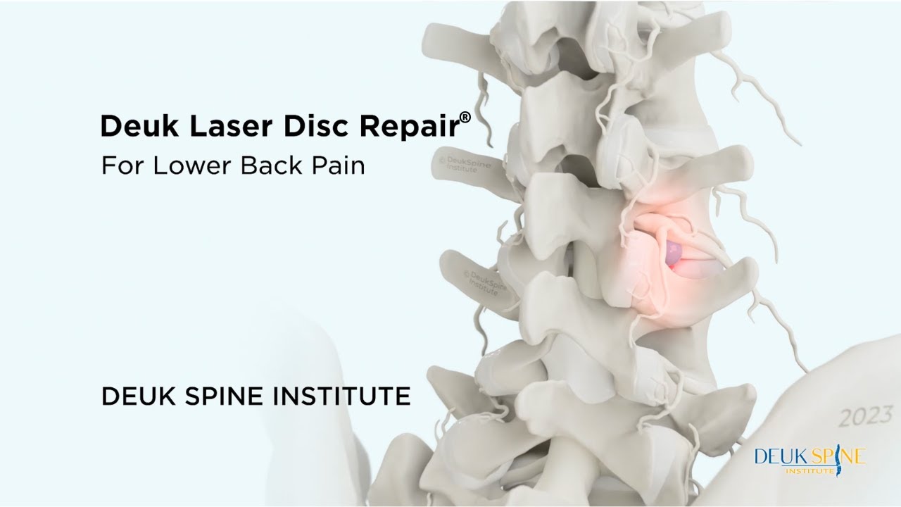
Full-endoscopic lumbar decompression is the modern, ultra-minimally-invasive treatment for a symptomatic L1-L2 disc bulge or contained herniation in appropriately selected patients. ⁸ ⁹
How it works
- A 7 mm skin incision is made under fluoroscopic guidance.
- A tubular dilator is passed between the paraspinal muscles. Muscles are spread, not cut.
- An HD endoscope with a working channel is advanced to the disc under continuous saline irrigation.
- Under magnified live visualization, the surgeon uses micro-instruments and a side-firing holmium laser to ablate the exact offending disc tissue.
- The lamina, facet joints, ligaments, and the conus itself are never manipulated. No implants are placed.
- The wound is closed with a single suture or skin adhesive.
Because no bone is cut and no structural element is removed, the procedure is motion-preserving. There is no fusion, no hardware, and no biomechanical liability transferred to adjacent levels.
What the evidence shows
- Randomized and prospective studies of full-endoscopic lumbar decompression report clinical success rates in the 85–95% range, with outcomes equivalent or superior to open discectomy and significantly less blood loss, shorter hospital stays, and faster return to work. ⁸ ⁹
- Reported major complication rates are on the order of 1–3%, comparable to or lower than open lumbar surgery. ⁹
- The endoscopic corridor is particularly advantageous at L1-L2 because the surgeon can approach the disc without ever crossing the plane of the conus medullaris.
Deuk Laser Disc Repair® for L1-L2
Deuk Laser Disc Repair® is our proprietary endoscopic-laser decompression, refined by Dr. Deukmedjian over more than 15 years of dedicated endoscopic practice. For a symptomatic L1-L2 disc bulge or contained herniation, DLDR® uses a side-firing holmium laser through a 7 mm working channel to ablate the offending disc tissue and decompress the affected nerve under local anesthesia, as an outpatient, in under an hour. Across more than 2,700 procedures, DLDR® has a 99% pain relief for treated pain sources and a 0.01% complication rate, and the vast majority of patients return to normal activity within 72 hours.
Who Can DLDR® Help?
Best-suited candidates:
- Symptomatic L1-L2 disc bulge or contained herniation with L1 or L2 radiculopathy
- Discogenic upper lumbar back pain confirmed by imaging and diagnostic injection
- Failure of 6–12 weeks of appropriate conservative care
- MRI-confirmed disc pathology matching clinical symptoms
- No radiographic instability, conus syndrome, or acute fracture
Less-appropriate candidates:
- Acute conus medullaris syndrome (requires emergency open decompression)
- Documented segmental instability
- T12 or L1 unstable compression fracture
- Multi-level severe degenerative disease
- Tumor or infection
Patients in the second group may still require a more traditional decompression or a fusion. An honest surgeon will tell you which category you fall into.
How to Choose the Right Surgeon for an L1-L2 Disc Problem
Endoscopic spine surgery is strongly surgeon-dependent, and endoscopic work at L1-L2 is even more so because of the anatomy. Before consenting to any procedure, ask:
- Are you board certified and fellowship trained in neurological or orthopedic spine surgery?
- How many endoscopic lumbar decompressions have you personally performed in the last 12 months and how many at L1-L2 specifically? Recent, level-specific volume matters more than lifetime totals.
- What is your published success rate and complication rate? A high-volume endoscopic surgeon can quote their own outcome data.
- Why do you recommend this procedure over the alternatives? The plan should be proportional to the pathology.
- What will you not do? A surgeon who defaults to fusion for every disc problem is not the right surgeon for a focal L1-L2 disc bulge.
- Have you gotten a second opinion? If a fusion has been recommended, get one. A phone call can prevent an irreversible surgical decision.
Treat the L1-L2 disc without fusing your spine.
L1-L2 sits at the transition of the spine near where the spinal cord ends — an anatomically sensitive level where fusion carries added risk and rarely fits the underlying problem. Before you consent to a fusion, send your MRI for a free review by Dr. Deukmedjian and learn whether Deuk Laser Disc Repair® can decompress the exact disc, bone spur, or ligament compressing your nerve through a 7 mm incision — no bone removal, no hardware, and your natural spinal motion preserved.
- 99%
- Pain relief
- 0.01%
- Complication rate
- 7mm
- Incision, outpatient
FAQs
What does an L1-L2 bulging disc feel like?
Most patients describe a deep ache across the upper lumbar region combined with pain, burning, or numbness across the groin, front of the hip, or upper anterior thigh. Because these areas are commonly attributed to hip, hernia, or urologic conditions, an L1-L2 disc problem is frequently misdiagnosed. ⁴
Can an L1-L2 disc bulge heal on its own?
The disc itself does not “unbulge,” but the inflammation irritating the nerve typically settles. The majority of patients with lumbar radiculopathy improve within 6–12 weeks of non-operative care. ⁶ Asymptomatic disc bulges do not require any treatment.
Is an L1-L2 disc problem serious?
It can be. Because the conus medullaris typically sits at or near L1-L2, a large central herniation at this level can compress the spinal cord itself and produce conus medullaris syndrome, which is a surgical emergency. ³ ¹⁰ Any sudden bowel or bladder change, saddle numbness, or bilateral leg weakness warrants immediate evaluation.
What is the difference between an L1-L2 herniation and a lower lumbar herniation?
L1-L2 sits at the thoracolumbar junction next to the terminal end of the spinal cord, while L4-L5 and L5-S1 sit below the cord and only affect nerve roots. This means L1-L2 herniations are less common but carry a higher potential for cord/conus injury and require different surgical approaches. ³
Do I need surgery for an L1-L2 disc problem?
Most patients do not. NASS guidance recommends 6–12 weeks of conservative care in the absence of red flags. ⁶ Surgery becomes appropriate when conservative care fails, weakness is progressing, pain is disabling, or any conus/cauda-equina symptoms emerge. Even then, surgery does not have to mean fusion.
Is fusion necessary for an L1-L2 disc herniation?
Almost never, unless there is documented instability, an unstable compression fracture, severe multi-level disease, or a tumor. A focal disc problem calls for a focal decompression, not permanent hardware — and at L1-L2, fusion also transfers load into the already-vulnerable thoracolumbar junction. ⁷ If a fusion has been recommended for an isolated L1-L2 disc bulge, get a second opinion.
How successful is endoscopic surgery for an L1-L2 disc problem?
Published outcomes for full-endoscopic lumbar decompression report meaningful improvement in 85–95% of appropriately selected patients, with results equivalent to or better than open discectomy and significantly less blood loss, shorter stays, and faster recovery. ⁸ ⁹ Deuk Laser Disc Repair® reports a 99% pain relief for treated pain sources across more than 2,700 procedures.
How long is recovery after endoscopic L1-L2 decompression?
Most patients are discharged the same day, walking within hours, and back to desk work within 3–7 days. Return to full activity typically takes 4–6 weeks. ⁸
Does insurance cover endoscopic lumbar decompression?
Most major U.S. insurance plans, Medicare, and workers’ compensation cover medically necessary endoscopic spine procedures, though coverage for specific advanced techniques varies by carrier. Deuk Spine Institute verifies benefits during a free MRI review.
Can I make an L1-L2 disc problem worse?
Yes. Heavy axial loading, deep flexion under load, high-impact sports during the acute phase, and smoking all accelerate disc degeneration or delay recovery. Correcting posture and lifting mechanics is essential.
Sources
View Sources
- Fardon et al. Lumbar disc nomenclature v2.0. Spine J. 2014.
- Sanderson et al. Characteristics of upper lumbar disc herniations. Neurosurgery. 2004.
- Soleiman et al. MRI of conus medullaris termination. Spine. 2005.
- Tarulli & Raynor. Lumbosacral radiculopathy. Neurol Clin. 2007.
- Brinjikji et al. Imaging findings in asymptomatic populations. AJNR. 2015.
- Kreiner et al. NASS guideline: lumbar disc herniation with radiculopathy. Spine J. 2014.
- Radcliff et al. Adjacent segment disease after lumbar surgery. Spine J. 2013.
- Ruetten et al. Full-endoscopic vs. microsurgical lumbar discectomy. Spine. 2008.
- Ahn Y. Endoscopic spine discectomy. Int Orthop. 2019.
- Brouwers et al. Conus medullaris & cauda equina syndrome. Spinal Cord. 2017.
- Battié et al. Twin Spine Study on disc degeneration. Spine J. 2009.