
By Dr. Ara Deukmedjian, MD
Board-Certified Neurosurgeon, Deuk Spine Institute
Medically reviewed on January 22, 2026
Medical disclaimer: This content is for educational purposes only and does not constitute medical advice. Individual results may vary. Always consult with your healthcare provider about your specific condition and treatment options.
Key Takeaways
- Nerve root impingement occurs when spinal nerve roots (next to the spinal cord) become compressed or irritated, causing pain, numbness, and weakness that radiates along the nerve pathway
- While similar to general nerve compression, nerve root impingement specifically involves irritation at the point where nerves exit the spinal cord, often requiring different treatment approaches
- Daily activities, including poor posture, repetitive overhead work, prolonged driving, and heavy lifting, significantly contribute to nerve root impingement development
- The L5-S1 and C6-C7 spinal segments are the most vulnerable areas for nerve root impingement due to their high mobility and weight-bearing demands
- Research shows that over 85% of acute herniated disc cases with nerve impingement improve within 6-12 weeks, though proper diagnosis and treatment significantly accelerate recovery1
- Ultra Minimally Invasive Endoscopic Spine Surgery offers curative treatment with 95% success rates, allowing patients to return home within one hour

Understanding Nerve Root Impingement
The spinal cord serves as the body’s central communication highway, transmitting vital signals between the brain and every part of the body. At each vertebral level, nerve roots branch from the spinal cord and exit through small openings called neuroforamina. When these nerve roots become compressed, pinched, or chemically irritated, the result is nerve root impingement, also known as radiculopathy.
Unlike general nerve compression that can occur anywhere along a nerve’s pathway, nerve root impingement specifically affects the nerve at or near its origin point from the spinal cord. This distinction is important because the location of compression determines both the symptoms experienced and the most effective treatment approach.
When nerve root impingement occurs, the affected areas typically extend along the entire pathway supplied by that nerve. This explains why neck problems can cause arm pain, and lower back issues can trigger leg symptoms. Research indicates that approximately 85 out of 100,000 adults experience nerve root impingement each year, with a peak incidence between ages 30 and 50.2
Nerve Root Impingement vs. Nerve Compression: Understanding the Differences
While the terms are often used interchangeably, understanding the distinction between nerve root impingement and general nerve compression helps clarify treatment options and expected outcomes.
Nerve root impingement specifically refers to compression or irritation at the point where the nerve root exits the spinal cord. This compression typically occurs within the spinal canal, lateral recess, or neural foramen. The causes are primarily spinal and include: Herniated discs, bone spurs, facet joint hypertrophy, or spinal stenosis. Because the compression occurs at the nerve’s origin, symptoms tend to follow specific dermatomal patterns and often involve both sensory and motor deficits.
General nerve compression, on the other hand, can occur anywhere along a nerve’s pathway, from the spine all the way to the extremities. Common examples include carpal tunnel syndrome (compression of the median nerve at the wrist) and cubital tunnel syndrome (compression of the ulnar nerve at the elbow). As discussed in our comprehensive guide to pinched nerves, these peripheral nerve compressions often result from various mechanisms and may require targeted treatment.
The treatment implications of this distinction are significant. While both conditions may initially respond to conservative care, nerve root impingement more often requires spinal interventions when symptoms persist, whereas peripheral nerve compression may benefit from localized decompression or behavioral modifications.
How Daily Activities Contribute to Nerve Root Impingement
One of the most overlooked aspects of nerve root impingement is the role of everyday activities and occupational factors in its development. Research has identified several modifiable risk factors that significantly increase the likelihood of developing this condition.3
Occupational and Postural Factors
Prolonged Sitting and Poor Ergonomics: Extended periods of sitting, particularly with improper posture, increase pressure on intervertebral discs by approximately 40% compared to standing.4 For office workers and those who work from home, slouching forward compresses the discs and can accelerate degenerative changes that lead to nerve root impingement. The head, which typically weighs 10-12 pounds, adds an additional 10 pounds of pressure per inch of forward lean, placing excessive strain on the cervical spine and increasing the risk of nerve compression.
Repetitive Overhead Work: Activities that require sustained work above shoulder height significantly increase cervical spine stress. Construction workers, painters, electricians, and other trades professionals face elevated risk due to repetitive neck extension and shoulder elevation. Studies have shown that these occupational exposures are associated with higher rates of surgically treated cervical radiculopathy.3
Heavy Lifting and Improper Technique: While isolated heavy lifting may not directly cause nerve root impingement, repeated lifting with poor form creates cumulative trauma to spinal structures. Lifting while twisting, failing to engage core muscles, or lifting objects away from the body’s center of gravity places asymmetric loads on intervertebral discs, accelerating degeneration and increasing the risk of herniation.
Prolonged Driving: Professional drivers and those with long commutes face unique risks. The combination of sustained sitting, whole-body vibration, and limited opportunity for position changes creates an environment conducive to disc degeneration. Research indicates that driving more than 1,000 miles weekly can increase nerve root pain risk significantly.5
Biomechanical Considerations
Static Postures and Muscle Imbalance: Maintaining static positions for extended periods restricts blood flow and increases pressure on nerves at common entrapment sites.6 Additionally, certain postures cause adaptive muscle shortening, which can secondarily compress nerves. For example, prolonged computer work often leads to forward head posture and rounded shoulders, creating a cascade of muscular imbalances that increase the risk of cervical nerve root compression.
Vibrating Tools and Equipment: Workers who regularly use vibrating tools face increased risk of nerve compression syndromes. The combination of sustained awkward postures, repetitive motions, and mechanical vibration creates conditions that promote both peripheral nerve compression and spinal nerve root irritation.
Lifestyle Factors
Tobacco Use: Smoking accelerates disc dehydration, diminishes nutrient delivery to spinal structures, and magnifies inflammatory signaling. Research demonstrates that smoking not only increases the likelihood of developing symptomatic disc disease but also prolongs recovery time when nerve root impingement occurs.3
Physical Inactivity: A sedentary lifestyle contributes to core muscle weakness, reduced flexibility, and poor spinal mechanics. Without regular strengthening and stretching, the spine becomes more vulnerable to injury from routine activities. Interestingly, while heavy manual work isn’t a proven risk factor for nerve root pain, lack of fitness and conditioning increases vulnerability when physical demands do occur.
Sleep Position and Support: The static positioning during sleep can contribute to nerve compression, particularly when sleeping in awkward postures or with inadequate pillow support. Poor sleeping positions can maintain neck flexion or extension for hours, potentially irritating already sensitive nerve roots.
The Anatomy Behind Nerve Root Impingement
Understanding spinal anatomy helps explain why certain areas are more vulnerable to nerve root impingement and why symptoms manifest in specific patterns.
The spinal cord extends from the base of the brain down to approximately the L1 vertebra in the lumbar spine. From there, nerve roots continue downward in a bundle called the cauda equina (Latin for “horse’s tail”).
Each nerve root exits the spinal column through a small opening called the intervertebral foramen. This space is bounded by the vertebral body anteriorly, the facet joint posteriorly, the disc below, and the pedicle above. Any structure that narrows this space can potentially compress the nerve root.

High-Risk Spinal Segments
The L5-S1 Segment: This junction between the lumbar spine and sacrum represents one of the most stressed areas of the entire spine. As detailed in our L4-L5 herniation article, similar principles apply to high-motion segments throughout the spine. The L5-S1 level experiences maximum mechanical stress due to the transition from the forward lumbar curve to the backward sacral curve, combined with bearing the weight of the entire upper body. Research confirms that approximately 95% of lumbar disc herniations occur at either L4-L5 or L5-S1.1
The C6-C7 Segment: In the cervical spine, the C6-C7 level marks the transition between the mobile neck and the more stable thoracic spine. It bears significant weight from the skull and upper cervical vertebrae while maintaining high mobility for head turning and tilting. Studies show the C7 nerve root is the most frequently affected in cervical radiculopathy.3
The Thoracic Spine Exception: The thoracic spine experiences relatively few disc herniations and nerve root impingements due to stabilization provided by the rib cage and the more limited range of motion in this region.
The Nerve Root Canal
Each nerve root travels through a confined space with limited room for expansion. This “confined space” means even small changes in surrounding structures can create significant compression. The nerve canal contains not only the nerve root but also blood vessels, fat, and cerebrospinal fluid. Conditions that cause space-occupying changes, such as disc herniations, bone spur formation, ligament thickening, or inflammatory swelling, reduce the available space and increase pressure on the nerve root.7
Common Causes of Nerve Root Impingement
Herniated Discs: The Primary Culprit
Herniated discs represent the most common cause of nerve root impingement. Each intervertebral disc consists of a tough outer layer (annulus fibrosus) and a gel-like inner core (nucleus pulposus). When the outer layer develops tears or weakens, the inner material can protrude into the spinal canal or neural foramen.
The herniated disc material can mechanically compress the nerve root, but research has also revealed an equally important chemical component. The nucleus pulposus contains inflammatory substances, including phospholipase A2 and cytokines. When these substances contact nerve roots, they trigger an inflammatory cascade that causes nerve irritation even without direct mechanical compression.8 This explains why some patients experience severe radicular pain despite relatively small disc herniations.
For comprehensive information about disc-related conditions, see our article on herniated disc causes and treatments.
Degenerative Changes
Facet Joint Hypertrophy: The facet joints, which guide spinal motion and provide stability, can enlarge due to osteoarthritis. Hypertrophy narrows the neural foramen, reducing the space available for exiting nerve roots.
Bone Spurs (Osteophytes): As discs degenerate and lose height, the spine attempts to stabilize itself by forming new bone. These bone spurs can project into the neural foramen or spinal canal, directly impinging on nerve roots. Our guide to understanding bone spurs provides detailed information about this condition.
Spondylosis: Age-related degenerative changes in the spine, collectively termed spondylosis, create multiple potential compression points. The combination of disc height loss, facet joint arthritis, ligament thickening, and bone spur formation can progressively narrow the spaces through which nerve roots travel.
Spinal Stenosis: This condition involves the narrowing of the spinal canal, often affecting multiple nerve roots simultaneously. For detailed information about this condition, refer to our comprehensive spinal stenosis guide.
Other Contributing Factors
Ligamentum Flavum Thickening: The ligaments that connect vertebrae can thicken with age and degeneration, encroaching on the spinal canal and neural foramina from the posterior aspect.
Spondylolisthesis: When one vertebra slips forward relative to the one below, it can narrow the neural foramen and compress exiting nerve roots.
Tumors and Cysts: Though rare, space-occupying lesions can compress nerve roots at any level of the spine.
Trauma: Acute injuries, including fractures, dislocations, or severe soft tissue damage, can cause immediate nerve root compression.
Comprehensive Symptom Guide: Location-Specific Presentations
Nerve root impingement symptoms vary dramatically based on which nerve root is affected. Understanding these patterns helps with both self-assessment and communication with healthcare providers.
Lumbar Nerve Root Impingement
L5-S1 Nerve Root Symptoms
The L5 nerve root, typically compressed at the L4-L5 level, and the S1 nerve root, compressed at L5-S1, produce the most common lower extremity radiculopathy patterns.
L5 nerve root compression causes:
- Weakness in ankle and great toe dorsiflexion (difficulty walking on heels)
- Numbness along the outer calf and top of the foot
- Pain radiating down the outer thigh and leg
- Diminished sensation in the web space between the first and second toes
S1 nerve root compression causes:
- Weakness in ankle plantarflexion (difficulty standing on toes or performing single-leg heel raises)
- Numbness along the outer foot and little toe
- Loss of ankle (Achilles) reflex
- Pain radiating down the posterior thigh and calf
- Buttock pain that may extend to the sole of the foot
These symptom patterns, commonly referred to as sciatica or lumbar radiculopathy, can significantly affect daily activities. Research shows that symptoms often worsen with sitting, bending, or sudden movements, while many patients find temporary relief when lying down.
Cervical Nerve Root Impingement
C6-C7 Nerve Root Symptoms
The C6 and C7 nerve roots, which are commonly affected in cervical radiculopathy, produce distinct upper-extremity symptom patterns.
C6 nerve root compression causes:
- Weakness in the biceps and wrist extensors
- Numbness and tingling in the thumb and index finger
- Pain radiating from the neck down the arm to the thumb
- Diminished biceps reflex
C7 nerve root compression causes:
- Weakness in the triceps and wrist flexors
- Numbness in the middle finger
- Pain shooting from the neck through the shoulder blade and down the arm
- Diminished triceps reflex
- Difficulty with pushing movements
Cervical radiculopathy often intensifies when the head is tilted toward the affected side (positive Spurling’s test), while many patients experience relief when raising the arm overhead or supporting it.3
Thoracic Nerve Root Impingement
Though less common, thoracic radiculopathy poses unique diagnostic challenges, as symptoms may mimic cardiac, pulmonary, or abdominal conditions. Patients typically experience:
- Band-like pain wrapping around the chest or abdomen
- Numbness along the rib line
- Pain that may worsen with deep breathing or trunk rotation

Diagnosis: From Physical Examination to Advanced Imaging
Accurate diagnosis of nerve root impingement requires a comprehensive approach combining clinical assessment with appropriate diagnostic testing.
Physical Examination
The diagnostic process begins with a detailed history and thorough physical examination. Physicians evaluate:
Pain Distribution: Mapping pain patterns helps identify which nerve root is affected. Pain that follows specific dermatomes (areas of skin supplied by single nerve roots) strongly suggests radiculopathy.
Motor Strength Testing: Systematic evaluation of muscle groups helps identify weakness patterns corresponding to specific nerve roots. Research shows that muscle weakness is a particularly concerning finding that may warrant earlier surgical intervention.
Reflex Testing: Diminished or absent reflexes at specific locations indicate nerve root compromise at predictable levels.
Sensory Examination: Testing for numbness, tingling, or altered sensation in dermatomal distributions helps confirm the affected nerve level.
Provocative Tests: Maneuvers like the straight leg raise (for lumbar radiculopathy) or Spurling’s test (for cervical radiculopathy) can reproduce symptoms and support the diagnosis. Studies demonstrate that combining multiple positive findings significantly increases diagnostic accuracy.9
Imaging Studies
MRI Scanning: Magnetic resonance imaging remains the gold standard for evaluating nerve root impingement. MRI provides exceptional detail of soft tissues, including discs, ligaments, and nerve roots. Research confirms substantial inter-reader reliability for disc morphology classification and good reliability for identifying nerve root compression.10
Before reviewing your MRI results, we recommend reading our guide on terms you should know before reading your MRI report to better understand the findings.
CT Scanning and Myelography: When MRI is contraindicated or provides insufficient detail, CT scanning with or without contrast myelography offers an alternative for visualizing bony structures and nerve root compression.
X-Rays: While they cannot show soft-tissue detail, X-rays help identify bony abnormalities, alignment issues, and overall spinal structure.
Electrodiagnostic Studies: EMG (electromyography) and nerve conduction studies can confirm nerve root dysfunction and differentiate radiculopathy from peripheral nerve problems. These studies show denervation patterns in affected muscles, typically becoming positive 2-3 weeks after nerve injury.
Correlation of Clinical and Imaging Findings
A critical aspect of diagnosis involves correlating imaging findings with clinical symptoms. Research emphasizes that the key issue in managing nerve root impingement is precisely this correlation between what images show and what patients experience.11 Not all anatomic abnormalities seen on imaging cause symptoms, and conversely, some patients have significant symptoms despite mild imaging findings.
Treatment Options: From Conservative Care to Advanced Surgery
While some risk factors for nerve root impingement cannot be modified (age, genetic factors), many lifestyle and behavioral changes significantly reduce risk.
Conservative Treatment
Research demonstrates that over 85-90% of patients with acute herniated disc and nerve root impingement experience significant improvement within 6-12 weeks without surgical intervention.1 This favorable natural history supports an initial trial of conservative care for most patients.
Medication Management:
- Non-steroidal anti-inflammatory drugs (NSAIDs) address both pain and inflammation
- Short-term oral corticosteroids for severe acute inflammation
- Neuropathic pain medications for nerve-related symptoms
- Muscle relaxants for associated muscle spasm
Physical Therapy: Structured therapy programs focus on:
- Core strengthening to support the spine
- Flexibility exercises to reduce nerve tension
- Postural training and ergonomic education
- Manual therapy techniques to improve mobility
Studies show that combining multiple conservative approaches provides better outcomes than any single treatment modality.3
Activity Modification: Avoiding or modifying activities that exacerbate symptoms while maintaining overall activity levels promotes recovery without prolonging disability.
Epidural Steroid Injections: For persistent symptoms that do not respond to oral medications and therapy, targeted corticosteroid injections can reduce inflammation around compressed nerve roots. These serve as a bridge treatment while natural healing occurs.
When Surgery Becomes Necessary
While most cases resolve with conservative care, certain situations warrant surgical consultation:
Immediate Surgical Indications:
- Cauda equina syndrome (progressive leg weakness, saddle numbness, bowel/bladder dysfunction)
- Rapidly progressive neurological deficit
- Severe muscle weakness
Relative Surgical Indications:
- Persistent disabling pain after 6-12 weeks of conservative treatment
- Recurrent episodes significantly impacting quality of life
- Persistent weakness despite conservative care
Research from the landmark Spine Patient Outcomes Research Trial (SPORT) showed that both surgical and non-surgical groups achieved substantial improvement over 2 years, though surgical patients experienced faster relief and better outcomes at 3 months and 1 year.12
Ultra Minimally Invasive Endoscopic Spine Surgery: The Deuk Spine Difference
At Deuk Spine Institute, we’ve revolutionized the treatment of nerve root impingement through our proprietary Ultra Minimally Invasive Endoscopic Spine Surgery techniques.
Real Patient Success: Indiana Patient Overcomes Two Disc Herniations
Consider the experience of one of our Indiana patients who suffered from nerve root impingement at two levels: L1-L2 and L5-S1. After years of debilitating pain that prevented him from working, he underwent Deuk Laser Disc Repair® and experienced remarkable relief. His story demonstrates how even complex, multi-level nerve root impingement can be successfully treated with our Ultra Minimally Invasive approach, allowing patients to return to their careers and normal activities. Watch his full testimonial here.
Deuk Laser Disc Repair® (DLDR): This procedure represents a paradigm shift in the treatment of disc-related nerve root impingement. Unlike traditional discectomy that removes disc tissue, DLDR® repairs the damaged disc while decompressing the nerve root. Key advantages include:
- Outpatient procedure with patients returning home within one hour
- No general anesthesia required
- Preservation of disc height and spinal mobility
- 99.6% success rate in eliminating pain
- No opioids needed for recovery
- Minimal tissue disruption with virtually no blood loss
The procedure has been medically validated in peer-reviewed literature, demonstrating safety and efficacy superior to traditional approaches.13
Why Choose Ultra Minimally Invasive Endoscopic Spine Surgery?
Endoscopic spine surgery techniques have demonstrated safety and effectiveness for treating disc herniation, spinal stenosis, and foraminal stenosis across all spinal regions.14 The ultra minimally invasive approach offers several advantages:
- Faster recovery and return to activities
- Reduced postoperative pain
- Lower infection risk
- Preservation of spinal structures
- No weakening of the spine
- Curative rather than palliative treatment
Research on patient outcomes shows that factors predicting surgical success include younger age, shorter symptom duration, and better preoperative physical activity levels. Our approach optimizes these factors by enabling earlier intervention before chronic changes develop.
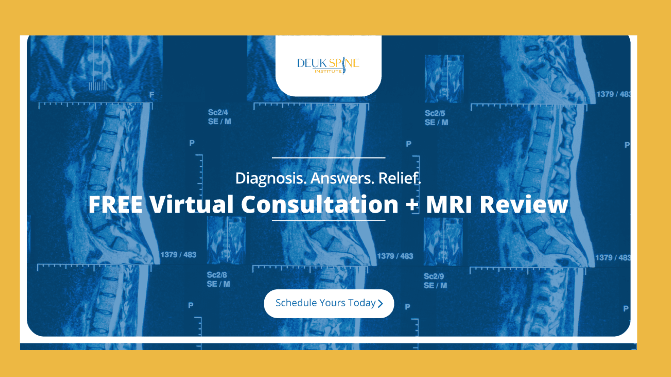
Searching for Ultra Minimally Invasive Spine Surgery?
Upload your latest MRI for a free virtual consultation and review with Dr. Ara Deukmedjian, a pioneer of minimally invasive techniques, Deuk Laser Disc Repair®.
Recovery Timeline and Expectations
Understanding the recovery process helps set realistic expectations and promotes optimal outcomes.
Conservative Treatment Recovery
For patients pursuing non-surgical management, symptoms typically improve progressively over 6-12 weeks. However, nerve healing follows a specific timeline: peripheral nerves regenerate at approximately 1.4mm per day, meaning complete recovery from nerve irritation may take several months even after mechanical compression is relieved.
During recovery:
- Acute pain often improves within 2-4 weeks
- Numbness and tingling may persist for 6-12 weeks
- Muscle strength gradually returns over 3-6 months
- Full functional recovery may require 6-12 months
Surgical Recovery
Patients undergoing Ultra Minimally Invasive Endoscopic Spine Surgery typically experience:
- Immediate relief of nerve compression symptoms
- Walking around within 1 hour of procedure completion
- Gradual return to light activities within days
- Progressive increase in activity over 4-6 weeks
- Full recovery, with lifting restrictions for one year, within 8-12 weeks
Prevention Strategies: Protecting Your Spine
Treatment for nerve root impingement follows a graduated approach, beginning with conservative measures and progressing to more invasive interventions when necessary.
Workplace Ergonomics
Optimize Your Workstation:
- Position monitors at eye level to prevent neck flexion
- Ensure chairs provide lumbar support and allow feet to rest flat
- Keep keyboards and mice at elbow height with shoulders relaxed
- Take micro-breaks every 30-60 minutes to change positions
Research confirms that ergonomic interventions, when combined with physical therapy and active rehabilitation, significantly reduce work-related musculoskeletal pain.
For Manual Labor Professions:
- Use proper lifting technique: bend at knees, keep load close, avoid twisting
- Employ mechanical aids when available
- Rotate tasks to avoid sustained awkward postures
- Request ergonomic assessments if experiencing symptoms
Lifestyle Modifications
Maintain healthy body weight: Excess weight increases mechanical stress on the spine, accelerating degenerative changes and increasing the risk of disc herniation.
Regular Exercise Program:
- Core strengthening exercises stabilize the spine
- Flexibility work reduces nerve tension and maintains mobility
- Weight-bearing activities promote bone and disc health
- Cardiovascular exercise improves tissue nutrition
Smoking cessation: Given smoking’s proven negative effects on disc health and recovery, quitting represents one of the most impactful preventive measures.
Proper Sleep Ergonomics:
- Choose supportive mattresses that maintain spinal alignment
- Use pillows that keep the neck in a neutral position
- Avoid prolonged stomach sleeping, which hyperextends the neck
Posture Awareness
Maintaining proper cervical posture during daily activities is crucial for preventing nerve root irritation. Key principles include:
- Keep ears aligned over shoulders
- Avoid sustained neck flexion during screen use
- Rotate the trunk rather than twisting the neck when changing direction
- Take regular breaks from static postures
Frequently Asked Questions (FAQs)
Q: How do I know if I have nerve root impingement or just a pinched nerve?
A: Nerve root impingement is a specific type of pinched nerve that occurs where the nerve exits the spinal cord. The key distinguishing feature is the pattern of symptoms: nerve root impingement typically causes pain, numbness, and weakness that follow a specific dermatomal pattern (along the nerve’s path from the spine to the extremity). For example, L5 nerve root impingement causes symptoms down the outer leg to the top of the foot, while C7 nerve root impingement affects the arm and middle finger. General pinched nerves at other locations (like carpal tunnel syndrome) cause more localized symptoms. A thorough examination by a spine specialist and MRI imaging can definitively identify nerve root impingement.
Q: Can nerve root impingement heal on its own?
A: Yes, research shows that 85-90% of acute nerve root impingement cases from herniated discs improve significantly within 6-12 weeks without surgery. The body’s natural healing processes include reabsorption of herniated disc material and resolution of inflammation around the nerve root. However, nerves heal slowly (approximately 1.4mm per day), so complete resolution of all symptoms may take several months. Factors that promote natural healing include maintaining appropriate activity levels, avoiding aggravating positions, managing inflammation, and allowing adequate time for recovery.
Q: What’s the difference between radiculopathy and nerve root impingement?
A: These terms are essentially synonymous. Radiculopathy is the medical term for nerve root impingement or compression. Both describe a condition where a spinal nerve root is compressed or irritated at or near its exit point from the spinal cord, causing pain, numbness, tingling, or weakness along the nerve’s distribution. “Cervical radiculopathy” and “cervical nerve root impingement” mean the same thing, as do “lumbar radiculopathy” and “lumbar nerve root impingement.”
Q: How long does it take for nerve root impingement to heal after surgery?
A: Recovery timeline varies based on the surgical technique and severity of nerve compression before surgery. With traditional surgery, patients typically require several weeks to months for full recovery. However, with Ultra Minimally Invasive Endoscopic Spine Surgery, such as Deuk Laser Disc Repair®, patients experience immediate relief of compression and can return home within one hour. Most patients report significant symptom improvement within the first week, with progressive recovery over 8-12 weeks. Because nerves heal slowly, some numbness or tingling may persist for several months even after successful decompression.
Q: Can poor posture really cause nerve root impingement?
A: While poor posture alone doesn’t directly cause nerve root impingement, it significantly accelerates the degenerative changes that lead to this condition. Poor posture increases mechanical stress on intervertebral discs, promotes premature disc degeneration, and can precipitate disc herniation in already weakened discs. Research shows that forward head posture adds 10 pounds of pressure per inch of forward lean, dramatically increasing cervical spine stress. Similarly, slouched sitting increases lumbar disc pressure by approximately 40% compared to standing. Over time, these sustained stresses contribute to the structural changes that ultimately compress nerve roots.
Q: What are the warning signs that I need surgery for nerve root impingement?
A: Immediate surgical consultation is warranted for progressive leg weakness, loss of bowel or bladder control, saddle numbness (numbness in the area that would contact a saddle), or rapidly worsening neurological symptoms. These could indicate cauda equina syndrome, a surgical emergency. Relative indications for surgery include: disabling pain that hasn’t improved after 6-12 weeks of conservative treatment, recurrent episodes significantly impacting quality of life, persistent muscle weakness despite conservative care, or symptoms that prevent you from working or performing essential daily activities. A spine specialist can evaluate whether your specific situation would benefit from surgical intervention.
Q: Is nerve root impingement the same as a herniated disc?
A: Not exactly, though they’re closely related. A herniated disc is a structural problem where the inner disc material protrudes through a tear in the outer disc wall. Nerve root impingement refers to compression or irritation of a nerve root, which can be caused by a herniated disc or by conditions such as bone spurs, spinal stenosis, or facet joint hypertrophy. Essentially, a herniated disc is the most common cause of nerve root impingement, but not all herniated discs cause nerve impingement, and not all nerve impingement comes from herniated discs.
Q: Will physical therapy help nerve root impingement, or do I need surgery?
A: Most cases of nerve root impingement initially respond well to conservative treatment, including physical therapy. Research shows that 85-90% of acute cases improve without surgery. Physical therapy helps by strengthening core muscles to better support the spine, improving flexibility to reduce nerve tension, correcting postural problems that contribute to symptoms, and teaching proper body mechanics. However, therapy works best when started early and followed consistently. If symptoms persist beyond 6-12 weeks despite appropriate conservative care, or if you develop progressive weakness, surgical consultation becomes appropriate. The key is correlating the severity of imaging findings with your symptoms and functional limitations.
Q: What activities should I avoid if I have nerve root impingement?
A: During the acute phase, avoid activities that increase pain or neurological symptoms: heavy lifting (especially with twisting), prolonged sitting without position changes, repetitive forward bending, high-impact activities like running or jumping, and contact sports. However, complete bed rest is not recommended; maintaining gentle activity promotes recovery. Once acute symptoms improve, gradually return to activities while avoiding positions that reproduce symptoms. Long-term, modify occupational exposures by using proper ergonomics, taking regular breaks from static postures, employing correct lifting techniques, and maintaining core strength. Your spine specialist or physical therapist can provide specific guidance tailored to your situation.
Q: Can nerve root impingement cause permanent damage?
A: If left untreated for extended periods, severe nerve root compression can cause permanent nerve damage. Warning signs include progressive muscle wasting (atrophy), persistent severe weakness, or complete loss of sensation in affected areas. The time to permanent damage varies, but most experts recommend surgical evaluation if significant weakness persists beyond 3-6 months or progressively worsens. However, with appropriate, timely treatment, whether conservative care or surgery, most patients recover fully without permanent deficits. The key is recognizing concerning symptoms early and seeking specialized evaluation when conservative measures aren’t providing adequate improvement.
Take the Next Step Toward Pain-Free Living
At Deuk Spine Institute, we specialize in Ultra Minimally Invasive Endoscopic Spine Surgery and comprehensive treatment approaches designed to cure, not just manage, nerve root impingement. Our world-class physicians are personally invested in every patient’s well-being and utilize techniques and technologies unavailable anywhere else.
Start your journey to recovery by submitting your MRI online for a free virtual consultation to determine your candidacy for our advanced surgical procedures. You can also schedule an in-person appointment at one of our locations by calling patient services at 1-800-FIX-MY-BACK (1-800-349-6922).
Don’t let nerve root impingement control your life. Experience the Deuk Spine difference and discover why our high success rate and one-hour outpatient procedures are revolutionizing spine care.
Sources
1: https://www.ncbi.nlm.nih.gov/books/NBK560878/
2: https://accidentcarechiropractic.com/symptoms/nerve-root-impingement/
3: https://www.ncbi.nlm.nih.gov/books/NBK441828/
4: https://spinehealth.org/article/spine-posture-workplace-ergonomics/
5: https://www.nhsfife.org/media/vztk8kti/nerve_root_pain_v7.pdf
6: https://pubmed.ncbi.nlm.nih.gov/9188034/
7: https://www.ncbi.nlm.nih.gov/books/NBK230871/
8: https://www.sciencedirect.com/topics/medicine-and-dentistry/nerve-root-compression
9: https://pmc.ncbi.nlm.nih.gov/articles/PMC2978791/
10: https://pubmed.ncbi.nlm.nih.gov/18427321/
11: https://pubmed.ncbi.nlm.nih.gov/24825130/
12: https://pubmed.ncbi.nlm.nih.gov/17119140/