


By Dr. Ara Deukmedjian, MD
Board-Certified Neurosurgeon, Deuk Spine Institute
Medically reviewed and updated on January 16, 2026
Medical Disclaimer: This content is for educational purposes only and does not constitute medical advice. Individual results may vary. Always consult with your healthcare provider about your specific condition and treatment options.
Key Points
✓ T7–T8 herniated discs are rare, accounting for less than 0.1% to 5% of all disc herniations, but they are frequently misdiagnosed as muscle strain, costochondritis, gallbladder disease, cardiac issues, or anxiety because thoracic nerves wrap around the chest and abdomen, producing band-like pain that mimics other conditions.
✓ The hallmark symptoms include deep, mechanical mid-back pain near the lower shoulder blades, radiating chest or rib pain that wraps around the torso, numbness or tingling along the chest wall, and in advanced cases, leg weakness, balance problems, or coordination issues caused by spinal cord compression (thoracic myelopathy).
✓ A thoracic MRI is the gold standard for diagnosis, but subtle findings are routinely missed by radiologists not specifically trained to evaluate the thoracic spine, which is why expert MRI review is essential before accepting any treatment plan, especially fusion.
✓ Traditional thoracic spine surgery (open thoracotomy, costotransversectomy, posterior decompression with fusion) carries higher risks than cervical or lumbar surgery due to the proximity of the spinal cord and limited working space, often requiring lengthy hospital stays, months of recovery, and the loss of natural spinal motion.
✓ Deuk Laser Disc Repair® treats the actual T7–T8 pain generator through a tiny incision with no chest opening, no rib removal, no fusion, and no hardware, removing only the 5–10% of damaged disc material causing symptoms, with a 99.6% success rate, same-day discharge, and return to work within one week.
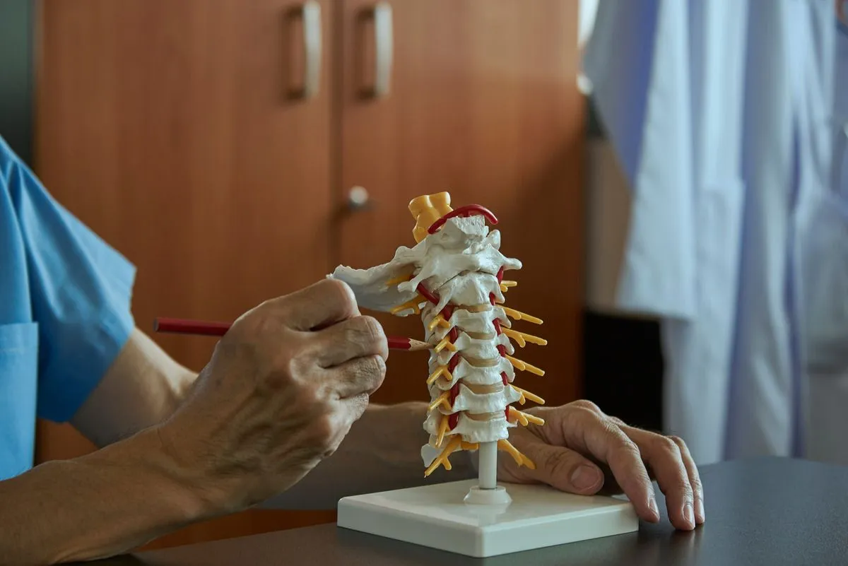
Understanding the Thoracic Spine and the T7–T8 Disc
The thoracic spine consists of 12 vertebrae, labeled T1 through T12, located in the middle of the back between the cervical spine (neck) and the lumbar spine (lower back). Between each vertebra sits an intervertebral disc that functions as a shock absorber and allows controlled movement.
Each disc consists of two distinct components:
- Annulus fibrosus: The tough, fibrous outer ring composed of multiple layers of collagen fibers arranged in a crisscross pattern
- Nucleus pulposus: The soft, gel-like inner core that provides cushioning and distributes compressive forces
The T7–T8 disc is located around the lower shoulder blade region, roughly at the midpoint of your thoracic spine. Unlike the cervical and lumbar areas of the spine, the thoracic spine is stabilized by the rib cage, which limits motion and reduces wear. This stability explains why disc herniations in this region are considerably less common than in the more mobile cervical and lumbar regions.
When a disc at T7–T8 herniates, the soft inner portion (nucleus pulposus) can protrude outward through a tear in the annulus fibrosus and compress nearby spinal nerves or, in more serious cases, the spinal cord itself. The pattern and severity of symptoms depend largely on what structure is being compressed and the degree of compression.
Clinical Insights from Dr. Deukmedjian, MD
Over my two decades performing spine surgery, I’ve encountered hundreds of patients with thoracic disc herniations who spent months being treated for conditions they didn’t have. One patient underwent extensive gastrointestinal testing, because they often report a burning sensation wrapping around the mid-back and into the upper abdomen that can worsen with extended periods of sitting.
This is the challenge with thoracic disc herniations. People simply don’t expect a disc problem in the mid-back, and neither do many physicians who aren’t spine specialists. However, when symptoms develop, they are very real and often progressive.
Why T7–T8 Herniated Discs Are Frequently Missed
Thoracic disc herniations present unique diagnostic challenges that often lead to delayed or incorrect diagnosis. Several factors contribute to this difficulty.
The Rarity Factor
Because thoracic disc herniations are not common, they’re often not the first condition considered during evaluation. The vast majority of disc problems occur in the lumbar spine (95%) or cervical spine (4%), making thoracic involvement statistically unlikely. This statistical reality can work against patients, as physicians naturally consider more common causes first.
Atypical Symptom Presentation
Unlike cervical and lumbar disc herniations, which follow relatively predictable symptom patterns, thoracic disc herniations can present with symptoms that mimic entirely different conditions. The thoracic nerves wrap around the chest and abdomen, so compression at T7–T8 can cause pain that seems unrelated to the spine.
Common misdiagnoses include:
- Muscle strain: The most frequent misdiagnosis, particularly in patients with desk jobs or poor posture
- Costochondritis: Inflammation of the cartilage connecting the ribs to the sternum
- Gastrointestinal disorders: Gallbladder disease, stomach ulcers, or pancreatitis when pain radiates to the upper abdomen
- Cardiac concerns: Chest wall pain is sometimes mistaken for heart-related problems
- Anxiety-related chest discomfort: Particularly when symptoms include chest tightness or difficulty breathing
- Shingles: When there’s band-like pain around one side of the torso
- Kidney problems: When pain radiates toward the flank
The medical literature confirms that thoracic disc herniations are rare and are often diagnosed late, particularly when symptoms are atypical or referred to other areas of the body.1
Limited Clinical Awareness
Many primary care physicians, and even some specialists, have limited experience with thoracic disc problems because they’re so uncommon. During medical training, emphasis is placed on cervical and lumbar spine pathology, with thoracic conditions receiving comparatively little attention. This educational gap can contribute to delayed diagnosis.
Common T7–T8 Herniated Disc Symptoms
Symptoms vary significantly depending on whether the disc is compressing a thoracic nerve root or the spinal cord, and on the degree of compression.
Mid-Back Pain
The most common symptom is persistent pain in the middle back, typically at the T7–T8 level near the lower shoulder blades. Patients often describe this pain as:
- Deep and aching in nature
- Sharp during twisting or bending movements
- Worse with prolonged sitting or standing
- Intensified by certain positions, particularly rotation or extension
- Unrelieved by rest alone
Unlike simple muscular pain, which typically improves with rest and responds well to over-the-counter pain relievers, disc-related pain persists and may worsen over time. The pain often has a mechanical quality, meaning it changes with position and movement, but doesn’t completely resolve regardless of what position you try.
Radiating Chest or Rib Pain
One of the most distinctive and misunderstood features of T7–T8 disc herniation is radiating pain that follows the distribution of the compressed thoracic nerve. Because thoracic nerves wrap around the chest and abdomen following the path of the ribs, compression at T7–T8 can cause pain that:
- Wraps around the rib cage from back to front
- Radiates into the chest wall
- Extends toward the upper abdomen
- Creates a band-like sensation around the torso
- May be felt on one side or both, depending on the herniation location
This type of pain is frequently mistaken for heart, lung, or digestive problems, leading patients to seek emergency or gastrointestinal evaluations before spinal imaging is ever performed. I’ve seen patients undergo extensive cardiac workups, including stress tests and even cardiac catheterization, before anyone considered their thoracic spine as the source of chest pain.
The Cleveland Clinic explains that thoracic nerve compression may cause band-like pain around the torso, a hallmark feature that distinguishes it from other conditions.2
Numbness or Tingling
A T7–T8 herniated disc may cause sensory changes that follow the dermatome distribution of the T7 or T8 nerve roots. These sensory disturbances include:
- Tingling sensations along the ribs or around the torso
- Numbness in patches of the chest or upper abdomen
- Heightened sensitivity (hyperesthesia) or reduced sensitivity (hypoesthesia) to touch
- Burning sensations in the affected distribution
- Pins-and-needles feelings that may be constant or intermittent
These symptoms may be subtle initially and become more noticeable as compression increases. Patients often describe these sensory changes as “strange” or “hard to pinpoint,” which can make them difficult to communicate to healthcare providers who aren’t familiar with the patterns of thoracic radiculopathy.
Weakness or Coordination Issues
In more advanced cases where the herniated disc compresses the spinal cord, a condition known as thoracic myelopathy, symptoms may appear far removed from the mid-back:
- Leg weakness or stiffness that progressively worsens
- Difficulty walking or changes in gait
- Balance problems and frequent stumbling
- Coordination difficulties with fine motor tasks
- Muscle spasms in the legs
- In rare and severe cases, changes in bowel or bladder function
This is one of the most challenging aspects of thoracic disc herniation diagnosis. When the spinal cord is involved, symptoms manifest in the lower extremities rather than at the level of the problem. A patient may complain only of leg weakness and balance problems without significant back pain, making the connection to a T7–T8 disc herniation far from obvious.
Dr. Deukmedjian emphasizes that when the spinal cord is involved, symptoms may appear far from the mid-back. This is why careful imaging and expert interpretation are essential.
The Mayo Clinic provides comprehensive information about herniated disc symptoms across all spinal regions.3
Additional Symptoms
Beyond the cardinal symptoms, patients may experience:
- Intercostal neuralgia: Sharp, stabbing pain between the ribs
- Difficulty breathing deeply: Not due to lung problems, but because deep breathing causes pain
- Postural changes: Leaning to one side or adopting unusual postures to minimize discomfort
- Sleep disturbances: Pain intensity often increases at night, particularly when lying flat
- Muscle spasms: Protective spasms in the paraspinal muscles
- Referred shoulder blade pain: Although the disc is at T7–T8, pain may be felt higher in the scapular region
What Causes a T7–T8 Herniated Disc?
While thoracic disc herniations are less common than cervical or lumbar herniations, when they do occur, several factors typically contribute to their development.
Degenerative Disc Changes
As discs age, they undergo natural degenerative changes that make them more vulnerable to herniation. The nucleus pulposus loses hydration and elasticity, while the annulus fibrosus develops small tears and weaknesses. This process, known as degenerative disc disease, typically begins in the third or fourth decade of life and progresses gradually.
In the thoracic spine, this degeneration occurs more slowly than in the cervical or lumbar regions due to the stabilizing effect of the rib cage. However, once degenerative changes reach a critical threshold, the disc becomes susceptible to herniation even with relatively minor stress.
Trauma or Injury
Acute traumatic events can cause immediate disc herniation. Common scenarios include:
- Falls: Particularly falls onto the back or from a height
- Motor vehicle accidents: Especially those involving sudden deceleration or rotational forces
- Sports injuries: Contact sports or activities involving sudden twisting motions
- Heavy lifting: Especially when combined with twisting or awkward positioning
- Workplace accidents: Industrial injuries or falls in occupational settings
The thoracic spine’s relative rigidity can actually make it more vulnerable to certain types of trauma. When force is applied to a relatively immobile segment, the energy must be absorbed by the discs rather than dispersed through motion.
Repetitive Mechanical Stress
Even without a single traumatic event, cumulative mechanical stress over years or decades can lead to disc herniation. Contributing factors include:
- Poor posture: Particularly prolonged slouching or forward head position
- Prolonged sitting: Especially in occupations requiring extended computer work
- Repetitive twisting motions: Common in certain occupations or sports
- Asymmetric loading: Consistently carrying heavy bags on one shoulder
- Inadequate core strength: Weak abdominal and back muscles place increased stress on discs
Genetic Predisposition
Research increasingly recognizes that genetic factors play a significant role in disc degeneration and herniation susceptibility. Some individuals experience disc degeneration earlier and more severely due to inherited structural factors, collagen abnormalities, or enzyme variations that affect disc composition.
If multiple family members have experienced disc problems at relatively young ages, genetic factors may be contributing to your risk.
For more information about the causes and mechanisms of disc herniation, visit the Deuk Spine Institute’s comprehensive resource.4
Diagnosing a T7–T8 Herniated Disc
Accurate diagnosis requires a systematic approach combining clinical evaluation with advanced imaging. Because thoracic disc herniations are so easily misdiagnosed, working with a spine specialist experienced in thoracic pathology is crucial.
Clinical Evaluation
The diagnostic process begins with a detailed medical history and physical examination. A thorough evaluation should include:
Medical History Assessment:
- Detailed description of symptom onset and progression
- Location and character of pain
- Factors that worsen or alleviate symptoms
- Impact on daily activities, work, and sleep
- Previous treatments and their effectiveness
- History of trauma or injury
- Presence of other medical conditions
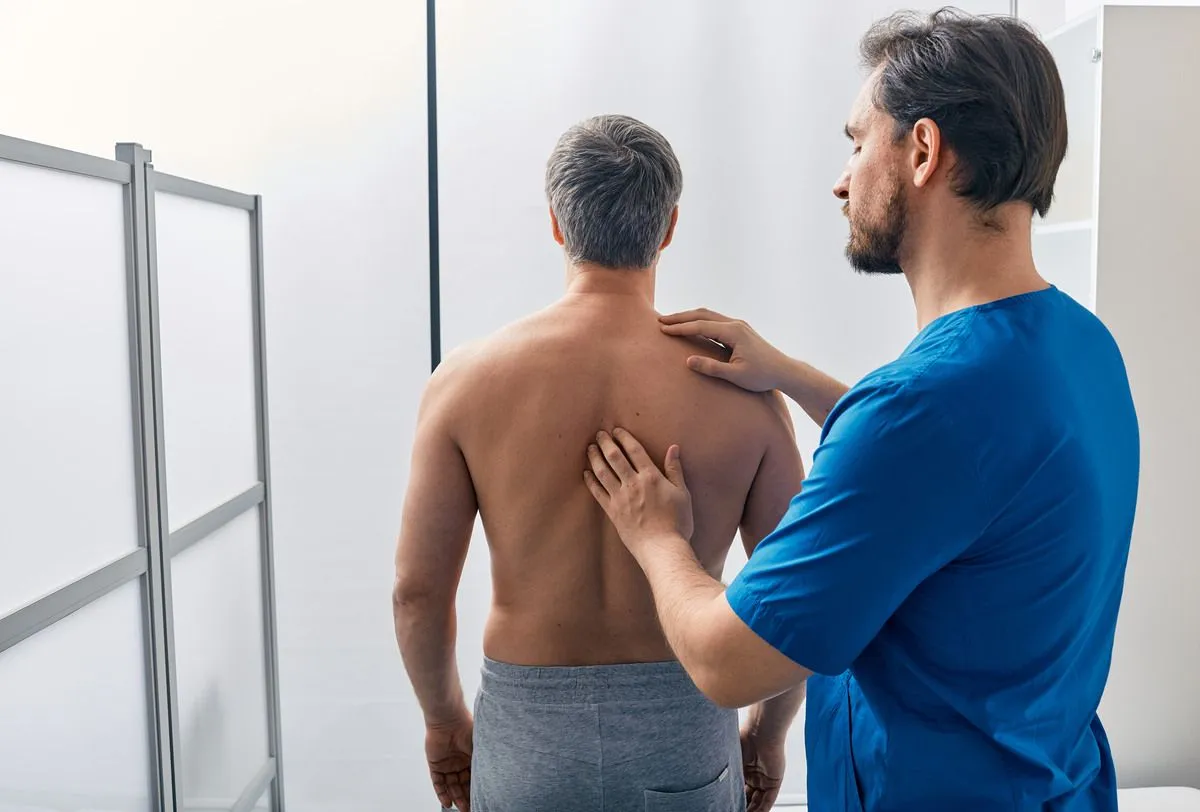
Physical Examination:
A comprehensive neurological examination focusing on:
- Inspection: Posture, spinal alignment, and any visible deformities
- Palpation: Tenderness along the thoracic spine and paraspinal muscles
- Range of motion: Thoracic spine flexion, extension, rotation, and lateral bending
- Neurological testing: Sensory examination of the chest and abdominal wall, motor strength testing, coordination assessment, and gait evaluation
- Special provocative tests: Positions or maneuvers that reproduce symptoms
MRI Imaging
Magnetic resonance imaging of the thoracic spine is the gold standard for identifying disc herniation and assessing nerve or spinal cord compression. MRI provides detailed visualization of soft tissues, including the intervertebral discs, spinal cord, nerve roots, and surrounding structures.
A thoracic MRI can reveal:
- The exact location and size of the disc herniation
- Whether the herniation is central, lateral, or foraminal
- The degree of spinal cord or nerve root compression
- Spinal cord signal changes that might indicate myelopathy
- Other degenerative findings or abnormalities
Dr. Deukmedjian notes that thoracic MRIs require careful imaging technique and experienced interpretation, as subtle findings are easily missed. I’ve reviewed countless outside MRIs in which significant thoracic pathology was overlooked or underappreciated because the radiologist wasn’t looking for it or didn’t recognize its significance.
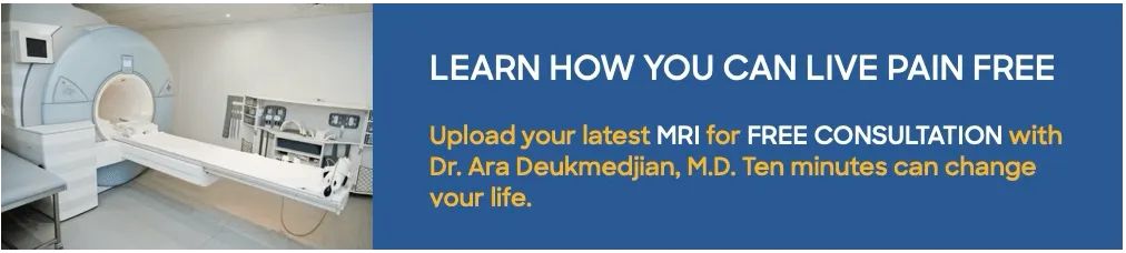
At Deuk Spine Institute, we offer a free consultation and MRI review for patients who want expert evaluation of their imaging studies. This second opinion can be invaluable, particularly for thoracic conditions that are so frequently misdiagnosed.
Seeking a Second Opinion?
Upload your latest MRI for a free consultation and MRI review with Dr. Ara Deukmedjian.
Additional Imaging Studies
While MRI is the primary diagnostic tool, other imaging modalities may provide complementary information:
X-rays:
- Assess overall spinal alignment
- Identify disc space narrowing, suggesting degeneration
- Reveal bony changes such as osteophytes
- Rule out fractures or structural abnormalities
CT Scan:
- Provides excellent bone detail
- Can identify calcified disc herniations
- Useful when MRI is contraindicated
- May be combined with myelography for enhanced soft tissue visualization
Electrodiagnostic Studies:
In select cases, electromyography (EMG) and nerve conduction studies may help confirm nerve involvement and rule out other neurological conditions, though they’re less commonly used for thoracic radiculopathy than for cervical or lumbar conditions.
Non-Surgical Treatment Options
Many patients with mild to moderate T7–T8 disc herniation symptoms improve with comprehensive conservative treatment. While recovery rates for thoracic disc herniations are less well-studied than for cervical or lumbar herniations, appropriate non-surgical management provides relief for a significant percentage of patients.
Physical Therapy
A structured physical therapy program forms the cornerstone of conservative treatment. An experienced physical therapist can design a program that includes:
Postural Training:
Thoracic disc problems are often associated with poor posture, particularly the forward-rounded shoulder position common with desk work. Postural retraining focuses on maintaining proper spinal alignment throughout daily activities.
Core Strengthening:
Strengthening the abdominal muscles, back extensors, and scapular stabilizers provides better support for the thoracic spine and reduces mechanical stress on the affected disc.
Flexibility Exercises:
Gentle stretching to improve thoracic spine mobility and reduce muscle tension can help alleviate symptoms. Thoracic extension exercises, in particular, can counteract the forward-flexed postures that stress the anterior disc.
Manual Therapy:
Skilled manual techniques, including soft tissue mobilization, joint mobilization, and myofascial release, may provide symptomatic relief, though aggressive manipulation should be avoided in the presence of a disc herniation.
Medications
Various medications can help manage symptoms during the healing process:
Anti-inflammatory Medications:
Non-steroidal anti-inflammatory drugs (NSAIDs) such as ibuprofen or naproxen reduce inflammation around the compressed nerve root. However, prolonged use carries risks of gastrointestinal and cardiovascular side effects.
Neuropathic Pain Medications:
Drugs such as gabapentin or pregabalin specifically target nerve pain and may be more effective than traditional pain relievers for radicular symptoms.
Muscle Relaxants:
Short-term use can help reduce painful muscle spasms that often accompany a disc herniation.
Oral Corticosteroids:
A short course (typically 5-7 days) of oral steroids may be prescribed to reduce acute inflammation, though evidence for long-term benefit is limited.
Activity Modification
Making intelligent adjustments to daily activities can facilitate healing and prevent symptom exacerbation:
- Avoid prolonged sitting, particularly in poor postural positions
- Take frequent breaks from static activities
- Use proper ergonomics at workstations
- Avoid heavy lifting and activities that require repetitive twisting
- Modify sleeping positions to reduce strain on the thoracic spine
Targeted Spinal Injections
These targeted injections can both diagnose and treat specific nerve root compression, though they’re technically more challenging in the thoracic spine than in the lumbar region.
It’s important to understand that injections typically provide only temporary relief, not definitive solutions. They may be valuable as a bridge to more definitive treatment or to confirm the source of the pain before surgery.
Harvard Health reports that many disc herniations improve over time with non-surgical care, although thoracic discs can be less predictable than lumbar or cervical discs.5
When Surgery Becomes Necessary
While many patients improve with conservative treatment, certain situations warrant surgical intervention. Understanding when surgery is appropriate can help you make informed decisions about your care.
Clear Indications for Surgery
Progressive Neurological Deficit:
If you’re experiencing worsening weakness, coordination problems, or balance difficulties despite conservative treatment, surgery should be considered promptly. Prolonged spinal cord compression can lead to permanent neurological damage.
Severe, Intractable Pain:
Pain that significantly impairs your quality of life and doesn’t respond to comprehensive conservative treatment (including physical therapy, medications, and injections) may require surgical intervention.
Spinal Cord Compression (Myelopathy):
Symptoms of thoracic myelopathy—including gait disturbance, leg weakness, coordination problems, or bowel/bladder changes—typically require surgical decompression. The spinal cord doesn’t tolerate compression well, and delays in treatment can result in permanent deficits.
Failed Conservative Treatment:
If you’ve undergone an appropriate trial of conservative care (typically 8-12 weeks for radiculopathy, shorter for myelopathy) without adequate improvement, surgery becomes a reasonable option.
The Challenge of Traditional Thoracic Spine Surgery
Traditional surgical approaches to thoracic disc herniation have historically been more challenging and carried higher risks than cervical or lumbar disc surgery. The proximity of the spinal cord, the limited working space, and the vascular structures in the thoracic region make these procedures technically demanding.
Conventional approaches have included:
Posterior Decompression:
Accessing the disc from the back of the spine, which may require removal of significant bone and carry risks of spinal cord injury.
Anterior Thoracotomy:
A major open chest surgery approaching the spine from the front, which requires entering the chest cavity and carries substantial risks and a lengthy recovery.
Costotransversectomy:
Removing a portion of the rib and the transverse process to access the disc laterally is a complex procedure with significant tissue trauma.
These traditional approaches can be highly invasive, require lengthy hospital stays, involve substantial postoperative pain, and carry significant complication risks.
Modern Minimally Invasive Surgical Options
At Deuk Spine Institute, treatment focuses on motion-preserving, minimally invasive approaches whenever appropriate. The goal is to address the herniated disc causing symptoms while minimizing tissue trauma and preserving normal spinal function.
Deuk Laser Disc Repair® for Thoracic Herniations
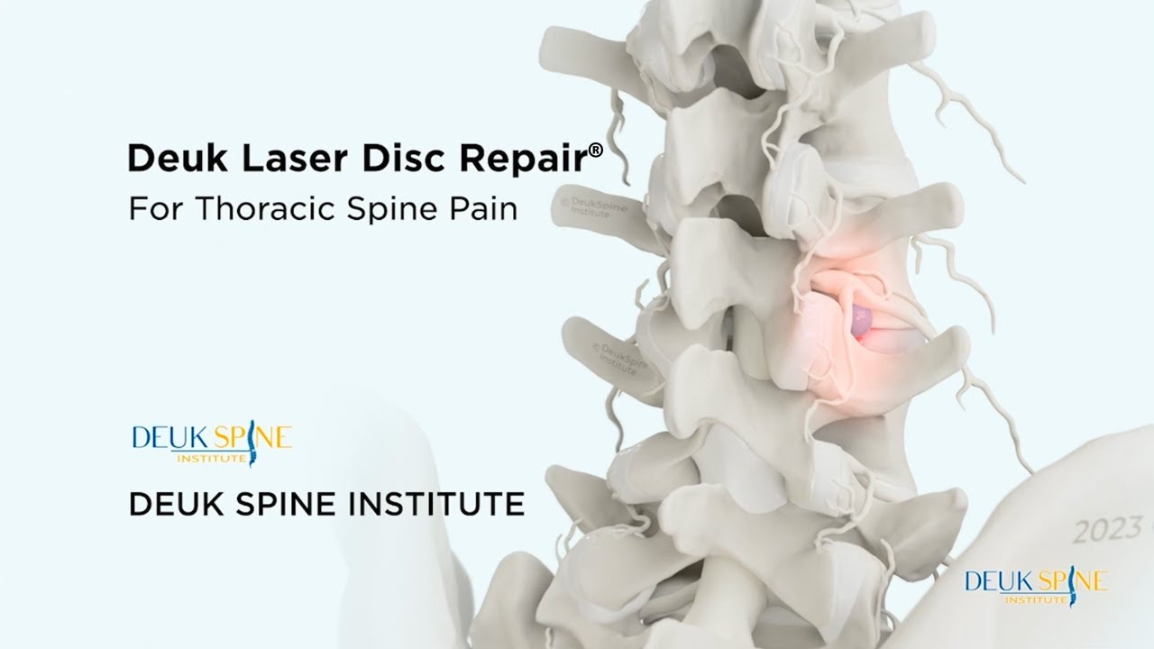
Deuk Laser Disc Repair® represents a revolutionary approach to treating thoracic disc herniations. Unlike traditional surgery that requires major incisions, extensive tissue dissection, and often spinal fusion or hardware placement, this advanced technique targets only the damaged portion of the disc while preserving healthy tissue and normal spinal mechanics.
How It Works:
The procedure is performed through a tiny incision. Through this minimally invasive opening, a specialized endoscope provides high-definition visualization of the surgical field. Using advanced laser technology, the surgeon can precisely target and remove only the damaged disc material causing symptoms, typically just 5-10% of the total disc.
Because most of the disc remains intact and functional, there’s no need for fusion, bone grafts, metal implants, or artificial disc replacement. The natural disc continues to provide cushioning and allow normal motion.
Recovery Experience:
Recovery from thoracic Deuk Laser Disc Repair® is remarkably quick:
- Day of surgery: Most patients notice significant pain relief as soon as they wake from anesthesia
- Same-day discharge: No hospital stay is required
- First week: The tiny incision heals within days and requires only a simple band-aid
- Return to work: Most patients return to desk work within 1 week
- Full recovery: Complete healing typically occurs within 2-3 weeks with minimal activity restrictions
Benefits Specific to Thoracic Treatment:
Dr. Deukmedjian explains that preserving natural spinal movement is especially important in the thoracic region, where stability and balance are critical. The thoracic spine provides the foundation for normal posture and respiratory mechanics. Fusion in this region can significantly impair these functions.
I’ve performed hundreds of thoracic disc procedures over my career, and the difference in recovery between traditional open approaches and minimally invasive endoscopic techniques is dramatic. Patients who would have spent days in the hospital, weeks on narcotic pain medications, and months in recovery can now return to normal life within days.
For detailed information about the benefits of thoracic Deuk Laser Disc Repair®, visit our thoracic spine treatment page.
Advantages Over Traditional Surgery
The benefits of this approach compared to traditional thoracic spine surgery are substantial:
- Minimal tissue trauma: No chest incision, rib removal, or extensive muscle dissection
- Preserved anatomy: No fusion means continued motion at the treated level
- No hardware: No implants that could migrate, fail, or cause complications
- Rapid recovery: Days instead of months
- Minimal pain: Most patients require only over-the-counter pain medication, if any
- Same-day discharge: No hospital stay required
- No adjacent segment disease: Because no fusion occurs, neighboring disc levels aren’t subjected to increased stress
- Excellent outcomes: High success rates with minimal complications
Living With and Recovering From a T7–T8 Herniated Disc
Recovery depends on symptom severity, treatment approach, and overall health. Whether you’re managing symptoms conservatively or recovering from minimally invasive surgery, following key principles can optimize your outcome.
Key Recovery Principles
Controlled Movement and Exercise:
While rest may be necessary during acute pain episodes, prolonged inactivity can actually worsen outcomes. Gentle movement, as tolerated, helps maintain flexibility, strength, and overall health. Work with a physical therapist or healthcare provider to determine appropriate activity levels.
Core and Postural Strengthening:
Strengthening the muscles that support your thoracic spine—including abdominal muscles, back extensors, and scapular stabilizers—provides better spinal support and reduces the likelihood of recurrent problems. These exercises should be progressive, starting gently and advancing as tolerated.
Avoiding Prolonged Inactivity:
Bed rest beyond a day or two is generally counterproductive. Extended inactivity leads to muscle weakening and stiffness and often worsens pain in the long run. Maintain as much normal activity as symptoms allow.
Ergonomic Awareness:
Pay attention to posture during daily activities. This is particularly important for individuals with desk jobs. Proper workstation setup, frequent position changes, and attention to posture can prevent symptom flare-ups and support healing.
Patient Perspective:
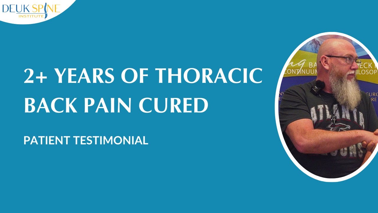
One of my patients who traveled from Georgia for treatment underwent thoracic Deuk Laser Disc Repair® for a T8–T9 herniated disc. He had suffered from debilitating back pain, tired legs, and rib pain for two years. The pain had become so severe that he described feeling hopeless and depressed, having been offered only fusion surgery by other physicians, an option he didn’t want to pursue.
After discovering Deuk Spine Institute through online research, he decided to travel to Florida for treatment. Just 12 hours after his procedure, he described immediate relief: “I feel no more tired legs, no more hurting ribs. I don’t know how to feel right now. I’ve had a hurting back for two years, been very depressed, felt hopeless.”
When I asked when he realized the pain was gone, he said, “Immediately after I woke up from surgery.” The next morning, he described getting out of bed without the struggle he’d experienced for two years: “I got straight up, and the other times I basically had to crawl out of bed. It’s been a long road. My body doesn’t know how to act right now.”
His advice to others: “If you’re having back problems and don’t know which way to turn, I advise you to do your research, find Deuk Spine. Dr. Deuk will fix your back, I guarantee it.” Watch his full testimonial.
Frequently Asked Questions
-
What does T7–T8 herniated disc pain feel like?
T7–T8 disc pain varies depending on what structures are being compressed and the severity of compression. The most common presentation is mid-back pain around the lower shoulder blade region, described as deep, aching, and mechanical in nature, meaning it changes with position and movement. Unlike muscular pain that typically improves with rest, disc-related pain persists and may worsen with certain movements, particularly twisting or prolonged sitting.
Many patients experience radiating pain that wraps around the rib cage from back to front, following the distribution of the compressed thoracic nerve. This pain can be sharp, burning, or electric-shock-like and may extend into the chest wall or upper abdomen. It’s this radiating pattern that often leads to misdiagnosis, as the pain mimics cardiac, pulmonary, or gastrointestinal conditions.
Some patients describe a band-like sensation around the torso, as if someone were tightening a belt. Numbness, tingling, or altered sensation in the same distribution may accompany the pain. In more advanced cases with spinal cord compression, pain may be less prominent than neurological symptoms like leg weakness or balance problems, which can make diagnosis particularly challenging.
-
Can a thoracic herniated disc heal on its own?
The question of whether thoracic disc herniations can heal spontaneously is complex. Some mild cases do improve with conservative treatment over time, as inflammation subsides and the body adapts to the disc changes. The immune system can recognize extruded disc material as foreign and initiate a process that helps resorb the herniated fragment, particularly with sequestered (completely separated) disc fragments.
However, thoracic disc herniations do not always resolve as predictably as lumbar disc herniations. Several factors influence the likelihood of spontaneous improvement, including the size and type of herniation, the degree of nerve or spinal cord compression, the presence of spinal stenosis, and patient-specific factors like age and overall health.
It’s important to understand that “healing” doesn’t mean the disc returns to its original, pre-injury state. Once the annulus fibrosus tears, permanent structural changes typically persist. While symptoms may resolve as inflammation decreases, the underlying disc abnormality often remains visible on imaging.
For patients with mild symptoms, a trial of conservative treatment is certainly reasonable. However, if symptoms persist beyond 8-12 weeks, progressively worsen, or include neurological signs like weakness or coordination problems, surgical evaluation is warranted. With modern minimally invasive approaches like Deuk Laser Disc Repair®, surgery no longer needs to be the dramatically invasive procedure it once was.
-
Is thoracic disc surgery dangerous?
The safety of thoracic disc surgery depends significantly on the specific approach used. Traditional open surgical techniques for thoracic disc herniation have historically carried higher risks than comparable cervical or lumbar procedures. This is due to several factors: the proximity of the spinal cord, the limited working space in the thoracic region, the presence of major vascular structures, and the need for extensive tissue dissection with traditional approaches.
Approaches like open thoracotomy (entering through the chest cavity) or extensive posterior decompressions involve significant surgical trauma and carry risks including spinal cord injury, vascular injury, pneumothorax (collapsed lung), significant blood loss, infection, prolonged recovery, and substantial postoperative pain.
However, modern minimally invasive techniques, such as Deuk Laser Disc Repair®, have dramatically changed the risk profile. By performing these procedures through tiny incisions with advanced endoscopic visualization, these procedures virtually eliminate the major risks associated with traditional surgery. The precision of the laser technology allows the surgeon to work around delicate neural structures without the collateral damage inherent in open approaches.
At Deuk Spine Institute, we’ve performed hundreds of thoracic Deuk Laser Disc Repair® procedures with a 99.6% success rate and zero surgical complications. The minimally invasive approach results in minimal blood loss (typically less than 3ml), no spinal cord manipulation, no hardware placement, same-day discharge, and rapid return to normal activities.
When performed by an experienced spine surgeon using advanced minimally invasive techniques, thoracic disc surgery can be remarkably safe and effective.
-
When should I seek medical attention for a suspected T7–T8 disc herniation?
You should seek prompt medical evaluation if you experience any of the following:
Immediate evaluation needed:
- Sudden onset of severe mid-back pain, especially following trauma
- Leg weakness that’s progressively worsening
- Difficulty walking or balance problems
- Numbness or tingling in both legs
- Bowel or bladder dysfunction
- Any combination of symptoms suggesting spinal cord compression
Evaluation within days to weeks:
- Persistent mid-back pain lasting more than a few weeks
- Pain that wraps around the rib cage or radiates to the chest or abdomen
- Numbness or tingling along the chest wall or abdomen
- Pain that significantly interferes with daily activities or sleep
- Symptoms that don’t improve with rest and over-the-counter pain medications
- Previous diagnosis of thoracic disc problems that have worsened
Remember that thoracic disc herniations are frequently misdiagnosed, so if you’ve been treated for other conditions without improvement, requesting a thoracic spine MRI may be appropriate.
-
How long does recovery take after T7–T8 disc surgery?
Recovery time varies dramatically depending on the surgical approach used. With traditional open thoracic disc surgery, including approaches like thoracotomy, costotransversectomy, or extensive posterior decompression, recovery is measured in months. Patients typically spend several days in the hospital, require narcotic pain medications for weeks, and may need 3-6 months before returning to full activities. These procedures involve significant tissue trauma, which necessitates prolonged healing.
In stark contrast, minimally invasive endoscopic procedures, such as Deuk Laser Disc Repair®, offer a dramatically faster recovery. Most patients experience immediate relief from arm or leg symptoms upon waking from anesthesia. They’re discharged home the same day, return to desk work within one week, achieve complete healing within 2-3 weeks, and require no long-term activity restrictions.
The difference stems from the fundamental approach. Traditional surgeries involve large incisions, significant muscle and bone removal, and often fusion or hardware placement. Minimally invasive endoscopic approaches work through tiny 4-7mm incisions, preserve normal anatomy, and cause minimal tissue trauma.
One of my thoracic surgery patients, an airline pilot, was particularly concerned about recovery time because of the strict medical requirements for his profession. After Deuk Laser Disc Repair® for his T7–T8 herniation, he returned to flying within three weeks with full medical clearance. He later said he couldn’t believe the dramatic difference between what he expected based on traditional surgery descriptions and his actual experience.
-
What happens if I don’t treat my T7–T8 disc herniation?
The natural history of untreated thoracic disc herniation varies significantly depending on the severity of the herniation and what structures are being compressed. For mild disc bulges causing minimal symptoms, some patients live indefinitely without significant progression. The symptoms may wax and wane with activities and stress levels, but remain manageable.
However, moderate to severe herniations, particularly those causing neurological symptoms, carry risks if left untreated. Prolonged nerve compression can lead to permanent nerve damage and chronic pain that becomes increasingly difficult to treat. Sensory changes may become permanent, and in cases of spinal cord compression (myelopathy), untreated progression can result in permanent weakness, gait disturbance, and coordination problems that don’t fully recover even after eventual treatment.
The challenge with thoracic disc herniations is their unpredictability. Unlike lumbar disc herniations, which often show spontaneous improvement, thoracic herniations can behave less predictably. Some remain stable for years, while others progressively worsen.
Based on my experience, I advise patients not to adopt a “wait and see” approach when symptoms significantly affect quality of life or when neurological signs are present. With modern minimally invasive options available, there’s no reason to suffer through months or years of symptoms, hoping for spontaneous resolution when definitive treatment is available with minimal risk and rapid recovery.
-
Can I prevent a T7–T8 disc herniation from recurring after treatment?
Prevention of recurrent disc herniation depends partly on the treatment approach used and partly on lifestyle modifications. In traditional fusion surgeries, recurrence at the treated level is eliminated because the segment no longer moves. However, fusion increases stress on adjacent disc levels, making herniation at neighboring levels more likely, a phenomenon called adjacent segment disease.
With motion-preserving procedures like Deuk Laser Disc Repair®, the treated disc continues to function normally, so theoretically, recurrence at the same level is possible. However, because the procedure removes only the damaged portion of the disc, recurrence rates are remarkably low. In our experience spanning hundreds of thoracic procedures, recurrence at the treated level is rare, typically occurring in less than 1% of cases.
Regardless of treatment approach, several strategies can reduce the risk of disc problems:
Maintain proper posture: Particularly during prolonged sitting or computer work
Strengthen core muscles: Strong abdominal and back muscles provide better spinal support
Maintain a healthy body weight: Excess weight increases mechanical stress on all spinal structures
Stay active: Regular exercise maintains disc nutrition and overall spinal health
Avoid smoking: Nicotine impairs blood flow to disc tissues and accelerates degeneration
Use proper ergonomics: At work, at home, and during recreational activities
Avoid repetitive stress: Particularly repetitive twisting motions with load
Key Takeaways: Understanding Your T7–T8 Disc Herniation
A T7–T8 herniated disc may be uncommon, but its effects can be life-altering. Persistent mid-back pain, chest-like symptoms, or unexplained neurological changes should never be ignored. Understanding your condition is the first step toward meaningful recovery.
The T7–T8 segment is located in the mid-thoracic spine and is subject to mechanical stress during daily activities. When the disc’s tough outer layer tears, allowing the soft inner material to protrude, it can compress the T7 or T8 nerve root—or in more severe cases, the spinal cord itself. This compression produces characteristic symptoms including mid-back pain, band-like pain wrapping around the rib cage, radiating chest or abdominal discomfort, numbness and tingling in dermatomal patterns, and, in advanced cases, leg weakness or coordination problems.
What makes thoracic disc herniations particularly challenging is their tendency to be misdiagnosed. Because they’re so uncommon (accounting for less than 1% of all disc herniations), and because symptoms often mimic cardiac, gastrointestinal, or pulmonary conditions, patients frequently undergo extensive testing and treatment for conditions they don’t have before anyone considers their thoracic spine.
While 75% of patients with mild symptoms improve with comprehensive conservative treatment, including physical therapy, medications, and activity modifications, those who don’t respond within 8-12 weeks or who develop progressive weakness should consider surgical intervention. The key is understanding that surgery doesn’t have to mean the dramatically invasive, high-risk procedures of the past.
Modern minimally invasive techniques, particularly Deuk Laser Disc Repair®, offer a superior alternative. By precisely removing only the damaged disc material (typically 5-10%) through a tiny incision, these procedures provide definitive relief while preserving normal spinal anatomy and function. With a 99.6% success rate, zero surgical complications, same-day discharge, and return to work within one week, patients can achieve lasting relief without the downsides of traditional surgery.
Dr. Deukmedjian often shares that many patients feel relief simply by learning there is a clear explanation for their symptoms. After months or years of being told their problems are muscular, anxiety-related, or psychosomatic, finally having concrete evidence of a structural problem that can be fixed provides hope. Understanding the source of pain is often the first step toward meaningful recovery.
If conservative treatments have not provided answers, or if you want expert insight into your imaging, consultation with a spine specialist experienced in thoracic conditions can help clarify next steps. At Deuk Spine Institute, we offer complimentary MRI reviews and virtual consultations. You can upload your latest MRI images securely through our website.
Don’t let a T7–T8 disc herniation control your life. Whether you ultimately choose conservative management or advanced minimally invasive treatment, making an informed decision based on accurate diagnosis and expert evaluation is essential. You deserve to understand your options and pursue the treatment approach that best fits your needs and goals.
Sources
1: https://pubmed.ncbi.nlm.nih.gov/40104307/
2: https://my.clevelandclinic.org/health/diseases/12768-herniated-disk
3: https://www.mayoclinic.org/diseases-conditions/herniated-disk/symptoms-causes/syc-20354095
4: https://deukspine.com/blog/herniated-disc/
5: https://www.health.harvard.edu/a_to_z/herniated-disk-a-to-z


