


By Dr. Ara Deukmedjian, MD
Board-Certified Spine Surgeon, Deuk Spine Institute
Medically reviewed on December 26, 2025
Medical Disclaimer: This content is for educational purposes only and does not constitute medical advice. Individual results may vary. Always consult with your healthcare provider about your specific conditions and treatment options.
If you’ve been told you have bone spurs, you’re not alone. These bony growths, medically known as osteophytes, are incredibly common, especially as we age. Think of them as your body’s way of trying to stabilize areas that have experienced wear and tear over the years. While the term “spur” might sound alarming, these smooth, rounded projections of extra bone tissue are actually your body’s attempt at self-repair.
Bone spurs typically develop near joints where two or more bones meet. They form gradually over time, often as a response to the breakdown of cartilage, the cushioning tissue that normally keeps bones from rubbing against each other. When cartilage wears down, your body tries to compensate by growing extra bone in an attempt to repair the damaged joint and increase its stability.
The interesting thing about bone spurs is that many people have them without even knowing it. They’re often discovered incidentally during X-rays or MRI scans performed for other reasons. However, when bone spurs do cause symptoms, they can significantly impact your quality of life, leading to pain, stiffness, and reduced mobility.
Who Develops Bone Spurs?
While anyone can develop bone spurs, certain factors increase your risk. Age is the most significant factor—most people who develop symptomatic bone spurs are over 60. However, younger individuals aren’t immune, particularly if they have a history of joint injuries or participate in high-impact activities.
Recent research from 2024 has shown that genetic factors may play a larger role than previously understood in the development of bone spurs.1 Individuals with a family history of osteoarthritis or other degenerative joint conditions are at higher risk. Additionally, obesity, repetitive stress on specific joints, poor posture, and previous joint injuries all contribute to the likelihood of developing bone spurs.
Athletes and individuals whose occupations involve repetitive movements or heavy lifting are particularly susceptible to developing bone spurs at a younger age. Construction workers, professional athletes, and others in physically demanding careers often develop bone spurs in their 40s or 50s rather than later in life.
Common Locations for Bone Spur Development
While bone spurs can technically form on any bone in your body, they most commonly appear in areas that experience significant stress or have undergone degenerative changes. The spine is a common site for bone spurs, particularly in the cervical (neck) and lumbar (lower back) regions. These spinal osteophytes can develop along the edges of vertebrae and within the facet joints, potentially narrowing the spinal canal or compressing nerve roots.
Beyond the spine, bone spurs often develop in weight-bearing joints such as the knees and hips. The feet are another common site, with heel spurs (calcaneal spurs) being particularly prevalent. These develop where the plantar fascia attaches to the heel bone and can cause significant discomfort with walking or standing.
Your hands and fingers can also develop bone spurs, often visible as small, hard lumps near the joints. These are particularly common in people with osteoarthritis and can affect grip strength and manual dexterity. The shoulders are another site where bone spurs may form, particularly around the rotator cuff, potentially contributing to impingement syndrome and reduced range of motion.
Spinal Conditions Associated with Bone Spurs
Bone spurs don’t exist in isolation—they’re often part of a larger picture of spinal degeneration. Understanding the conditions associated with bone spurs can help you better comprehend your diagnosis and treatment options.
Osteoarthritis of the Spine (Spondylosis)
Osteoarthritis is the most common condition associated with bone spur formation in the spine. Sometimes called degenerative arthritis or “wear and tear” arthritis, this condition develops when the protective cartilage within the facet joints begins to break down. As this cushioning deteriorates, your vertebrae experience increased friction during movement.
In response to this increased stress, your body produces extra bone tissue at the edges of vertebrae and within the facet joints themselves. While this is an attempt at stabilization, these bone spurs can actually contribute to further problems. They may narrow the spaces through which spinal nerves exit (foraminal stenosis) or reduce the diameter of the spinal canal itself.
Recent studies in 2024 have revealed that inflammatory markers play a more significant role in osteoarthritis progression than previously understood.2 This has led to new treatment approaches that target inflammation alongside structural changes.
Spinal Stenosis
Spinal stenosis is a narrowing of the spaces within your spine, most commonly in the lumbar (lower back) or cervical (neck) regions. Bone spurs are among the primary contributors to this narrowing. As osteophytes grow along the edges of vertebrae or within the facet joints, they gradually encroach upon the space available for the spinal cord and nerve roots.
When this happens in the central canal, it’s called central canal stenosis. When it occurs in the openings where nerves exit the spine, it’s called foraminal stenosis. Both types can cause significant symptoms, including pain, numbness, tingling, and weakness. In the lower back, spinal stenosis often causes a characteristic pattern of leg pain that worsens with walking and improves with rest, a condition called neurogenic claudication.3
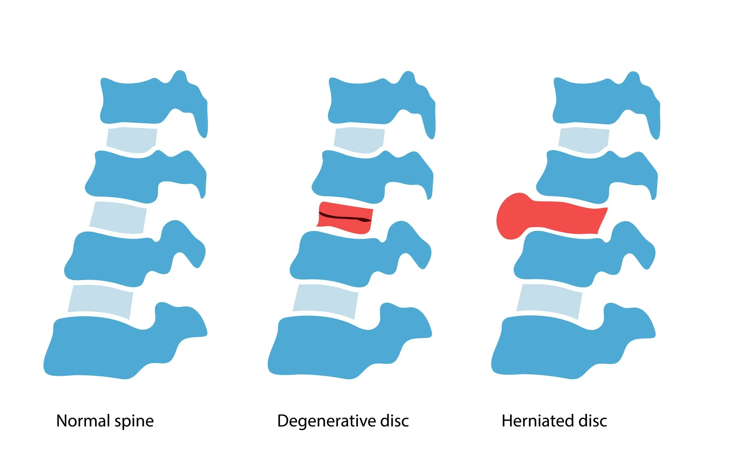
Degenerative Disc Disease
While the name suggests a disease, degenerative disc disease is actually a condition that describes the natural aging process of your spinal discs. These cushioning structures between your vertebrae lose water content over time, becoming thinner and less effective at absorbing shock. As the disc space narrows, it places additional stress on the vertebrae and facet joints.
This increased mechanical stress triggers the formation of bone spurs along the edges of vertebrae, a condition sometimes called vertebral endplate osteophytes. These bony growths attempt to increase the surface area between vertebrae to better distribute the load that’s no longer being absorbed by healthy discs.
The combination of disc degeneration and bone spur formation can lead to a range of symptoms, from localized back pain to radiating nerve pain if the osteophytes compress nerve structures.4
Facet Joint Hypertrophy
The facet joints are small joints located at the back of your spine that provide stability and guide spinal movement. Like other joints in your body, they’re covered with cartilage and can develop arthritis over time. When facet joints undergo arthritic changes, they often enlarge—a condition called facet joint hypertrophy.
This enlargement typically includes bone spur formation along the joint margins. As these osteophytes grow, they can contribute to spinal stenosis, narrow the neural foramina, and cause inflammation of surrounding soft tissues. Facet joint hypertrophy with associated bone spurs is particularly common in the lumbar spine, where these joints bear significant weight and undergo substantial mechanical stress during daily activities.
Herniated and Bulging Discs
While bone spurs and disc herniations are separate conditions, they often coexist and can compound each other’s symptoms. When a disc herniates or bulges, it creates an unstable segment in your spine. Your body may respond to this instability by forming bone spurs to stabilize the affected area.
Conversely, bone spurs that develop due to other degenerative processes can place additional stress on nearby discs, potentially contributing to disc bulging or herniation. When both conditions are present, they can create a complex clinical picture with multiple pain generators.5
Ankylosing Spondylitis
Unlike the degenerative conditions mentioned above, ankylosing spondylitis is an inflammatory arthritis that primarily affects the spine. This autoimmune condition causes inflammation of the vertebrae, which can eventually lead to fusion of spinal segments. As part of the inflammatory and healing process, bone spurs form between vertebrae, sometimes creating bony bridges called syndesmophytes.
While this condition is less common than degenerative causes of bone spurs, it’s important to distinguish it because the treatment approach differs significantly. Ankylosing spondylitis requires management of the underlying inflammatory process in addition to addressing symptoms.
Recognizing the Symptoms of Spinal Bone Spurs
The symptoms you experience from bone spurs depend entirely on their location and whether they’re pressing on nerves or other sensitive structures. Many people have bone spurs that never cause symptoms—these are sometimes called “silent” osteophytes and are only discovered during imaging for unrelated reasons.
However, when bone spurs do cause symptoms, they can range from mild and intermittent to severe and debilitating. Understanding these symptoms can help you recognize when it’s time to seek medical evaluation.
Pain and Discomfort
Localized pain is often the first symptom people notice. In the spine, this might manifest as neck pain if the bone spurs are in the cervical region, or lower back pain if they’re in the lumbar spine. The pain typically starts gradually and may worsen with activity or at the end of the day. Morning stiffness that improves with movement is also common.
What makes bone spur pain distinctive is that it often has both mechanical and inflammatory components. Mechanical pain arises from abnormal movement and stress in the affected area, while inflammation in surrounding soft tissues contributes to additional discomfort. This combination means that pain might not respond predictably to rest or activity.
Radiating Pain and Nerve Symptoms
When bone spurs compress or irritate nerve roots, the symptoms can extend far beyond the spine itself. In the neck, cervical bone spurs might cause pain that radiates into your shoulders, arms, or hands. You might notice sharp, shooting sensations or a persistent burning sensation along the path of the affected nerve.
In the lower back, bone spurs that compress nerve roots can cause sciatica—pain that travels down one or both legs. This can be accompanied by sensations of numbness, tingling, or a “pins and needles” feeling. Some people describe it as feeling like an electric shock running down their leg.
Research published in 2024 has shown that nerve irritation from bone spurs often involves a combination of mechanical compression and chemical inflammation.6 The growth factors and inflammatory mediators released during bone spur formation can sensitize nerves, even in the absence of direct physical pressure.
Numbness and Weakness
As nerve compression becomes more severe or prolonged, you might start to notice numbness in specific patterns corresponding to the affected nerve. In your arms, this might affect your thumb and index finger (C6 nerve root), or your ring and pinky fingers (C8 nerve root). In the legs, numbness might appear on the top of your foot, the sole, or along the outer leg.
Muscle weakness is a more concerning symptom that suggests significant nerve involvement. You might notice difficulty gripping objects, trouble lifting your foot (foot drop), or weakness when trying to stand from a seated position. These motor symptoms indicate that the nerve is not just being irritated, but that its function is actually being impaired.
Reduced Range of Motion and Stiffness
Bone spurs in the facet joints or along vertebral bodies can mechanically limit your spine’s movement. You might notice that you can’t turn your head as far as you used to, or that bending forward or backward has become more restricted. This stiffness tends to be worse in the morning and after periods of inactivity.
The reduced range of motion isn’t just from the physical presence of the bone spurs themselves. The surrounding soft tissues—muscles, ligaments, and joint capsules—often become tight and fibrotic in response to chronic irritation and altered movement patterns. This creates a cycle in which limited motion leads to increased stiffness, which further limits motion.
Additional Symptoms
Some people experience headaches when cervical bone spurs affect the upper neck, particularly near the base of the skull. These headaches typically start at the back of the head and may radiate forward. Balance problems can occur if bone spurs compress the spinal cord, though this is less common.
In severe cases of spinal cord compression from bone spurs (cervical myelopathy), you might notice coordination problems, difficulty with fine motor tasks like buttoning a shirt, or changes in your walking pattern. These symptoms require prompt medical evaluation as they indicate significant spinal cord involvement.
When Conservative Treatment Isn’t Enough: Signs You May Need Surgery
Most people with bone spurs don’t need surgery. In fact, conservative treatments successfully manage symptoms for the majority of patients. However, there are specific situations in which surgical intervention is necessary or advisable. Understanding these scenarios can help you make informed decisions about your care.
Progressive Neurological Symptoms
One of the clearest indicators that surgery may be needed is the development of progressive neurological symptoms. If you’re experiencing increasing numbness, spreading weakness, or worsening nerve pain despite conservative treatments, this suggests that the nerve compression from bone spurs is not resolving on its own.
Progressive weakness is particularly concerning because prolonged nerve compression can lead to permanent damage. If you notice that your grip is getting weaker, you’re having more frequent episodes of foot drop, or that numbness is spreading to involve larger areas, these are red flags that warrant surgical evaluation.
Spinal Cord Compression
When bone spurs in the cervical spine grow large enough to compress the spinal cord, this condition is called cervical myelopathy. Unlike nerve root compression, which affects specific limbs, spinal cord compression can affect function throughout your body below the level of compression.
Signs of myelopathy include difficulty with coordination, balance, and walking, changes in bowel or bladder function, and loss of fine motor control in your hands. This is a serious condition that often requires surgical intervention because the spinal cord has a limited capacity to recover once damage becomes severe.
Failed Conservative Management
If you’ve diligently pursued conservative treatments for 3-6 months without significant improvement, surgery may be a reasonable next step. This timeline is important—bone spurs and their associated conditions often improve with consistent non-surgical care, and rushing to surgery isn’t advisable for most patients.
However, “failed conservative management” doesn’t just mean that some pain remains. It means that your symptoms are significantly limiting your quality of life despite optimal non-surgical treatment. You might be unable to work, struggling with daily activities, or finding that pain is affecting your sleep and mental health.
Severe Spinal Stenosis
When imaging studies show severe narrowing of the spinal canal or neural foramen due to bone spurs, and this corresponds with your symptoms, surgery is often the most effective treatment. Severe stenosis has limited improvement potential with conservative care because the physical space cannot be increased without removing the offending bone.
Advanced imaging techniques developed in recent years allow surgeons to more accurately assess stenosis severity and predict which patients are most likely to benefit from surgical decompression.7
Significant Functional Impairment
If bone spurs are preventing you from performing essential daily activities despite other treatments, this represents a valid reason to consider surgery. This includes being unable to walk reasonable distances, difficulty caring for yourself, or the inability to work at a job that’s appropriate for your education and skills.
Quality of life matters, and while surgery carries risks, living with severe, unrelenting symptoms also has high costs to your physical and mental well-being. The decision to pursue surgery should involve a careful discussion with your surgeon about realistic expectations and the likely improvement from surgery.
Cauda Equina Syndrome
Though rare, cauda equina syndrome is a surgical emergency. This occurs when bone spurs, or other structures, compress the bundle of nerve roots at the base of the spinal cord. Symptoms include severe low back pain, numbness in the saddle area (inner thighs and buttocks), loss of bladder or bowel control, and weakness in both legs.
If you experience these symptoms, you should seek emergency medical care immediately. Decompression surgery within 24-48 hours offers the best chance of recovery from this condition.
Get Answers. Find Relief.
Upload your latest MRI for a free 10-minute virtual consultation and MRI review with neurosurgeon Dr. Ara Deukmedjian today.
Conservative Treatment Options: The First Line of Defense
Before considering surgery, most patients with bone spurs will try conservative treatments. These non-surgical approaches aim to reduce pain, improve function, and help you manage symptoms effectively. While they won’t remove existing bone spurs, they can often provide significant relief and prevent progression of symptoms.
Medication Management
Pain relief often begins with medication. Over-the-counter options like acetaminophen can help with mild pain, while non-steroidal anti-inflammatory drugs (NSAIDs) such as ibuprofen or naproxen address both pain and inflammation. These medications work by reducing the inflammatory response around bone spurs and affected joints, providing relief for many patients with mild to moderate symptoms.
For more significant pain, your doctor might prescribe stronger medications. Short-term use of prescription NSAIDs, muscle relaxants for associated muscle spasms, or nerve pain medications like gabapentin or pregabalin for radiating nerve symptoms may be appropriate. It’s important to use these medications under medical supervision, as they can have significant negative side effects with long-term use.
Recent advances in topical pain medications have also provided new options. Prescription-strength topical NSAIDs and lidocaine patches can provide localized relief without the systemic side effects of oral medications, making them particularly useful for patients who can’t tolerate traditional pain relievers.
Physical Therapy and Exercise
Physical therapy forms the cornerstone of conservative treatment for bone spurs. A skilled physical therapist can design a program that addresses your specific symptoms and limitations. This might include exercises to strengthen the muscles supporting your spine, stretches to improve flexibility, and techniques to improve your posture and movement patterns.
Core strengthening is particularly important for spinal bone spurs. When the muscles of your abdomen and back are strong and work together effectively, they reduce the load on your spine’s bony structures and joints. This can significantly decrease pain and may slow the progression of degenerative changes.
Manual therapy techniques, including joint mobilization and soft tissue work, can improve mobility and reduce pain. Your physical therapist might also teach you proper body mechanics for daily activities to minimize stress on affected areas. Consistency is key—the patients who see the best results are those who commit to their home exercise programs and make physical therapy an ongoing part of their lifestyle.
Lifestyle Modifications
Simple changes to your daily routine can make a surprising difference in managing bone spur symptoms. Weight management is crucial if you’re carrying extra pounds, as excess weight places additional stress on weight-bearing joints and the spine. Even modest weight loss of 5-10% of your body weight can lead to noticeable improvement in symptoms.
Ergonomic adjustments at work and home can reduce the mechanical stress that aggravates bone spurs. This might mean adjusting your desk setup, using supportive chairs, taking frequent breaks from prolonged sitting or standing, and being mindful of your posture throughout the day. For those with foot bone spurs, wearing well-cushioned, supportive shoes with proper arch support can provide significant relief.
Activity modification doesn’t mean becoming sedentary—quite the opposite. Finding the right balance of staying active while avoiding activities that exacerbate symptoms is important. Low-impact exercises like swimming, water aerobics, cycling, and walking are often well-tolerated and provide the benefits of exercise without excessive joint stress.
For most patients, a comprehensive conservative approach that combines these elements provides good symptom control and improved quality of life. The key is giving these treatments adequate time to work—typically at least 3-6 months of consistent effort before considering more invasive options.
Surgical Treatment Methods: When More Definitive Solutions Are Needed
When conservative treatments don’t provide adequate relief, or when there’s evidence of progressive neurological problems, surgery may be the most appropriate option. Modern spine surgery has evolved significantly in recent years, with new techniques offering better outcomes and faster recovery times than ever before.
Traditional Surgical Approaches
Laminectomy is one of the most common procedures for addressing bone spurs that cause spinal stenosis. During this operation, the surgeon removes part or all of the lamina—the back portion of the vertebra—to create more space for the spinal cord and nerve roots. Any bone spurs compressing neural structures are also removed during this procedure.
While effective at relieving nerve compression, traditional laminectomy is an open surgery requiring general anesthesia, muscle retraction, and a hospital stay. Recovery typically takes several weeks to months, and some patients experience ongoing back pain due to the disruption of the spine’s normal anatomy.
Discectomy involves removing a portion of an intervertebral disc and may be combined with bone spur removal when both structures contribute to nerve compression. This procedure can be performed through open or minimally invasive techniques. While effective for relieving acute nerve compression, traditional discectomy doesn’t address the underlying degenerative process and doesn’t repair the disc itself.
Spinal fusion is a more extensive procedure that permanently joins two or more vertebrae together. Surgeons may recommend fusion when bone spurs are associated with significant instability or when multiple levels need decompression. During fusion, bone spurs are removed, and the vertebrae are connected using bone grafts, plates, and screws.
While fusion can provide stability and pain relief, it also comes with significant drawbacks. The fused segments lose motion, placing increased stress on adjacent vertebrae—a condition called adjacent segment disease. Studies show that up to 25% of patients who undergo fusion will develop new problems at adjacent levels within 10 years, sometimes requiring additional surgery.
Minimally Invasive Surgical Techniques
The field of spine surgery has experienced a revolution with the development of minimally invasive techniques. These procedures use smaller incisions, specialized instruments, and often advanced imaging guidance to achieve the same goals as traditional open surgery, with less tissue disruption.
Minimally invasive laminectomy uses tubular retractors and small incisions to access the spine, allowing surgeons to remove bone spurs and decompress nerves while minimizing damage to surrounding muscles and ligaments. This approach typically results in less postoperative pain, shorter hospital stays, and faster recovery compared to open procedures.
Recent technological advances in 2024 have further refined these techniques with improved visualization systems and specialized instruments that allow even more precise bone removal while preserving important stabilizing structures.8
Endoscopic spine surgery represents an even less invasive approach, using small cameras and specialized tools inserted through incisions of less than an inch. For appropriate candidates, endoscopic techniques can remove bone spurs and decompress nerves with minimal tissue disruption. However, this approach has limitations and isn’t suitable for all types of bone spur problems.
Minimally invasive discectomy can be combined with bone spur removal through small incisions, often allowing patients to go home the same day. These procedures use specialized retractors that gently spread muscles rather than cutting them, preserving normal anatomy and function.
Artificial Disc Replacement
Total disc replacement (TDR) is an alternative to fusion that preserves motion at the treated level. During this procedure, the damaged disc is removed along with any associated bone spurs, and an artificial disc is implanted in its place. The artificial disc mimics the function of a healthy natural disc, allowing continued movement.
While this approach avoids the adjacent segment problems associated with fusion, it’s only appropriate for certain patients and specific conditions. Not all insurance plans cover artificial disc replacement, and it’s typically only performed at certain levels of the spine. Long-term outcomes are still being studied, though 10-year results have been promising for appropriately selected patients.
Radiofrequency Ablation (RFA)
For patients whose pain primarily comes from arthritic facet joints with bone spurs, radiofrequency ablation offers a minimally invasive option. This procedure uses heat generated by radio waves to interrupt pain signals from the affected joints. While RFA doesn’t remove bone spurs or address nerve compression, it can provide significant pain relief for patients with primarily facet-mediated pain.
The effects of RFA typically last 6-12 months, after which the nerves may regenerate, and pain can return. However, the procedure can be repeated. Recent refinements in RFA technique, including cooled radiofrequency and pulsed radiofrequency, have improved outcomes and duration of relief.
Deuk Laser Disc Repair®: A Revolutionary Approach
Deuk Laser Disc Repair® represents a paradigm shift in how we treat spinal conditions associated with bone spurs and disc degeneration. Unlike traditional approaches that remove large amounts of tissue or fuse vertebrae together, this procedure focuses on precisely treating the underlying problem while preserving as much normal anatomy as possible.
The procedure uses advanced laser technology to treat damaged discs that compress nerves. Through a small incision and using real-time imaging guidance, surgeons can access the affected area with remarkable precision. The laser’s energy enables controlled removal of problematic tissue, while minimizing bleeding and trauma to surrounding structures.
What makes Deuk Laser Disc Repair® unique is its tissue-preserving philosophy. Rather than removing entire segments of bone or fusing vertebrae, the procedure targets only the specific structures causing problems. This means patients maintain natural spinal motion and flexibility after surgery. Preserving normal anatomy also reduces the risk of adjacent-segment disease, which can occur after fusion procedures.
The minimally invasive nature of the procedure translates to significant benefits for patients. Procedures are performed as outpatient surgery, meaning you can go home the same day. Recovery time is substantially shorter than traditional spine surgery—many patients return to normal activities within weeks rather than months. The smaller incisions also mean less postoperative pain and a reduced risk of infection and other complications.
Recent outcome studies have demonstrated the effectiveness of this approach. Patients treated with Deuk Laser Disc Repair® show significant improvements in pain scores, functional ability, and quality-of-life measures. Preserving spinal motion and anatomy appears to yield more durable long-term results than some traditional surgical approaches.
How it Works
Watch this video for a short overview of our minimally invasive procedure, Deuk Laser Disc Repair®.
Deuk Plasma Rhizotomy®: Advanced Nerve Pain Relief
For patients whose symptoms primarily involve nerve pain from bone spurs and other compression, Deuk Plasma Rhizotomy® offers a highly effective solution. This procedure specifically addresses compressed and irritated nerves that cause radiating pain, numbness, and weakness.
Using advanced plasma technology, surgeons can precisely treat the affected nerve roots while removing any bone spurs or other tissues that are compressing them. The plasma device operates at much lower temperatures than traditional surgical tools, allowing for extremely precise tissue modification without damaging delicate nerve structures. This precision is crucial when working in the tight spaces of the spinal canal, where millimeters matter.
The procedure is particularly effective for treating conditions such as foraminal stenosis-related pain, in which bone spurs narrow the openings through which nerve roots exit the spine. By carefully removing the obstructing material and treating the inflamed nerve tissue, Deuk Plasma Rhizotomy® can provide immediate and lasting relief from radiating pain and neurological symptoms.
One of the key advantages of this approach is minimal tissue disruption. Plasma technology allows surgeons to work in confined spaces without the need for extensive bone removal or muscle retraction. This tissue-preserving technique reduces postoperative pain and accelerates recovery. Most patients notice immediate improvement in their nerve pain following the procedure.
The safety profile of Deuk Plasma Rhizotomy® is excellent. Because the procedure is performed through small incisions under real-time imaging guidance, the risk of complications is significantly lower than traditional open nerve decompression surgeries. The outpatient nature of the procedure also means lower overall healthcare costs and less disruption to your life.
Clinical outcomes from Deuk Plasma Rhizotomy® have been impressive. Patients report substantial reductions in radiating pain, improvements in sensation and strength, and a rapid return to normal activities. The procedure’s success rate for appropriately selected patients is high, with the majority experiencing significant long-term relief from their nerve-related symptoms.
How it Works
Watch this video for a short overview of our minimally invasive procedure, Deuk Plasma Rhizotomy®.
Both Deuk Laser Disc Repair® and Deuk Plasma Rhizotomy® represent the cutting edge of spine surgery—procedures that leverage advanced technology to provide effective relief while respecting the body’s natural anatomy. These approaches align with the modern understanding that less invasive doesn’t mean less effective, and that preserving normal spinal function leads to better long-term outcomes.
If you’re struggling with symptoms from bone spurs and haven’t found adequate relief with conservative treatments, it’s worth getting a professional evaluation to determine if you’re a candidate for these advanced procedures. The team at Deuk Spine Institute offers a free virtual consultation and MRI review to help you understand your options and determine the best path forward.
FAQs
Q: Can bone spurs be dissolved or removed without surgery?
A: Unfortunately, there’s no medication, supplement, or non-surgical treatment that can dissolve or remove bone spurs once they’ve formed. Despite various claims you might see online, bone is a solid tissue that cannot be broken down by topical treatments, oral supplements, or physical therapy. However, the good news is that many people with bone spurs never need surgery because conservative treatments can effectively manage symptoms. Anti-inflammatory medications, physical therapy, lifestyle modifications, and injections can reduce the pain and inflammation associated with bone spurs, even though they don’t remove the spurs themselves. Surgery is only necessary when bone spurs cause significant nerve compression, progressive neurological symptoms, or severe pain that doesn’t respond to conservative care.
Q: How long does recovery take after surgery for bone spurs?
A: Recovery time varies significantly depending on the type of surgery performed. Traditional open procedures like laminectomy or fusion may require 3-6 months for full recovery, with patients typically spending 1-3 days in the hospital and needing 6-12 weeks before returning to normal activities. In contrast, minimally invasive procedures such as Deuk Laser Disc Repair® or Deuk Plasma Rhizotomy® are often performed as outpatient surgeries, with patients going home the same day. Most people who undergo these minimally invasive procedures return to light activities within 1-2 weeks and normal activities within 4-6 weeks. Your surgeon will provide specific guidelines based on your procedure, overall health, and the nature of your work and activities. Following your post-operative instructions carefully is crucial for optimal recovery, regardless of which procedure you have.
Q: Will bone spurs grow back after they’re removed surgically?
A: Bone spurs that are surgically removed typically don’t grow back in the exact same location. However, if the underlying condition that initially caused them—such as osteoarthritis or degenerative disc disease—continues to progress, new bone spurs can form in other areas or adjacent segments of the spine. This is why addressing the underlying degenerative process, maintaining a healthy weight, staying active, and following proper body mechanics remain important even after successful surgery. Procedures that preserve spinal motion and anatomy, like Deuk Laser Disc Repair®, may reduce the risk of new bone spurs forming in adjacent segments compared to fusion surgery, which places increased stress on neighboring vertebrae. Your surgical team will discuss strategies to minimize the risk of developing new symptomatic bone spurs after your procedure.
Q: Are bone spurs related to calcium deposits or too much calcium in the diet?
A: This is a common misconception. While bone spurs are made of bone tissue, which contains calcium, they’re not caused by consuming too much calcium or by calcium deposits forming randomly in your body. You don’t need to avoid calcium-rich foods or supplements (in fact, adequate calcium intake is important for bone health). Bone spurs form as a response to mechanical stress, joint instability, and cartilage breakdown—your body actively grows this extra bone tissue in an attempt to stabilize damaged joints. They’re the result of a biological process rather than calcium “settling” in the wrong places. If you’re concerned about your calcium intake or bone health, discuss it with your healthcare provider, but don’t worry that eating dairy products or taking calcium supplements will cause or worsen bone spurs.
TLDR: Bone Spurs on Your Spine and What You Can Do About Them
Bone spurs, or osteophytes, are smooth bony growths that commonly develop near joints, particularly in the spine, as a natural response to aging, arthritis, and wear and tear on your body. While many people have bone spurs without symptoms, they can cause pain, stiffness, nerve compression, and reduced mobility when they grow large enough to affect surrounding structures.
Spinal bone spurs are associated with several conditions, including osteoarthritis, spinal stenosis, degenerative disc disease, and facet joint arthritis. They can compress nerves, causing radiating pain, numbness, weakness, and other neurological symptoms. Diagnosis involves a physical examination and imaging studies such as X-rays, CT scans, or MRIs.
Most people with bone spurs can manage their symptoms effectively with conservative treatments, including medications, physical therapy, lifestyle modifications, and targeted injections. Surgery is reserved for cases with progressive neurological symptoms, failed conservative management, severe nerve or spinal cord compression, or significant functional impairment.
Surgical options range from traditional open procedures like laminectomy and fusion to advanced minimally invasive techniques. Modern approaches like Deuk Laser Disc Repair® and Deuk Plasma Rhizotomy® offer effective relief while preserving spinal anatomy and motion, resulting in faster recovery times and better long-term outcomes compared to traditional surgery.
If you’re experiencing symptoms from bone spurs, start with conservative treatments under medical guidance. If these don’t provide adequate relief after several months, or if you develop worrisome neurological symptoms, consult with a spine specialist to discuss whether advanced surgical options might be appropriate. The goal is always to relieve your pain, restore function, and help you return to the activities you enjoy.
Sources
- https://www.sciencedirect.com/science/article/pii/S1063458424014456
- https://www.arthritis.org/research
- Cervical Spinal Stenosis : Causes, Treatments
- Degenerative Disc Disease: Causes, Symptoms, And Treatment
- Bulging Disc Vs Herniated Disc: Key Differences
- https://www.ncbi.nlm.nih.gov/books/NBK441828
- https://jss.amegroups.org/article/view/6138
- https://pmc.ncbi.nlm.nih.gov/articles/PMC12446114/


