
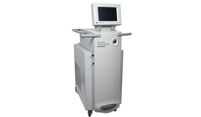
Where would laser spine surgery be without lasers? Deuk Spine Institute uses a top-of-the-line surgical Holmium: YAG laser manufactured in Yokneam, Israel and imported into the United States by Boston Scientific. FDA approved and patented, the VersaPulse PowerSuit is known for its established safety profile in healthcare applications. With a unique blend of 2100 nm wavelength electromagnetic energy and cavitation displacement of liquid, this laser is the perfect surgical instrument for the Deuk Laser Disc Repair® procedure. Zero laser associated complications have occurred at Deuk Spine Institute in nearly 20 years and over 5,000 laser disc repairs performed.
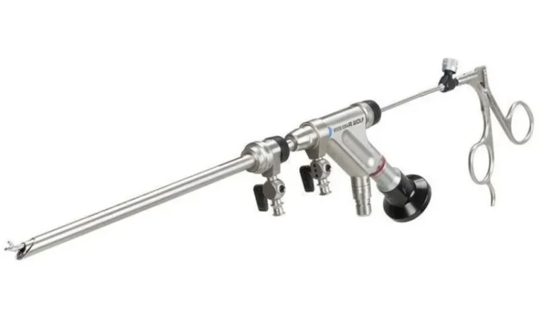
Laser energy is delivered to the tip of an 8 ft long and 1 mm wide FDA approved optical fiber passing through an endoscopic channel with a distal aperture located within the painful posterior annular tear(s) of the injured spinal disc being treated. Laser energy is delivered from the optical fiber tip to damaged parts of the painful disc while under the direct control of a trained Deuk Spine Institute surgeon. The spine surgeon uses endoscopic magnification and illumination to visualize microscopic painful tissue within the annular tear and direct the laser’s therapeutic energy to remove this damaged tissue. When combined with high-definition real time spinal endoscopy, the laser has precision unrivaled in medicine. No other surgical instrument can match the laser’s dynamic therapeutic range of 0.1 mm precision in the human body and yet fit so easily through the slim hull of the endoscope.
When you think of endoscopic surgery you may think “less invasive, smaller incision, less bleeding, faster recovery, less scar tissue, greater safety, less pain”, and you would be correct. These are some of the benefits of endoscopic spine surgery. Deuk Spine Institute has used the same spinal endoscopes for nearly 20 years. FDA approved and manufactured by Richard Wolf in Germany, our spinal endoscopes are considered to have the highest quality optics and remain functionally superior to other brands.
Endoscopes provide both light illumination and a magnified view of the area within an injured disc being operated on. In addition, the endoscope has channels that run the length of the scope and they can be used for irrigation, suction and injecting medication. The main working channel of the endoscope allows the surgeon to insert instruments directly into target areas within the injured disc. Laser spine surgery is only possible when the laser is used in combination with the endoscope to visualize and remove the herniated disc material scarred to the walls of the posterior annular tear. Endoscopy makes areas of the spine that were once hard to get to, more accessible. A well-planned endoscopic spine surgery can be performed through a 4 – 7 millimeter skin incision. The same surgery performed “open” is done through a 40 – 400 millimeter incision with far more bleeding and surgical trauma to surrounding tissues. Surgical trauma and complications are greatly reduced with endoscopic spine surgery. Common spine problems such as herniated discs, spinal stenosis and radiculopathy can now be treated endoscopically without having to “open” up the patient’s body. Recovery from endoscopic spine procedures is fast, without bleeding, scar tissue or the use of dangerous opioid painkillers. Endoscopic spine surgery is one of the most important recent advances in spine surgery and still only available at a few spine centers worldwide.
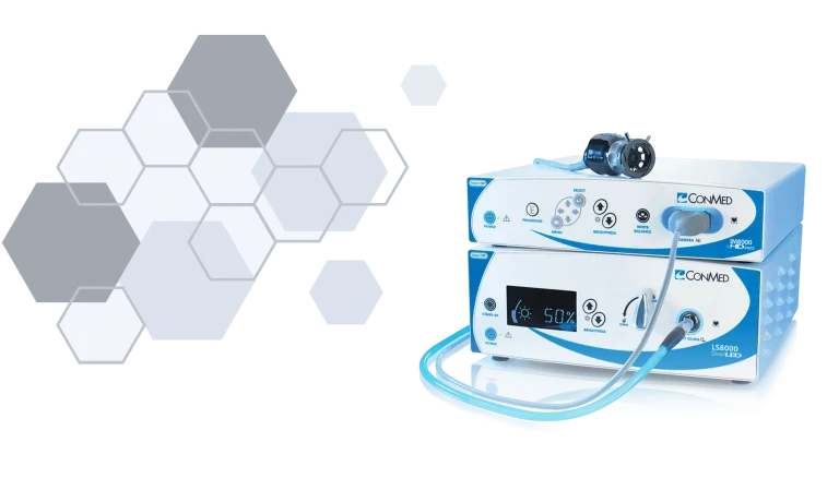
“Lights, Camera and Action!” Combining the endoscope with a high-definition camera system allows the spine surgeon to see minute details within the injured spinal disc. Annular tears, the most common source of back pain, are too small to be seen with the naked eye. Their location within the back wall of an injured spinal disc makes annular tears even harder to find and treat without a surgical camera and endoscope system. Because annular tears are the most common cause of chronic back and neck pain, it is essential for the surgeon to visualize their full extent for repairs during surgery. Deuk Laser Disc Repair® uses a laser to repair painful annular tears only visible with the aid of an endoscope and camera system.
The high-definition camera system projects real-time video images from the tip of the endoscope onto a television screen in the operating room for the surgeon to see and treat all of the pain generators located within the injured disc. High-definition endoscopic camera systems are not only an essential part of the equipment package needed to perform Deuk Laser Disc Repair®, they are also needed to record and livestream broadcast our spine surgeries to the world. Every week Deuk Spine Institute livestreams its spine surgeries unedited on our YouTube, Facebook, X/Twitter and LinkedIn platforms for the public to watch on computers and mobile devices. Viewers can ask questions to the surgeon in real-time and get answers during the surgery livestream. No other spine center in the world has offered unedited, real-time livestream high-definition spine surgery with Q & A. Deuk Spine Institute has provided this educational service to the public since 2014 on an ongoing basis and free of charge. Why? Our founder, neurosurgeon Dr. Ara Deukmedjian MD, believes in these two principles: “Knowledge is power” and “Power to the people.”
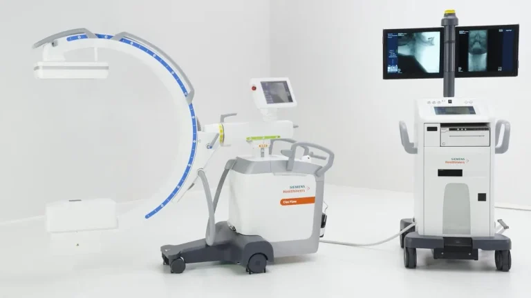
How does a spine surgeon know where to cut, how deep to go and in what direction? Whether the surgical target is a herniated disc, bulging disc, spinal stenosis, facet arthropathy, sacroiliac joint or piriformis muscle, the surgeon relies on navigational equipment to reach it. Target identification, localization, surgical path planning and ultimately navigation are all highly complex tasks completed in the operating theatre by the spine surgeon every time an operation is performed. The most important navigational tool for spine surgery is a multiplanar fluoroscope. There are many fluoroscopes available today, both new and old, but only one stands out as the best for spine surgery and it is manufactured by Siemens in Germany. Deuk Spine Institute only uses Siemen’s fluoroscopes because they provide the highest quality images possible used to identify, plan, localize and navigate areas in the spine and joints. With lower quality or older fluoroscopes, important details are absent from the images the surgeon sees which in turn results in inaccurate targeting and navigation.
Let’s take a moment to understand the value of the information provided to the surgeon by the intraoperative fluoroscope. The spine is located deep within the body and cannot be seen directly with the naked eye and rather than “digging around” looking for a specific part of the spine that needs treatment, the surgeon uses a machine that allows non-invasive target localization with the patient on the operating table. Spine surgeons do not have “X-ray vision” so they use a machine that does, the multiplanar fluoroscope. Once the target in the spine is identified in real time while the patient is on the operating table, the surgeon can plan a safe path from the skin down to the target located in the spine. Using real time patient specific anatomical information gathered from the fluoroscope, the surgeon can identify the best starting point on the skin, surgical corridor, target approach and capture. Equipment can make or break the outcome of spine surgery. With the best equipment, the surgical plan can be optimized by the surgeon to get the best result for the patient having the procedure. “The ideal surgical plan takes into consideration both the safety and effectiveness of the procedure being performed” says founding neurosurgeon, Dr Ara J Deukmedjian MD. “Only as strong as the weakest link.” Deuk Spine Institute’s core philosophy is to provide our patients with the best spine care possible on earth. To this end we use only the best equipment sourced from around the world to perform our spinal diagnostics and treatments. Using the best fluoroscope in the world means better results for our patients and it allows Deuk Spine Institute to fulfil its mission of being the best spine center in the world. In a time when many doctors’ offices and hospitals are cutting back on spending by using inexpensive, outdated equipment; Deuk Spine Institute spares no expense to continue to provide our patients with the best.
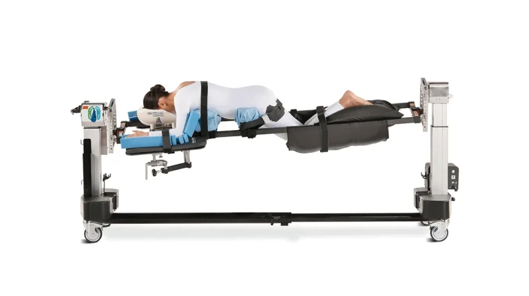
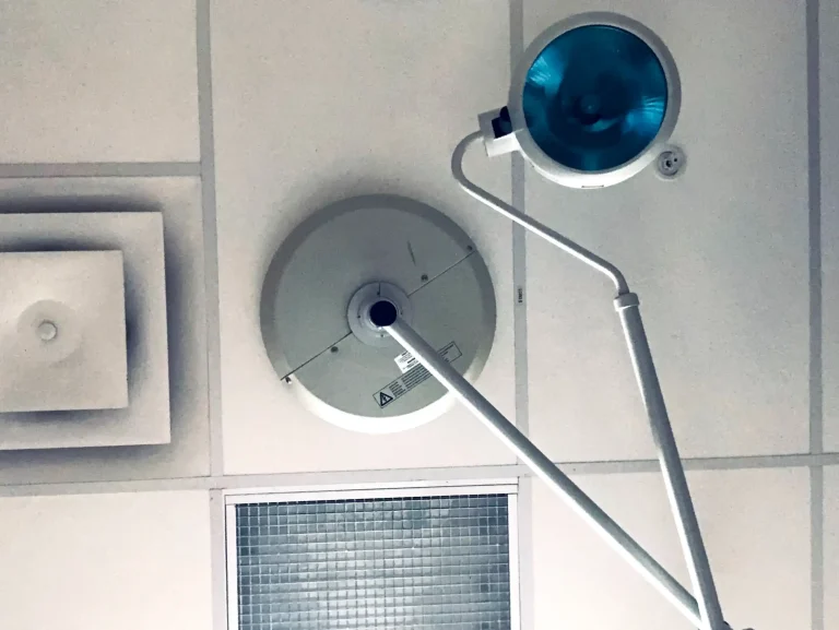
Spine surgery is performed with the patient asleep on a specialized table in the operating theatre. Operating tables range in quality and cost from $10,000 to over $100,000. The best operating tables provide safety and functionality for the spine surgery being performed. Deuk Spine Institute uses the Jackson Spinal Surgical Table for its procedures. This top-of-the-line operating table allows for the best patient positioning and access of surgical equipment needed to perform our proprietary procedures including Deuk Laser Disc Repair® and Deuk Plasma Rhizotomy®. Carbon fiber table components provide clear, unobstructed fluoroscopic views of the spine during the localization and navigation portions of spine surgery.
Abdominal pressure is reduced with the patented table design and this feature results in less bleeding during spine surgery. Specialized body pads hold the patient in a stable fixed position during surgery while sensitive areas are protected from injuries commonly seen with less advanced operating tables. Deuk Spine Institute is firmly committed to giving our patients the world class care they deserve and this is made possible by providing their surgeons with the best equipment and support.
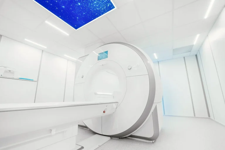
Deuk Spine Institute employs state of the art medical imaging modalities to aid in the diagnosis and treatment of spine conditions such as back pain, sciatica, radiculopathy, neck pain, herniated disc, spinal stenosis, spondylosis and neurogenic claudication. For these conditions, spinal MRI is the most important medical imaging test available to identify structural damage to the spine. MRI uses powerful magnetic fields to excite water molecules in bodily tissues. When the magnetic field is removed, energy is immediately released from these different tissues and their “signals” are captured by the MRI detector. The MRI images we see are actual averages of signal energy detected in a small area usually 1-3 mm thick. These volume averages cannot be thought of as actual slices of tissue but as an approximation of true structural composition.
“MRI never tells you where pain is coming from, only the patient does that.” This is one of Deuk Spine Institute founder Dr. Ara Deukmedjian’s favorite sayings and it couldn’t be truer. MRI scans show structural abnormalities such as herniated discs and spinal stenosis. “Pain is a physiological abnormality, not seen on MRI or any other test”, another famous saying of Dr. Ara Deukmedjian MD. “There are millions of people with abnormal MRI scans and zero pain.” The reason is simple, pain involves molecular level chemical transmitters not visible on MRI scans, yet. In light of this fact, Deuk Spine Institute developed the Deuk Spine Exam® to aid in the diagnosis of the source of back and neck pain also called the “pain generator”. There are 30 potential pain generators for back pain and another 30 for neck pain. Each patient’s specific source of back pain or neck pain can only be discovered using a specialized physical exam, called the Deuk Spine Exam®, in combination with a high-quality MRI or CT scan. When nerve damage is found on the exam an EMG/NCS is ordered to ascertain which nerves are affected and to what condition. Image guided diagnostic injections of suspect spinal joints or muscles are helpful at times. Combining information gleaned from the physical exam, MRI, diagnostic blocks and EMG/NCS the actual pain generator(s) can be identified with 99.5% accuracy.
World-class equipment. World-class surgeons. World-class results.