Understanding L4-L5 Disc Herniation: Your Complete Guide to Relief and Recovery

Picture of Dr. Ara J. Deukmedjian, MD
Dr. Ara J. Deukmedjian, MD

Board-Certified Neurosurgeon, CEO & Founder of Deuk Spine Institute

View Profile
Published: January 19, 2026
Last updated: June 2, 2026
7 min read
Share this article:
3D illustration of the human spine with the L4-L5 segment highlighted in the lower lumbar region.
Medical Disclaimer: This content is for educational purposes only and does not constitute medical advice. Individual results may vary. Always consult with a qualified spine specialist about your specific condition and treatment options. 

Key Points 

✓ L4-L5 is the most mechanically stressed segment in the lumbar spine — and the most common site for disc herniation. ¹ 

✓ The hallmark symptom is sharp, shooting pain down the outer leg to the top of the foot, often with foot weakness or numbness. These findings, not imaging alone, establish the diagnosis. ² 

✓ 75–90% of patients improve with 8–12 weeks of comprehensive conservative care. Most herniations do not require surgery. ³ 

✓ Bowel or bladder dysfunction with bilateral leg weakness signals cauda equina syndrome — a surgical emergency. Go to an emergency room immediately. ⁵ 

✓ Not all surgical options are equal. Fusion eliminates motion permanently and carries a 5–18% adjacent segment disease rate at 4–14 years. ⁶ 

✓ Deuk Laser Disc Repair® uses a 4–7 mm incision, no fusion, no hardware, no hospital stay — with a 99% success rate across 2,000+ procedures. ⁸ 

✓ MRI findings do not always match symptoms. Accurate diagnosis requires clinical correlation, not imaging alone. 


Diagnosis. Answers. Relief.

FREE Virtual Consultation + MRI Review

Submit your MRI for a free expert review by Dr. Ara Deukmedjian, M.D. — board-certified neurosurgeon. No obligation. Real answers.

Schedule Yours Today 2,000+ procedures · Zero major complications · No cost, no obligation

What “L4-L5 Disc Herniation” Actually Means 

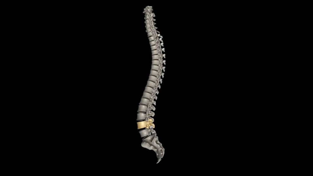
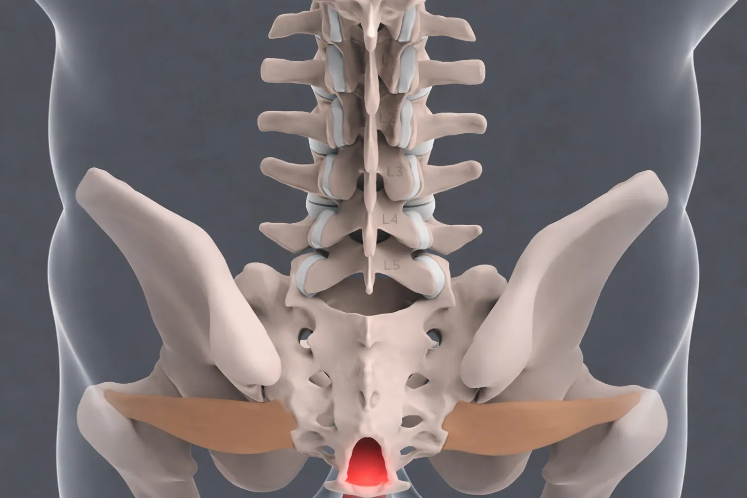
The lumbar spine consists of five vertebrae, L1 through L5, stacked between the thoracic spine above and the sacrum below. The L4-L5 segment sits at the bottom of that stack, at the point where the mobile lumbar spine transitions into the relatively fixed pelvis. Every time you bend, lift, twist, or sit, that segment absorbs a disproportionate share of the mechanical load. 

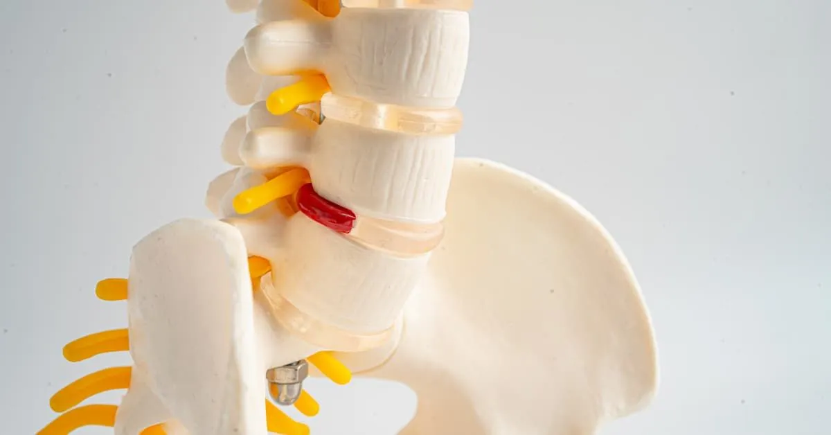
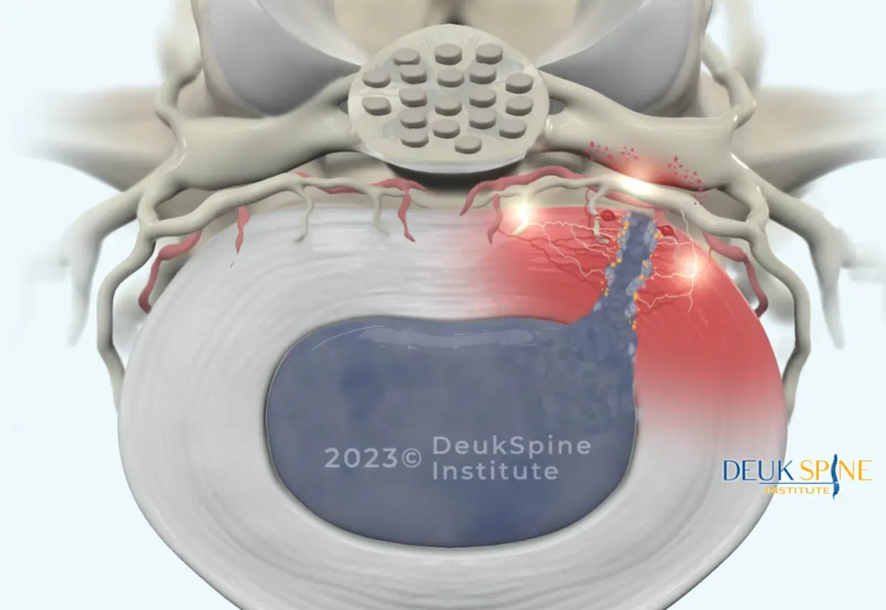
The intervertebral disc between L4 and L5 is a two-layer structure: a firm outer ring of collagen called the annulus fibrosus, and a soft, gel-like inner core called the nucleus pulposus. A herniation occurs when the annulus develops tears or weakens enough to let the nucleus push through. When that displaced material contacts and compresses a nearby nerve root, it causes the inflammation and neurological symptoms that bring most patients to a spine specialist. 

The specific nerve most commonly affected at this level is the L5 nerve root, which provides sensation to the outer leg and top of the foot and controls muscles responsible for lifting the foot and extending the big toe. This anatomy explains the predictable symptom pattern that physicians use to identify L4-L5 as the pain source. Without needing to guess. 

The Four Types of Disc Displacement at L4-L5 

Not all disc herniations are the same, and the type affects both natural history and treatment planning: 

Disc bulge: The disc extends symmetrically beyond the vertebral margin. The annulus is intact. This represents early degenerative change and may be asymptomatic. 

Disc protrusion: The nucleus pushes against an intact but weakened annulus, creating a focal outpouching. The base of the protrusion is wider than the displaced portion. 

Disc extrusion: The nucleus breaks fully through the annulus but remains connected to the parent disc. This typically causes more significant nerve compression and paradoxically, extruded fragments are more likely to reabsorb naturally because the immune system identifies them as foreign tissue. ⁴ 

Disc sequestration: A free fragment of disc material separates completely and migrates within the spinal canal. This is the most severe displacement but, counterintuitively, may show greater spontaneous reabsorption over time. 

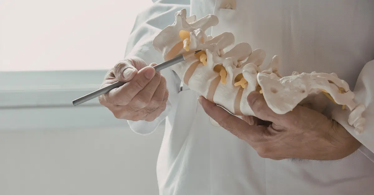
Each vertebra in your lumbar spine has specific characteristics and potential issues:4

Doctor using a spine model to demonstrate lumbar vertebrae and nerve structures during a medical consultation.

What Causes L4-L5 Disc Herniation 

Understanding causation helps explain why the same segment is so frequently affected and why some patients are more vulnerable than others. 

Illustration of the lower human spine and pelvis with highlighted lower vertebrae.

Age-related degeneration is the dominant driver. Discs lose water content progressively beginning in the third or fourth decade of life, making the annulus stiffer and more prone to tearing. This is not pathology in the clinical sense. It is universal but it creates the substrate for herniation under load. 

Acute trauma can herniate an otherwise healthy disc. Motor vehicle accidents, falls, and improper lifting with heavy loads can generate forces that exceed the disc’s structural tolerance in a single event. ⁹ 

Construction workers in safety gear handling tools on a high steel beam.

Cumulative occupational stress: heavy lifting, prolonged sitting, vibration exposure can cause cumulative annular damage over months and years. Each individual stress is minor; the aggregate effect is not. 

Genetic predisposition is increasingly recognized as a contributor. Studies show that disc degeneration has a hereditary component, with some individuals showing accelerated disc aging at relatively young ages regardless of activity level. 

Modifiable Risk Factors That Affect Prognosis 

Several factors within a patient’s control affect both the risk of initial herniation and the likelihood of symptom improvement: 

Smoking reduces disc nutrition by impairing vascular supply, increases systemic inflammation, and creates repetitive spinal stress through chronic coughing. Smokers consistently show worse disc degeneration and poorer treatment outcomes across studies. 

Obesity increases compressive loads on lumbar discs and is associated with chronic systemic inflammation that may impair disc metabolism. 

Prolonged sitting increases intradiscal pressure by approximately 40% compared with standing. A figure from biomechanical research that explains the near-universal experience of sitting-intolerant sciatica. ¹⁰ This single data point should inform every acute management plan. 

Sedentary lifestyle and weak core musculature remove the active stabilization that reduces disc loading during daily movement. 

Recognizing L4-L5 Disc Herniation: Symptoms

The distribution of symptoms at L4-L5 is specific enough that an experienced clinician can often identify the correct level before any imaging is ordered. Understanding this distribution helps patients recognize whether their symptoms fit the pattern. 

The Hallmark: L5 Radiculopathy 

herniated-disc-shown.jpg

When the herniated disc at L4-L5 compresses the L5 nerve root, it produces a characteristic pain that travels from the lower back or buttock down the outer side of the leg, continuing to the top of the foot and the space between the first and second toes. This is not generalized leg pain. It follows a specific anatomical path. 

The pain is typically described as sharp, shooting, or electric feeling dull, aching back pain. It can be severe enough to stop movement mid-stride or make sitting in a car or chair nearly impossible. 

Neurological Findings That Confirm Back Pain At L5

Beyond pain, L5 nerve root compression produces predictable neurological deficits that a physician can test at the bedside.

Medical illustration of a spinal disc herniation with highlighted nerve irritation.

Weakness in ankle dorsiflexion: Difficulty lifting the front of the foot, which can progress to foot drop. The inability to clear the foot during walking. This is one of the more serious functional consequences of L4-L5 compression. 

Weakness in Big toe extension: Inability to push the big toe up against resistance. 

Numbness and tingling in the L5 dermatome distribution: outer leg, top of foot, and the first two toes. 

Trendelenburg gait: Hip dropping on the affected side during single-leg stance, indicating gluteus medius weakness. Recent clinical guides specifically identify this as a 2025-confirmed finding in L5 radiculopathy. ² 

These specific findings are clinically valuable because they help distinguish L4-L5 compression from L5-S1 compression (which typically affects the heel and outer foot via the S1 nerve root) and from peripheral nerve disorders. 

Symptoms That Require Emergency Evaluation 

One symptom cluster demands immediate emergency care, not a scheduled appointment: 

Bowel or bladder dysfunction. Difficulty urinating, urinary retention, or loss of bowel control; combined with saddle anesthesia (numbness in the groin and inner thighs) and bilateral leg weakness indicates cauda equina syndrome. This occurs when a massive herniation compresses multiple nerve roots simultaneously. Without urgent surgical decompression, permanent loss of bowel, bladder, and lower extremity function can result. ⁵ 

If you develop any combination of these symptoms, go to an emergency room immediately. This is the one situation in which waiting for conservative treatment is not appropriate. 


Diagnosis. Answers. Relief.

FREE Virtual Consultation + MRI Review

Submit your MRI for a free expert review by Dr. Ara Deukmedjian, M.D. — board-certified neurosurgeon. No obligation. Real answers.

Schedule Yours Today 2,000+ procedures · Zero major complications · No cost, no obligation

How L4-L5 Disc Herniation Is Accurately Diagnosed 

Accurate diagnosis is the prerequisite for successful treatment. Many patients arrive after months of failed therapy not because their treatments were wrong, but because their diagnosis was incomplete or imprecise. 

The Clinical Examination: What Physicians Are Looking For 

A comprehensive clinical examination should include: 

Straight leg raise test: The examiner lifts the extended leg while the patient lies supine. Radicular pain below 60 degrees of elevation is a positive finding, suggesting nerve root compression. 

Motor strength testing: Specific muscle groups controlled by L5 are evaluated ankle dorsiflexion, great toe extension, hip abduction and any asymmetry between sides is documented. 

Sensory testing: The examiner maps areas of reduced or altered sensation against the known L5 dermatome distribution. 

Gait analysis: Walking on heels (L4-L5 function) and tiptoes (L5-S1 function) can identify deficits efficiently. 

Imaging: What MRI Shows and What It Doesn’t 

MRI is the diagnostic gold standard for L4-L5 disc herniation. It provides detailed soft-tissue visualization of the disc, nerve roots, spinal cord, and ligaments. None of which appear on X-ray. MRI identifies the size and location of the herniation, the degree of nerve compression, whether the fragment is contained or extruded, and the presence of concurrent conditions such as stenosis or facet arthropathy. 

However, the single most important caveat in lumbar spine diagnosis is this: imaging findings and symptoms frequently do not correlate. Population studies consistently document significant disc herniations in individuals with no back or leg pain whatsoever. A herniation on MRI is not, by itself, the diagnosis. The herniation must anatomically explain the patient’s specific symptoms and examination findings to be the diagnosis. 

This is where many diagnostic workups go wrong. Treating the MRI rather than the patient leads to surgeries that fail not because the operation was poorly performed, but because the operated disc was not the actual pain source. 

Electrodiagnostic studies (EMG/NCS) add value when the diagnosis is uncertain. They confirm whether a nerve root is physiologically compressed, identify which nerve root is involved, distinguish lumbar radiculopathy from peripheral nerve entrapment, and assess the severity and chronicity of nerve damage. 

MRI machine at Deuk Spine Institute

No cost · No obligation

Learn How You Can
Live Pain Free

Upload your MRI for a free expert review by Dr. Ara Deukmedjian, M.D. — board-certified neurosurgeon. Ten minutes can change your life.

Submit My MRI — It's Free 2,000+ procedures · Zero major complications

L4-L5 Disc Herniation Treatment: What the Evidence Supports 

Treatment follows a logical progression from the least invasive to more definitive options, calibrated to the patient’s symptom severity, trajectory, and response. 

Conservative Treatment: Effective for Most Patients 

The data supporting an initial conservative approach is strong. Studies show approximately 75–90% of patients with lumbar disc herniation improve meaningfully with comprehensive conservative care. ³ The operative word is “comprehensive.” Not all conservative treatments are equally effective, and a token prescription for NSAIDs plus a few physical therapy sessions is not the evidence-based trial that warrants surgery if it fails. 

Physical therapy forms the core of non-surgical management. A spine-specialized therapist should design a program including: 

  • Core stabilization (planks, bridges, bird dogs) to reduce mechanical load on the disc through improved segmental support 
  • McKenzie methodology, which uses repeated movements to “centralize” pain. Shifting it from the leg back toward the spine, a sign of neurological decompression 
  • Flexibility work for hamstrings and hip flexors, which, when tight, increase lumbar disc stress ⁹ 
  • Low-impact aerobic exercise (walking, swimming) to reduce inflammation and support healing 

Medications manage symptoms during the healing window. NSAIDs reduce nerve root inflammation; neuropathic agents such as gabapentin address nerve-specific pain that responds poorly to standard analgesics; short-course oral steroids may be appropriate for severe acute inflammation. 

Epidural steroid injections deliver anti-inflammatory medication directly to the compressed nerve root. Benefits are real but temporary, typically lasting weeks to months, and success rates are variable. Their value is primarily in managing severe acute pain while natural healing progresses. 

The 8–12 Week Decision Point 

The standard trial period for conservative treatment is 8–12 weeks. If a patient has received a genuinely comprehensive program. Not a passive, appointment-filling substitute and has not achieved meaningful improvement, continuing the same approach indefinitely is not supported by evidence. The decision about surgical evaluation should be active and deliberate, not a default. 

When Surgery Should Not Be Delayed 

Four clinical situations warrant surgical consultation without waiting for a full conservative trial: 

Progressive motor weakness: Increasing foot weakness or the onset of foot drop indicates ongoing nerve injury. Nerve tissue has limited regenerative capacity. Prolonged compression causes irreversible damage. 

Cauda equina syndrome: Emergency surgical decompression, not a trial of physical therapy. 

Severe intractable pain preventing sleep, work, or basic function despite adequate medical management. 

Failed comprehensive conservative trial: 8–12 weeks of structured, supervised care without meaningful improvement. 

Surgical Treatment: What You’re Choosing Between 

When surgery is indicated, understanding the differences between available procedures is essential. The gap between options is not a matter of incision size. It is a matter of what happens to the spine itself. 

Traditional Microdiscectomy 

The long-standing surgical standard for lumbar disc herniation. The surgeon accesses the spine from the back, removes a portion of the lamina to reach the disc, and extracts the herniated fragment pressing on the nerve. 

Success rates for leg pain relief are reported at 85–95%, with lower rates for axial back pain. Recurrent herniation occurs in 5–15% of patients. Recovery requires 3–6 months before return to full activity, and a hospital stay of 1–2 days is typical. 

The procedure is effective, but it requires bone removal to access the disc. Which is a structural trade-off, not a neutral act. 

Spinal Fusion 

When disc problems occur alongside spinal instability, some surgeons recommend fusion. Permanently joining two or more vertebrae with bone graft and metal hardware. 

Fusion can be appropriate for specific anatomical indications. It is over-applied for isolated disc herniation without instability. The long-term consequences are well-documented and not small: 

  • Adjacent segment disease occurs in 5–18% of patients within 4–14 years, driven by the biomechanical load redistribution that results from eliminating motion at one or more levels. ⁶ Many require reoperation. 
  • Hardware complications: screw loosening, cage migration, pseudoarthrosis. Are distinct surgical risks that persist for the patient’s lifetime. 
  • Permanent loss of motion at the fused segment is not a side effect. It is the intended outcome. Patients consenting to fusion should understand they are consenting to that permanently. 
  • Recovery is measured in months, not weeks: 6–12 months for full return to activity is typical. 

Deuk Laser Disc Repair®: A Structurally Different Operation 

Deuk Laser Disc Repair® (DLDR®) is not a smaller version of microdiscectomy or a marketing rebranding of fusion. It is a categorically different operation based on a different philosophy of treatment. 

The procedure is performed through a 4–7 mm incision about the diameter of a pencil eraser. A specialized endoscope provides high-definition visualization. Using precision laser technology, only the damaged disc material causing nerve compression is removed. Typically 5–10% of the total disc. The remaining healthy disc, the annulus, the lamina, the facet joints, and the surrounding musculature are left completely undisturbed. 

No bone is removed. No fusion is performed. No hardware is implanted. The procedure takes approximately one hour under conscious sedation. Patients walk out of recovery the same day. 

Published outcomes: 

  • 94.6% average symptom resolution in a peer-reviewed consecutive case series ⁸ 
  • 99% success rate across 2,700+ procedures over 20+ years of institutional data ⁸ 
  • Zero reported major complications 
  • No adjacent segment disease; because no segment is fused 
  • Return to desk work within 3–5 days 

The reason these numbers differ from fusion data is not the incision size. It is that no motion is eliminated, no adjacent segments are placed under redistributed load, and no hardware is introduced to fail. The cascade of complications that drives the long-term reoperation rate after fusion does not have a mechanism to occur after DLDR®. 

Side-by-Side: Treatment Comparison at L4-L5 

Metric 

Microdiscectomy 

Spinal Fusion 

Deuk Laser Disc Repair® 

Incision size 

1–2 inches 

2–4 inches 

4–7 mm 

Bone removal 

Yes (laminotomy) 

Yes 

No 

Hardware implanted 

No 

Yes 

No 

Motion at treated level 

Preserved 

Eliminated permanently 

Preserved 

Hospital stay 

1–2 days 

2–4 days 

None (outpatient) 

Return to desk work 

4–6 weeks 

3–6 months 

3–5 days 

Full recovery 

3–6 months 

6–12 months 

2–3 weeks 

Recurrence rate 

5–15% 

N/A (disc removed) 

1.5% ⁸ 

Adjacent segment disease risk 

Low 

5–18% at 4–14 years ⁶ 

None 

Reversibility 

No bone returned 

None 

No structural alteration 

MRI machine at Deuk Spine Institute

No cost · No obligation

Learn How You Can
Live Pain Free

Upload your MRI for a free expert review by Dr. Ara Deukmedjian, M.D. — board-certified neurosurgeon. Ten minutes can change your life.

Submit My MRI — It's Free 2,000+ procedures · Zero major complications

What These Numbers Don’t Tell You 

  1. The Most Important Variable Is Whether the Right Operation Was Chosen

A 95% successful procedure applied to the wrong diagnosis has a 0% chance of helping the patient. The most common reason spine surgery fails is not technical execution. It is a mismatch between the procedure performed and the actual pain source. A patient with discogenic pain from a contained herniation is not made better by fusion regardless of how cleanly the screws are placed. A patient with true mechanical instability from spondylolisthesis is not made better by an endoscopic discectomy regardless of how elegant the technique. 

The diagnostic process; clinical correlation of symptoms, examination, and imaging. Is not administrative. It is the most consequential step in the entire treatment pathway. 

  1. “Minimally Invasive” Describes the Access, Not the Operation

This distinction matters when evaluating any surgical recommendation. A minimally invasive fusion is still a fusion. The smaller incision spares some muscle and shortens early recovery. It does not change what happens to the vertebrae, the disc, the hardware, or the adjacent segments over the following decade. Patients consenting to any spine surgery should understand what is being done to their spine beneath the skin. Not just how large the incision is. 

  1. Delaying Necessary Surgery Has Real Costs

The appropriate framing is not “avoid surgery as long as possible.” It is “give conservative treatment a genuine, time-limited trial, then make a deliberate decision.” Prolonged compression of the L5 nerve root can cause: 

  • Irreversible nerve damage: Muscle atrophy from denervation may not fully recover even after successful surgical decompression. The window for optimal neurological recovery is not unlimited. 
  • Central sensitization: Extended severe pain alters the nervous system’s pain processing, making pain harder to treat even after the structural source is addressed. 
  • Disability cascade: Job loss, deconditioning, depression, and opioid dependence are documented consequences of undertreated chronic radiculopathy. 

Doctor assessing a patient’s lower back pain during a clinical spine examination.

Living with L4-L5 Disc Herniation: Evidence-Based Self-Management 

While pursuing evaluation or treatment, practical management strategies can reduce symptom burden and prevent worsening. 

Sleep position: Back sleeping with a pillow under the knees is optimal. It maintains the lumbar curve and minimizes disc pressure. Side sleeping with a pillow between the knees is acceptable. Stomach sleeping increases lumbar extension and disc loading and should be avoided entirely. 

Sitting: Given the 40% intradiscal pressure increase that sitting produces compared with standing, ¹⁰ taking a 2–3 minute standing break every 30 minutes is not a comfort preference. It is evidence-based management. Lumbar support that maintains the natural lordosis is important when sitting cannot be avoided. 

Lifting: Bend at the knees, not the waist. Keep objects close to the body. Avoid twisting during the lift. Ask for help with heavy objects. 

Exercise: Walking, water exercise, and stationary cycling are appropriate for most patients in the subacute phase. High-impact activities, heavy deadlifts and squats, and sports involving twisting should be avoided until symptoms are controlled. 

Smoking cessation: One of the few modifiable variables with documented impact on disc healing and surgical outcomes.


Diagnosis. Answers. Relief.

FREE Virtual Consultation + MRI Review

Submit your MRI for a free expert review by Dr. Ara Deukmedjian, M.D. — board-certified neurosurgeon. No obligation. Real answers.

Schedule Yours Today 2,000+ procedures · Zero major complications · No cost, no obligation

You’ve Done the Research. Now Get a Real Answer.

Most patients who find this article have already spent months. Sometimes years trying to fix their back pain. You’ve done the physical therapy. You’ve had the injections. You may have been told that fusion is your only option, or worse, that you simply need to “live with it.”

You don’t.

The purpose of this guide isn’t to push you toward surgery. It’s to make sure you have complete information before making any decision. At Deuk Spine Institute, the first step has no commitment attached to it: submit your MRI and receive a direct review from Dr. Ara Deukmedjian, a board-certified neurosurgeon who has performed over 2,000 cervical and lumbar procedures with zero major complications.

If you’re a candidate for a motion-preserving alternative to fusion, you’ll know. If you’re not, you’ll know that too and you’ll have a clear picture of your options either way.

That review is free. The conversation has no obligation. And for many patients, it’s the first honest answer they’ve received.

MRI machine at Deuk Spine Institute

No cost · No obligation

Learn How You Can
Live Pain Free

Upload your MRI for a free expert review by Dr. Ara Deukmedjian, M.D. — board-certified neurosurgeon. Ten minutes can change your life.

Submit My MRI — It's Free 2,000+ procedures · Zero major complications

Frequently Asked Questions About L4-L5 Disc Herniation

What are the most common symptoms of L4-L5 disc herniation?

The hallmark symptom of L4-L5 disc herniation is sciatica, sharp, shooting pain that radiates from your lower back or buttock down the outer leg, potentially extending to the top of your foot. This occurs because the herniated disc typically compresses the L5 nerve root.

Additional symptoms include numbness and tingling in the outer leg, top of foot, and between the first and second toes; weakness in lifting your foot or toes (dorsiflexion), which can lead to foot drop; difficulty walking on your heels; localized lower back pain that worsens with bending, sitting, or twisting; and muscle spasms in the lower back.

In rare but serious cases, a massive disc herniation can cause cauda equina syndrome, characterized by bowel or bladder dysfunction, saddle anesthesia (numbness in the groin area), and bilateral leg weakness. This is a medical emergency requiring immediate treatment.

It’s important to note that imaging findings don’t always correlate with symptoms; you can have a disc herniation visible on MRI without pain, or experience significant symptoms with relatively mild imaging findings.

Diagnosis involves a combination of clinical examination and imaging studies.

Clinical examination: Your physician will take a detailed history of your symptoms and perform specific tests including the straight leg raise test (lifting your leg while you lie on your back—pain radiating down the leg suggests nerve compression), motor strength testing of specific muscle groups controlled by the L5 nerve, sensory examination to check for numbness or altered sensation, and reflex testing.

Imaging studies: MRI is the gold standard for diagnosing disc herniation, as it provides detailed visualization of discs, nerve roots, and soft tissues.1 X-rays can show disc space narrowing and bone spurs, but cannot visualize the disc herniation itself. CT scans provide excellent bony detail and may be used when an MRI is contraindicated. Electrodiagnostic studies (EMG/NCS) can confirm nerve compression and identify which specific nerve root is affected.

The key is comprehensive clinical correlation—your symptoms, examination findings, and imaging must all align to ensure accurate diagnosis and appropriate treatment planning.

Yes, many L4-L5 disc herniations can improve significantly without surgical intervention. Research indicates that approximately 75-90% of patients experience substantial improvement with appropriate conservative treatment.

The natural healing process involves several mechanisms: acute inflammation around the nerve root subsides over time, reducing pain even if some compression persists; the immune system can gradually resorb some herniated disc material, particularly with disc extrusions where material has escaped beyond the disc boundaries; the nervous system can adapt to chronic compression through neuroplastic mechanisms; and the disc may gradually desiccate and retract somewhat from neural structures.

However, “healing” doesn’t mean the disc returns to its pre-injury state. Once the annulus fibrosus develops tears, permanent structural changes typically persist. While symptoms may resolve, underlying disc degeneration remains.

Key factors influencing whether conservative treatment will succeed include the size and type of disc pathology, the degree of nerve compression, your age and overall health, and adherence to treatment recommendations.

If symptoms persist beyond 6-12 weeks of comprehensive conservative treatment, or if progressive weakness develops, surgical evaluation is warranted.

Recovery time varies dramatically depending on which surgical procedure is performed.

Traditional microdiscectomy: Hospital stay of 1-2 days is typical. Return to light activities takes 4-6 weeks. Full recovery requires 3-6 months. Physical therapy is usually needed for several months. Some activity restrictions may be permanent.

Spinal fusion: Hospital stay of 2-4 days. Bone fusion takes 3-6 months to solidify. Return to work takes 3-6 months. Full recovery can take up to a year. Permanent loss of motion at the fused segment. Risk of adjacent segment disease requiring future surgery.

Deuk Laser Disc Repair® (minimally invasive): Same-day outpatient procedure with no hospital stay. Most patients experience immediate relief from leg pain. Return to desk work within 3-5 days. Complete healing within 2-3 weeks. No prolonged activity restrictions. Full range of motion preserved. No risk of adjacent segment disease.

The dramatic difference stems from the fundamental approach; traditional surgeries involve significant tissue trauma and often permanent alteration of spinal anatomy, while minimally invasive endoscopic approaches preserve healthy tissue and maintain normal biomechanics.

Conservative (non-surgical) treatment aims to reduce inflammation, manage symptoms, and allow natural healing to occur. It includes physical therapy and exercise to strengthen supporting muscles, medications to manage pain and inflammation, activity modifications and ergonomic improvements, epidural steroid injections for temporary relief, and typically requires 8-12 weeks to assess effectiveness.

Conservative treatment is appropriate as the first-line approach for most patients. However, it doesn’t directly remove the herniated disc material—it relies on natural healing processes.

Surgical treatment directly addresses the structural problem by removing the herniated disc material and decompressing the nerve. Options range from traditional microdiscectomy (removing bone to access and remove herniated disc material) and spinal fusion (permanently joining vertebrae when instability is present) to advanced minimally invasive procedures like Deuk Laser Disc Repair® (precise removal of only damaged disc material while preserving healthy structures and natural motion).

Surgery should be considered when conservative treatment has failed after an appropriate trial, progressive muscle weakness is developing, severe pain significantly impairs quality of life despite conservative care, or cauda equina syndrome is present (medical emergency).

The key is matching treatment to your specific situation based on symptom severity, functional impact, and response to conservative approaches.

This depends on your treatment approach and individual situation.

With conservative treatment success: If symptoms resolve with conservative care, you can typically return to most activities with appropriate precautions. You may need to permanently modify activities that place excessive stress on the lumbar spine, such as heavy lifting, high-impact sports, or prolonged sitting without breaks. Maintaining core strength, proper body mechanics, and a healthy weight becomes a lifelong commitment.

After traditional fusion surgery: Fusion eliminates motion at the treated segment, and certain activities may need permanent modification, including high-impact activities, sports requiring extensive spinal flexibility, heavy physical labor, and activities with repetitive twisting. You’ll also face an increased risk of adjacent segment disease as other levels compensate for the fused segment.

After Deuk Laser Disc Repair®: Because DLDR® preserves natural spinal motion and doesn’t fuse vertebrae, most patients can return to full activities within weeks with no long-term restrictions. The procedure maintains normal biomechanics, reducing the risk of future problems at adjacent levels. Patients typically return to work, sports, travel, and all normal activities without the limitations imposed by fusion.

The bottom line: motion-preserving surgical techniques offer the best opportunity for returning to unrestricted activity, while fusion surgery typically requires permanent activity modifications.

Absolutely. Getting a second opinion is highly advisable for anyone diagnosed with a lumbar disc herniation, particularly if surgery has been recommended.

Here’s why: treatment approaches vary dramatically among surgeons, with some primarily performing fusion procedures and others offering motion-preserving alternatives. The stakes are high—spinal surgery can profoundly impact your quality of life for years or decades. Many patients discover treatment options they didn’t know existed when consulting specialists with different training and expertise. You deserve to understand all available options before committing to any intervention.

A meaningful second opinion comes from a provider with different capabilities from your first consultation. If you saw a fusion surgeon, seek an opinion from a motion-preservation specialist. If you have consulted a pain management physician, consider seeing a spine surgeon to determine whether definitive treatment is appropriate.

At Deuk Spine Institute, we offer a complimentary MRI review and virtual consultation. You can upload your imaging studies through our secure portal to receive an expert assessment with no cost or obligation. Many patients who thought fusion was their only option discovered they were candidates for motion-preserving procedures that offered superior outcomes with dramatically faster recovery.

While conservative treatment is appropriate as a first-line approach, unnecessarily delaying surgery when it’s genuinely needed carries several significant risks:

  • Irreversible nerve damage: Prolonged nerve compression can cause permanent damage to the nerve fibers. If you’re experiencing progressive weakness, delaying surgery may result in muscle atrophy and functional loss that won’t fully recover even after successful decompression. Nerve tissue has limited regenerative capacity, and there’s often a “window of opportunity” for optimal recovery.
  • Chronic pain sensitization: Extended periods of severe pain can cause changes in your nervous system called central sensitization, where the nervous system becomes hyperreactive to pain signals. This can make pain more difficult to treat, even after the underlying structural problem is addressed.
  • Disability and quality of life impact: Months or years of severe symptoms can lead to job loss and financial stress, social isolation and depression, loss of physical fitness and muscle conditioning, and development of unhealthy pain coping mechanisms, including excessive medication use.
  • Muscle atrophy: Muscles that aren’t used due to pain or nerve damage begin to waste away. Once significant atrophy has occurred, full strength may not return even after successful nerve decompression.
  • Cauda equina syndrome: If you develop bowel or bladder dysfunction, saddle anesthesia, or bilateral leg weakness, this is a surgical emergency. Delaying treatment for cauda equina syndrome can result in permanent loss of bowel/bladder control and permanent leg weakness.

The key is finding the right balance—giving conservative treatment an adequate trial (typically 8-12 weeks) while remaining vigilant for signs that surgical intervention should not be delayed further. An experienced spine specialist can help you determine when you’ve reached that point.

Success rates vary depending on the treatment approach and how “success” is defined.

Conservative treatment: Studies show that approximately 75-90% of patients with lumbar disc herniation experience significant improvement with comprehensive conservative care. However, improvement doesn’t always mean complete symptom resolution, and some patients have recurrent episodes.

Traditional microdiscectomy: Success rates of 85-95% for leg pain relief are commonly reported. However, success for back pain is lower (60-70%). Recurrent herniation occurs in approximately 5-15% of patients. Patient satisfaction varies widely depending on expectations and activity level.

Spinal fusion: While fusion can be effective for specific indications, it carries a 10-15% risk of adjacent segment disease requiring reoperation. Fusion doesn’t always eliminate pain, particularly if the disc itself was the main pain generator rather than instability.

Deuk Laser Disc Repair®: Our findings show a 99% success rate in eliminating disc-related pain with zero surgical complications in over 2,00 patients treated. Patient satisfaction is exceptionally high due to rapid recovery and preservation of natural motion.

It’s important to understand that “success” means different things to different people. For some, any reduction in pain represents success. For others, only complete elimination of symptoms and return to all activities qualifies as success. Discuss your specific goals and expectations with your treatment provider to ensure you’re aligned on what constitutes a successful outcome.

Sources

  1. Mayfield Clinic. Anatomy of the Spine. https://mayfieldclinic.com/pe-anatspine.htm
  2. National Library of Medicine. L4-L5 Disc Herniation — Clinical Findings. Stat Pearls. https://www.ncbi.nlm.nih.gov/books/NBK554393/ 
  3. Deuk Spine Institute. Herniated Disc Symptoms. https://deukspine.com/blog/herniated-disc-symptoms/ 
  4. Frontiers in Medicine. Spontaneous reabsorption of lumbar disc herniation — mechanisms review. July 2025. https://www.frontiersin.org/journals/medicine/articles/10.3389/fmed.2025.1633762/full
  5. AANS. Cauda Equina Syndrome. https://www.aans.org/en/Patients/Neurosurgical-Conditions-and-Treatments/Cauda-Equina-Syndrome
  6. Hashimoto K et al. Adjacent segment degeneration after fusion spinal surgery: a systematic review. International Orthopaedics. 2019. https://pmc.ncbi.nlm.nih.gov/articles/PMC11605282/ 
  7. Deuk Spine Institute. The True Cost of Spinal Fusion. https://deukspine.com/blog/true-cost-of-spinal-fusion 
  8. Deukmedjian AJ, Cutright J, Cianciabella A, Deukmedjian A. Deuk Laser Disc Repair® is a safe and effective treatment for symptomatic cervical disc disease. Surgical Neurology International. 2013;4:68. https://pubmed.ncbi.nlm.nih.gov/23776754/
  9. AANS. Lumbar Spinal Stenosis — Patient Information. https://www.aans.org/en/Patients/Neurosurgical-Conditions-and-Treatments/Lumbar-Spinal-Stenosis
  10. National Library of Medicine. Biomechanics of the Lumbar Spine. StatPearls. https://www.ncbi.nlm.nih.gov/books/NBK560878/
  11. PMC. AI-assisted prognosis in lumbar disc herniation — 2025 outcomes study. https://pmc.ncbi.nlm.nih.gov/articles/PMC12180165 
  12. Journal of Minimally Invasive Spine Surgery and Technique. Endoscopic discectomy outcomes 2024. https://jmisst.org/journal/view.php?viewtype=pubreader&number=349
Share this article:
Table of Contents

If you have been told you have a herniated disc, a bulging disc, a disc protrusion, a slipped disc, or…

By Dr. Ara Deukmedjian Board-Certified Neurosurgeon Medically reviewed on May 28, 2026 Medical Disclaimer: This content is for educational purposes…

By Dr. Ara Deukmedjian Board-Certified Neurosurgeon Medically reviewed on May 27, 2026 Medical Disclaimer: This content is for educational purposes…