By Dr. Ara Deukmedjian
Board Certified Neurosurgeon
Reviewed on July 29, 2026
Disclaimer: This information is for educational purposes only and is not intended to replace the advice of a physician. Results may vary from person to person. Consult your doctor regarding your particular situation.
Key Points
✓ Disc herniation from T1 to T2 is an uncommon pathology. And occurs in less than 1% of all surgical disc herniations. ¹ ²
✓ T1 nerve root compression causes grip weakness, medial forearm numbness. And loss of hand dexterity. ³ ⁴
✓ Horner syndrome (drooping eyelid, small pupil, facial dryness) is the signature finding at this level. ⁵ ⁶
✓ Diagnosis is frequently delayed because standard cervical MRI stops at C7-T1 and misses this level. ³ ⁵
✓ ~69% of T1-T2 herniations are posterolateral, compressing the nerve root rather than the cord. ⁷
✓ Central herniations can compress the spinal cord, causing myelopathy. ⁷ ⁸
✓ 11–37% of asymptomatic adults show thoracic disc herniations on MRI. Imaging alone does not justify surgery. ⁹
✓ Most patients improve with 6–12 weeks of conservative care; surgery is reserved for progressive deficits. ¹⁰
✓ Deuk Laser Disc Repair®: 99.6% success, 0.01% complication rate, 2,700+ procedures. Motion-preserving, outpatient.
What Is a T1-T2 Herniated Disc?
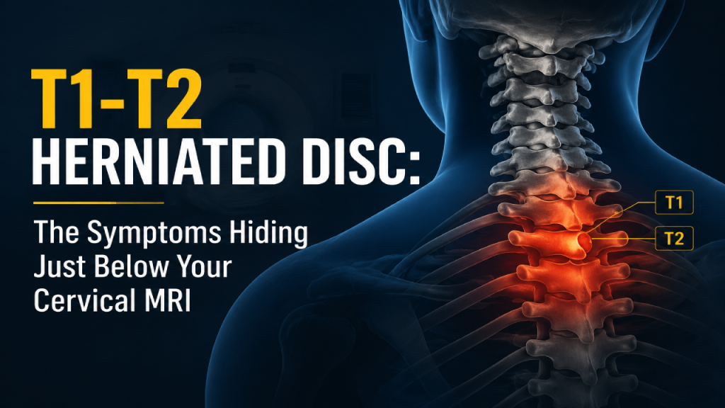
The T1-T2 disc sits at the junction between the base of the neck and the upper back, right where the cervical spine transitions into the thoracic spine. T1 is the first thoracic vertebra and T2 is the second. The disc between them cushions these two vertebrae and allows limited movement at this level.
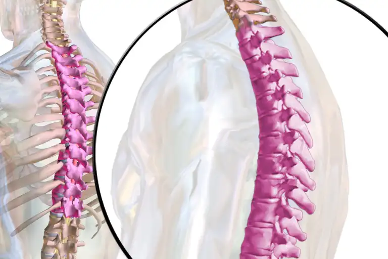
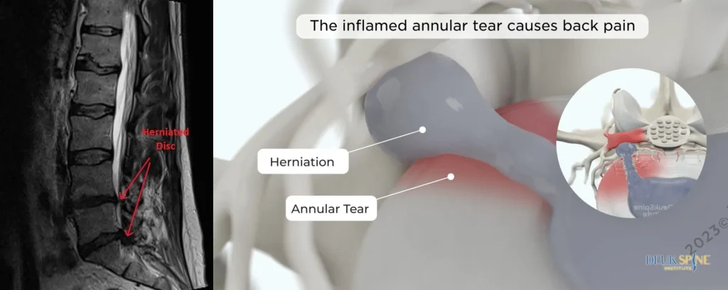
Herniation of the disc between the T1-T2 segments is characterized by the displacement of the soft inner part of the disc (nucleus pulposus) through a tear of the outer hard covering of the disc (annulus fibrosus). In such a case, the displaced substance may put pressure on the T1 nerve roots, the spinal cord, or the sympathetic nerve chain responsible for controlling functions of the eye and face. ¹ ⁵
This level is anatomically unusual for two reasons. First of all, the thoracic vertebrae are supported by the rib cage, which decreases motion and, consequently, the mechanical stresses that result in herniated discs in cervical and lumbar vertebrae. ² Secondly, the T1-T2 vertebral level is situated in a transitional area of anatomy that is typical for both cervical and thoracic spines; that is the very reason why symptoms at this level tend to resemble those of cervical radiculopathy. ³
How rare is a T1-T2 herniated disc?
Disc herniations of the thoracic spine constitute 0.25%-0.75% of all symptomatic spinal disc herniations. ¹¹ Disc herniations in the thoracic spine tend to occur mostly in the lower region, with T11-T12 being the most common level. ² Herniations in the upper thoracic spine, specifically those of T1-T2, make up the least common group. There were 39 total cases of disc herniation of the T1-T2 levels described in a literature review in JAAOS Global Research & Reviews. ⁵ 36 cases were documented in another review in Surgical Neurology International. ⁷
Herniated Disc from T1 to T2 Symptoms
The symptoms of a herniated disc from T1 to T2 can vary depending on which structure is compressed: the T1 nerve root, spinal cord, or the sympathetic chain. Many patients present with a combination.
1. T1 Radiculopathy: Pain and Weakness in the Arm and Hand
This particular nerve supplies the intrinsic muscles of the hand, which are the small muscles that spread fingers apart, clench and perform other delicate functions. ⁴ If the T1-T2 intervertebral disc is herniated, the following symptoms are exhibited by the patient:
- Medial arm and forearm pain. A deep, aching, or burning pain that radiates from the upper back or base of the neck down the inside (medial) aspect of the arm and forearm.
- Ring and little finger numbness and tingling. Dermatome of T1 extends to the medial part of the forearm and to the medial antecubital fossa (inner part of the elbow joint). Paresthesia (tingling sensation) is commonly experienced in the inner part of the forearm, the fourth, and fifth fingers. ³ ⁵
- Grip weakness and loss of hand dexterity. Because T1 innervates the hand intrinsic muscles via the ulnar nerve, patients lose the ability to spread their fingers apart (finger abduction), pinch forcefully, or perform fine motor tasks like buttoning a shirt. ⁴ ⁵
- Periscapular pain. Pain between or around the shoulder blades is one of the most commonly reported initial symptoms in published case reports. ³ ⁵ ⁷
In a review of 21 T1-T2 herniated disc cases in the literature, the most frequently reported signs and symptoms of T1 nerve root compression included hand weakness, medial arm/forearm pain and paresthesias, and grip deficits. ⁵
Why it mimics cervical radiculopathy
The overlap is significant. The C8 nerve root compressed by a C7-T1 disc herniation and the T1 nerve root produce nearly identical symptoms: ulnar-sided hand pain, intrinsic hand weakness, and medial forearm numbness. ³ This is why a patient with a T1-T2 herniated disc is often initially diagnosed with a cervical disc problem. The distinction is critical because cervical imaging alone will not reveal the T1-T2 level, and the wrong surgical target means no relief.
2. Horner Syndrome: The Signature Finding
Horner syndrome is the most distinctive and diagnostically important symptom of a T1-T2 herniated disc. It occurs because the sympathetic nerve pathway the nerves that control involuntary functions like pupil dilation and sweating. Originates in the hypothalamus and synapses in the spinal cord at the C8-T2 levels. A disc herniation at T1-T2 can directly disrupt this pathway. ⁵ ⁶
Horner syndrome presents as a triad of:
- Ptosis — drooping of the upper eyelid on the affected side
- Miosis — constriction (small size) of the pupil on the affected side
- Anhidrosis — decreased or absent sweating on the affected side of the face
Of the 39 T1-T2 disc herniation cases identified in one literature review, seven presented with Horner syndrome. ⁵ In a separate review of 36 cases, Horner syndrome was again identified as a distinguishing clinical feature of upper thoracic disc disease. ⁷
Horner syndrome combined with upper extremity radicular pain should always prompt imaging that includes the upper thoracic spine. ⁵ ⁶
3. Thoracic Myelopathy: Spinal Cord Compression
If a herniated disc of T1-T2 type is centralized, it may cause direct compression of the thoracic spinal cord because of the narrowness of the spinal canal at this level compared to the spinal canal at the cervical level. Symptoms of myelopathy include:
- Leg weakness and spasticity. Difficulty walking, feeling of heaviness or stiffness in the legs, frequent tripping.
- Gait instability and balance problems. A wide-based, unsteady gait. Patients may report feeling as though they are “walking on cotton.”
- Upper motor neuron signs. Hyperactive reflexes in the legs, clonus (rhythmic involuntary muscle contractions), and a positive Babinski sign (upgoing great toe when the sole of the foot is stroked).
- Bowel and bladder dysfunction. Urinary urgency, hesitancy, retention, or incontinence; constipation. These symptoms indicate significant cord compression and are considered a surgical emergency.
- Sensory level. A band-like area of numbness or altered sensation across the chest or upper abdomen that corresponds to the T1-T2 level.
In the Surgical Neurology International review, two of four patients presented with paraparesis (weakness in both legs) from central disc herniations compressing the spinal cord. ⁷
4. Neck Pain Without a Cervical Source

A counterintuitive but frequently reported symptom is neck pain. In a review of 36 T1-T2 disc herniation cases, 24 out of 36 patients (67%) presented with neck pain as a primary complaint. ⁷ This makes clinical sense when you consider that the T1-T2 level is anatomically adjacent to the cervicothoracic junction, and referred pain from upper thoracic pathology radiates upward into the neck and trapezius region. It is also the reason this condition is so often misdiagnosed as a cervical spine problem.
Why T1-T2 Herniated Discs Are So Often Misdiagnosed
Diagnosis of T1-T2 disc herniations is difficult indeed, and according to the scientific literature, delayed diagnosis is the rule rather than the exception. ³ ⁵ ⁷ The reasons for this include:
1. The MRI often stops too early
Standard cervical MRI protocols typically image from C1 to C7-T1. The T1-T2 disc space is at or just below the inferior margin of most cervical studies. If the radiologist or ordering physician does not specifically request upper thoracic imaging, the T1-T2 level may be partially visualized or missed entirely. ³ ⁵
2. Symptom overlap with cervical disc disease
As described above, T1 radiculopathy and C8 radiculopathy produce nearly identical symptoms. Without imaging that extends below C7-T1, the clinician will attribute hand weakness and medial arm pain to a cervical source, especially if age-related cervical degenerative changes are present on the MRI.
3. Symptom overlap with peripheral nerve entrapment
Intrinsic weakness of the hands and ulnar nerve entrapment symptoms include:
- Cubital tunnel syndrome
- Carpal tunnel syndrome
- Thoracic outlet syndrome
An electromyography (EMG) and nerve conduction study can help distinguish peripheral entrapment from a nerve root lesion, but only if the clinician suspects the correct level. ⁴
4. Rarity creates unfamiliarity
Since upper thoracic disc herniations are rare in occurrence, T1-T2 is often not even considered when developing a differential diagnosis for a patient. This tendency towards more frequent conditions such as cervical radiculopathy, carpal tunnel, and thoracic outlet syndrome is the biggest challenge to diagnosing T1-T2.
What Causes a T1-T2 Herniated Disc?
While the T1-T2 level is protected by the rib cage, which limits motion and reduces repetitive mechanical stress, herniation can still occur from:

- Trauma. Motor vehicle accidents, falls, and heavy lifting are commonly reported causes in the case literature, particularly in younger patients. ³ ⁵
- Degenerative disc disease. Age-related wear and tear weakens the annulus fibrosus over decades, and while less common at thoracic levels, it still occurs, particularly at the cervicothoracic junction.
- Genetic predisposition. Research on twins demonstrates that 34–74% of disc degeneration may be genetically determined, regardless of spinal level. ¹²
- Congenital anomalies. Cervical ribs, conjoined nerve roots, and other anatomic variants at the cervicothoracic junction have been reported in association with T1-T2 herniations. ⁵
- Adjacent-level degeneration. Patients with pre-existing cervical disc disease or prior cervical fusion may develop accelerated degeneration at the T1-T2 level.
Diagnosing a T1-T2 Herniated Disc
The diagnostic sequence
- Detailed history and physical examination. The combination of medial arm/forearm pain, intrinsic hand weakness, and when present Horner syndrome should raise suspicion for T1 radiculopathy.
- MRI of the cervical AND upper thoracic spine. This is the single most important diagnostic step. A standard cervical MRI will miss the T1-T2 level. The ordering physician must explicitly request imaging that extends through T2-T3 at minimum. MRI is the imaging test of choice for soft-tissue pathology such as disc herniations and nerve root compression. ⁵ ⁷
- CT scan or CT myelogram. Useful when the herniation is calcified (which is more common in thoracic discs) or when MRI is contraindicated. CT provides superior bony detail. ⁷
- EMG and nerve conduction studies. Can localize the lesion to the T1 nerve root and exclude peripheral nerve entrapment (cubital tunnel, carpal tunnel, thoracic outlet). ⁴
- Pharmacologic pupil testing. When Horner syndrome is suspected, topical cocaine or apraclonidine eye drops can confirm the diagnosis and help localize the level of sympathetic chain disruption.
The MRI Trap
Thoracic disc herniations are found incidentally on MRI in 11–37% of people who have no symptoms whatsoever. ⁹ In one study of 90 asymptomatic individuals, 74% had thoracic disc lesions visible on MRI, and 29% demonstrated spinal cord deformation, yet none had symptoms. ¹³ This means imaging findings must match the clinical presentation before any surgical intervention is considered.
Non-Surgical Management of Herniated Disc at T1-T2 Level
In those who do not have myelopathy, weakness, or red flags, an organized plan of conservative management is advisable and effective most of the time.
Physical therapy
For physical therapy for upper thoracic disc herniation, emphasis should be on the flexibility of the thoracic spine, scapular stability, posture correction, and neural mobilization. This is because T1-T2 pathology is frequently associated with stiffness of the cervicothoracic junction.
Medications
- Non-steroidal anti-inflammatory drugs (NSAIDS; ibuprofen, naproxen) are used initially for inflammation and pain relief.
- Oral short term corticosteroids (dose pack of methylprednisolone) will help in alleviating acute inflammation of nerve roots and radiculitis.
- Medicines for neuropathic pain (gabapentin, pregabalin) can be useful for treatment of constant numbness and tingling sensation despite of having side effects such as drowsiness and cognitive slowness. ¹⁰
Epidural steroid injections
Epidural or selective nerve root injection at T1-T2 level under fluoroscopic guidance will help in alleviating pain temporarily, but more importantly it will help in confirming T1-T2 as pain generator. ¹⁰
Activity modification
Ergonomic adjustments, avoidance of heavy overhead lifting, and postural awareness (particularly reducing prolonged forward-head posture) can reduce mechanical stress at the cervicothoracic junction.
When conservative care is not enough
Non-surgical management should be reconsidered when:
- Symptoms persist or worsen after 6–12 weeks of appropriate conservative treatment
- Motor weakness is progressive (worsening grip strength, increasing hand clumsiness)
- Myelopathic signs develop (gait instability, hyperreflexia, bowel/bladder changes)
- Horner syndrome is present and worsening
Surgical Treatment Options for T1-T2 Herniated Discs
When surgery is indicated, the choice of surgical approach depends on the location of the herniation posterolateral versus central.
Posterior approach
For posterolateral T1-T2 herniations compressing the T1 nerve root in the foramen, a posterior approach allows direct access to the compressed nerve root. This is the most commonly used approach for lateral T1-T2 herniations and involves removing a small window of bone (lamina) and widening the foramen to decompress the nerve. ⁵ ⁷
Anterior approach
For central T1-T2 herniations compressing the spinal cord, an anterior approach is often required because retracting the spinal cord from a posterior approach is dangerous and poorly tolerated. Anterior surgery at the cervicothoracic junction is technically demanding due to the sternum, clavicle, and great vessels. ⁷
Endoscopic and minimally invasive approaches
Full-endoscopic decompression techniques have advanced significantly for thoracic disc herniations. These procedures use small incisions, tubular retractors, and high-definition endoscopic visualization to decompress the nerve root or spinal cord with minimal tissue disruption, reduced blood loss, shorter hospital stays, and faster recovery compared to open surgery. ¹⁴ ¹⁵
Deuk Laser Disc Repair® the motion-preserving alternative
Deuk Laser Disc Repair® is an ultra-minimally-invasive endoscopic laser procedure that treats the inflamed annular tear. The actual pain generator rather than removing the entire disc or fusing the segment. Through a 4–7 mm incision, a side-firing holmium laser ablates only the inflamed tissue inside the annular tear under live endoscopic visualization. No bone is cut. No hardware is implanted. The disc, lamina, facets, and ligaments are preserved. ¹⁶
For thoracic herniations, this approach eliminates the risks of open thoracotomy or complex cervicothoracic anterior surgery while preserving full motion at the treated level. Patients walk out within an hour and typically return to desk work within a week.
Results: 99.6% success rate, 0.01% complication rate across 2,700+ procedures.
Differential Diagnosis for C7–T1 Symptoms
Ulnar-sided hand pain, ring and little finger numbness, and grip weakness can arise from several distinct conditions. Here is how each one separates itself from a C7–T1 herniated disc.
The presence of Horner syndrome with upper extremity radicular symptoms should always prompt upper thoracic imaging and should raise concern for Pancoast tumor, which must be excluded. ⁵ ⁶
Treat the T1-T2 disc without fusion or spinal-cord risk.
Standard cervical MRI stops at C7-T1, which is exactly why T1-T2 disc herniations — hand weakness, medial arm pain, Horner syndrome — get misdiagnosed for months. If your workup missed this level or open surgery here feels disproportionate, send your MRI for a free review by Dr. Deukmedjian. Deuk Laser Disc Repair® can decompress the T1 nerve through a 7 mm incision — no fusion, no hardware, and none of the anterior-approach risk that makes T1-T2 open surgery so technically demanding.
- 99.6%
- Success rate
- 0.01%
- Complication rate
- 2,700+
- Procedures performed
FAQs
Is a T1-T2 herniated disc serious?
It depends on what the herniation compresses. A posterolateral T1-T2 herniation compressing only the T1 nerve root produces pain, numbness, and hand weakness. Uncomfortable and functionally limiting but not dangerous if treated appropriately. A central T1-T2 herniation compressing the spinal cord is serious and can cause progressive myelopathy with leg weakness, gait instability, and bladder dysfunction. Any myelopathic symptoms require urgent evaluation. ⁷ ⁸
Can a T1-T2 herniated disc heal on its own?
Many thoracic disc herniations stabilize or decrease in size over time. ⁹ Conservative treatment. Physical therapy, medications, and activity modification is effective for the majority of patients without neurological deficits. The inflamed annular tear that generates pain can heal when the inflammatory tissue is removed or subsides naturally. ¹⁰
Why was my T1-T2 herniated disc missed on my cervical MRI?
Standard cervical MRI protocols typically image from C1 through C7-T1. The T1-T2 disc space sits at or below the lower margin of most cervical studies. If the MRI was ordered as a standard cervical study without instructions to include the upper thoracic spine, T1-T2 was simply outside the field of view. ³ ⁵ If your symptoms suggest T1 radiculopathy, request imaging that extends through T3.
What does Horner syndrome look like with a T1-T2 herniated disc?
One eyelid droops slightly (ptosis), the pupil on that side appears smaller than the other (miosis), and sweating may be reduced on that side of the face (anhidrosis). These signs develop on the same side as the disc herniation and nerve compression. ⁵ ⁶
How is T1 radiculopathy different from C8 radiculopathy?
Both affect the hand and medial forearm, but T1 radiculopathy specifically weakens finger abduction (spreading the fingers apart) through the ulnar nerve, while C8 radiculopathy predominantly affects finger flexion (gripping). ⁴ The critical diagnostic difference is the location of the disc herniation on MRI: C7-T1 for C8, versus T1-T2 for T1.
Can Deuk Laser Disc Repair® treat a T1-T2 herniated disc?
Yes. Deuk Laser Disc Repair® is performed through the thoracic spine under endoscopic visualization. Because the procedure uses a 4–7 mm incision, no bone removal, and no hardware, it avoids the significant access-related morbidity of open thoracic surgery while preserving motion at the treated level.
When should I get a second opinion?
If you have been diagnosed with a cervical disc problem but treatment has not resolved your hand weakness, medial arm pain, or Horner syndrome, request an MRI that includes the upper thoracic spine and consider a second opinion from a spine specialist experienced with thoracic disc disease. If open thoracic surgery has been recommended, ask whether an endoscopic approach is appropriate for your specific herniation pattern.
Sources
View Sources
- Arce CA, Dohrmann GJ. Thoracic disc herniation. Improved diagnosis with computed tomographic scanning and a review of the literature. Surg Neurol. 1985;23(4):356-361.
- Quint U, et al. Thoracic disc herniation: Surgical treatment. Neurosurgery. 2002.
- Ozer E, et al. A rare case of T1-2 thoracic disc herniation mimicking cervical radiculopathy. Int J Spine Surg. 2017;11(4):30.
- Cohen-Gadol AA. T1 radiculopathy: Electrodiagnostic evaluation. PM&R. 2009.
- Schessler MJ, et al. T1-T2 disk herniation presenting with Horner syndrome: A case report with literary review. JAAOS Glob Res Rev. 2018;2(11):e068.
- Oliveira Santos BF, et al. T1-T2 herniated disk presenting with Horner syndrome. World Neurosurg. 2017;108:e517-e521.
- Asgari N, et al. T1–T2 disc herniation: Report of four cases and review of the literature. Surg Neurol Int. 2019;10:74.
- Shirzadi A, et al. Atypical presentation of thoracic disc herniation: Case series and review of the literature. Case Rep Orthop. 2013;2013:621476.
- Wood KB, et al. The natural history of asymptomatic thoracic disc herniations. Spine. 1997;22(5):525-530.
- Physio-pedia. Thoracic disc syndrome: Conservative management and surgical indications.
- Carson J, Gumpert J, Jefferson A. Diagnosis and treatment of thoracic disc herniation. BC Med J. 2019.
- Battié MC, et al. The Twin Spine Study: Contributions to a changing view of disc degeneration. Spine J. 2009;9(1):47-59.
- Defined in BC Medical Journal citation above: MRI study of 90 asymptomatic individuals with 74% thoracic disc lesions.
- Choi KY, et al. Endoscopic transforaminal thoracic foraminotomy and discectomy for the treatment of thoracic disc herniation. Asian Spine J. 2013.
- Ruetten S, et al. Full-endoscopic cervical foraminotomy RCT. Spine. 2008.
- Deuk Spine Institute. Deuk Laser Disc Repair® procedure overview.