By Dr. Ara Deukmedjian
Board Certified Neurosurgeon
Reviewed on Aug 5, 2026
Disclaimer: This information is for educational purposes only and is not intended to replace the advice of a physician. Results may vary from person to person. Consult your doctor regarding your particular situation.
Key Points
✓ Herniation of upper lumbar discs at L1-L2 and L2-L3 accounts for about 1% of lumbar disc herniations. ¹ ²
✓ Compression of the L2 nerve root leads to pain in the anterior thigh, weakness in hip flexors, and reduction in knee reflexes. ³ ⁴
✓ The spinal cord (conus medullaris) usually ends close to the level of L1-L2, and thus large L2-L3 herniations can cause symptoms related to the spinal cord and the nerve root. ⁵ ⁶
✓ Spinal canal is narrower and has lesser epidural fat at upper lumbar levels; thus, even small herniations can impinge on multiple nerve roots. ² ⁷
✓ Delayed diagnosis occurs because the symptoms resemble hip pathology, femoral neuropathy, or low lumbar disc disease. ³ ⁸
✓ 95% of lumbar disc herniations occur at L4-L5 or L5-S1, hence a possible neglect of the upper lumbar spine in the differential diagnosis. ⁹
✓ Up to 30% of asymptomatic individuals demonstrate lumbar disc herniations on MRI. Imaging alone does not justify surgery. ¹⁰ ¹¹
✓ Deuk Laser Disc Repair®: a minimally invasive endoscopic laser procedure that treats lumbar disc herniations. Including the L2-L3 level; through a tiny incision with same-day discharge.
What Is a L2-L3 Herniated Disc?
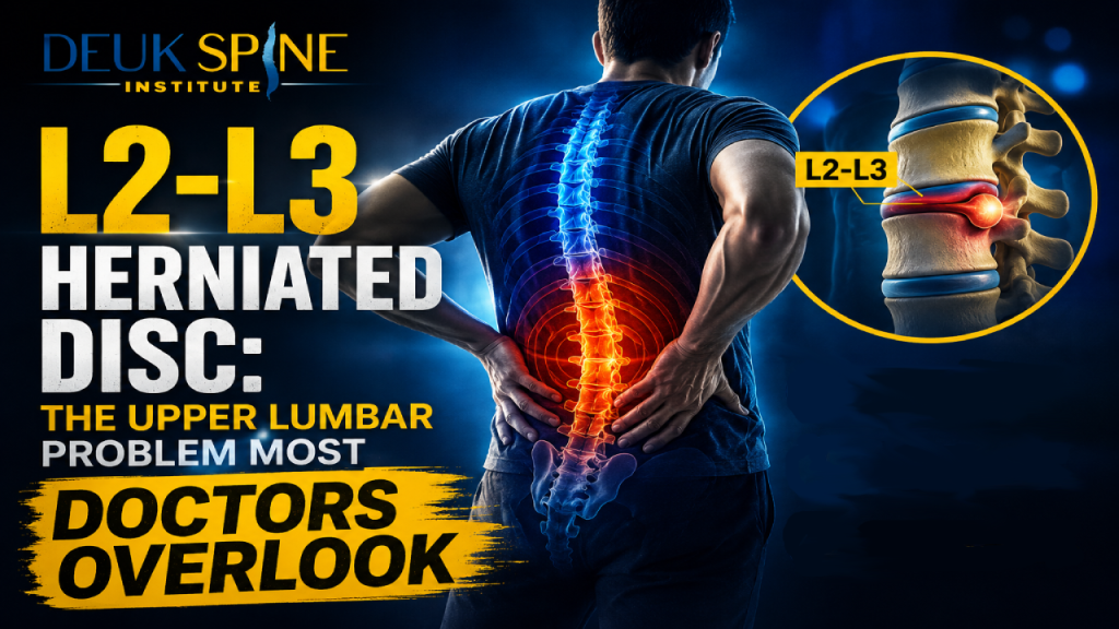
L2-L3 intervertebral disc is located at the upper area of the lumbar spine, between the second and third lumbar vertebrae. This spinal segment belongs to the region of “upper lumbar spine,” according to spine doctors’ terms; in this context, “upper lumbar spine” means either L1-L2, L2-L3, and sometimes even L3-L4. The disc in this particular area serves as a shock absorber, distributing the compression load along the spine, and allowing the flexion, extension, and rotation movements.
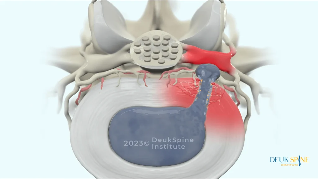
When herniation takes place, the soft gelatinous nucleus of the disc is pushed out by the tear in its tougher exterior. The herniated piece can press not only the L2 nerve root but other nerve roots going through the thecal sac and in severe cases even the bottom of the spinal cord. ¹ ⁵
What makes L2-L3 anatomically distinct from the more commonly herniated lower lumbar levels (L4-L5 and L5-S1) is its proximity to the conus medullaris. The terminal end of the spinal cord, which in most adults lies at approximately the L1 vertebral level. ⁵ ⁶ Because of this, a large disc herniation at L2-L3 does not simply compress a single nerve root. It can affect multiple roots within the cauda equina simultaneously, or even impinge upon the conus itself, producing a complex and potentially dangerous neurological picture. ² ⁷
Additionally, the spinal canal diameter at the upper lumbar levels is smaller than at L4-L5 or L5-S1, and the epidural space contains less protective fat. ² ⁷ The result is that even a moderate-sized herniation at L2-L3 is more likely to cause significant neural compression than a comparably sized herniation at a lower level.
How rare is a L2-L3 herniated disc?
The occurrence rate of upper lumbar disc herniation in the levels of L1-L2 and L2-L3 ranges between 1% of all surgically treated lumbar disc herniations. ¹ ² If, however, the definition of “upper lumbar” is extended to include L3-L4 as well, then the occurrence rate increases to between 1–3.8% of all lumbar herniations, although the L2-L3 itself still remains an unusual finding. ⁷
A series of 7,592 surgically treated cases reported only 51 patients who suffered from upper lumbar herniations in the region of L1-L2 or L2-L3, 38 out of whom had herniations in the L2-L3 level. ¹ A multi-centered case series found that of 141 patients operated on upper lumbar herniations within ten years only 21 cases occurred in the L2-L3 level. ⁸ The majority of lumbar disc herniations, 95%, are located in the regions of L4-L5 and L5-S1, which explains the reason why L2-L3 is usually overlooked in the initial medical examination. ⁹
Herniated Disc at L2-L3 Symptoms
The symptoms of a herniated disc at L2-L3 vary depending on which neural structures are compressed: the L2 nerve root exiting at that level, the traversing nerve roots within the thecal sac (L3, L4, L5, S1), or the lower spinal cord. Many patients present with a combination of findings, and the clinical picture is often more complex than that of a standard lower lumbar disc herniation. ³ ⁸
1. L2 Radiculopathy: Pain and Weakness in the Anterior Thigh
The L2 nerve root provides motor control of the hip flexor muscles (iliopsoas muscle), while being part of the hip adductor muscles, along with L3 and L4, in the formation of the quadriceps through the femoral nerve. ³ ⁴ As the nerve root becomes compressed by a L2-L3 disc herniation, the following occur:
- Anterior thigh pain. A deep, aching, or burning pain that radiates from the lower back or flank into the groin, anterior thigh, and sometimes the medial aspect of the upper leg. This pain pattern is distinct from the sciatic distribution (posterior thigh and calf) that characterizes lower lumbar herniations. ³ ⁴
- Groin numbness and paresthesias. The L2 dermatome innervates the anterior and medial aspect of the thigh including the groin area. There is often numbness, paresthesia, or a “crawling” sensation over the region. ⁴ ⁸
- Hip flexion weakness. Since the L2 nerve contributes to the formation of the iliopsoas muscle, there will be hip flexion weakness where the individual has difficulty raising his thigh against gravity or rising from sitting. ³ ⁴
- Hip flexor weakness. Because L2 is a primary contributor to the iliopsoas muscle, patients may notice difficulty lifting the thigh against gravity, climbing stairs, or getting out of a chair. ³ ⁴
- Diminished or absent knee jerk reflex. The patellar reflex is primarily mediated by L3-L4, but L2 contributes. A depressed knee reflex in the presence of anterior thigh symptoms should raise suspicion for upper lumbar pathology. ⁴ ⁸
In a review of upper lumbar disc herniations, the most common presenting complaints were back pain (reported in approximately 88% of patients) and leg pain (approximately 89%). The femoral nerve stretch test; rather than the straight leg raise was positive in 87% of patients with upper lumbar pathology. ⁸
Why it mimics hip or groin pathology
It should be noted that the pain distribution pattern in the anterior thigh and groin associated with L2 radiculopathy bears a striking resemblance to those observed in cases of hip joint arthritis, inguinal hernia, iliopsoas bursitis, and meralgia paresthetica (lateral femoral cutaneous nerve entrapment). ³ Since both the patient and the medical practitioner think of “disc problems” associated with posterior leg pain (sciatica), anterior thigh pain is automatically thought to be of hip origin.
2. Multi Root Compression: Polyradiculopathy
While herniations at other lumbar vertebrae affect usually only one nerve root, those at the higher level involve the presence of the cauda equina in the lumbar spinal canal, which is a collection of nerve roots that run from the conus medullaris to the sacrum. Due to the narrowed diameter of the canal and limited space within it, even a moderate herniation may cause compression of several nerve roots (L2, L3, L4, and so on). ² ⁷
This multi root compression can manifest as:
- Combined anterior and posterior thigh symptoms. Pain, numbness, or weakness that spans both the femoral (anterior thigh) and sciatic (posterior thigh/calf) distributions.
- Bilateral leg involvement. Central or large paracentral herniations can compress roots on both sides, producing bilateral symptoms. ⁵ ⁷
- Remote radiculopathy. In rare documented cases, a large L2-L3 herniation has caused isolated L5 radiculopathy (foot drop) without any L2 or L3 symptoms, because the extruded disc material compressed the L5 nerve root as it traveled within the thecal sac. ¹³ This can lead to significant diagnostic confusion.
3. Conus Medullaris Syndrome: When the Spinal Cord Is Involved
The spinal cord terminates as the conus medullaris, which in the average adult lies at the level of the L1 vertebral body. However, there is normal anatomical variation, and in some individuals the conus extends as low as L2. ⁵ ⁶ A large central L2-L3 disc herniation in these patients or a large upward migrated extrusion. Can directly compress the conus medullaris, producing upper motor neuron findings that are not expected with a typical lumbar disc problem.
Conus medullaris compression can produce:
- Bowel and bladder dysfunction. Urinary retention, urinary incontinence, fecal incontinence, or loss of rectal tone. These are emergency findings. ⁵ ⁶
- Saddle anesthesia. Numbness in the perineal, perianal, and inner thigh regions (S2-S4 dermatomes).
- Bilateral lower extremity weakness. Weakness that may include both upper and lower motor neuron features. Brisk reflexes (upper motor neuron) combined with flaccid weakness (lower motor neuron) in a mixed pattern. ⁶
- Sexual dysfunction. Erectile dysfunction or loss of genital sensation.
- Extensor plantar response (Babinski sign). An upper motor neuron sign indicates cord-level compression. A finding that would not occur with a simple nerve root problem at lower lumbar levels.
A prospective study of upper lumbar disc herniations in the Indian population found that L2-L3 and L3-L4 herniations more frequently presented with gait imbalance, extensor plantar response, and bowel or bladder symptoms compared to L1-L2, reflecting epiconus-related neural compromise. ¹⁴
Any bowel or bladder symptoms, saddle anesthesia, or rapidly progressive bilateral leg weakness in the setting of a known or suspected L2-L3 disc herniation constitutes a surgical emergency requiring immediate evaluation and likely urgent decompression. ⁵ ⁶
4. Cauda Equina Syndrome
While cauda equina syndrome (CES) is most commonly caused by massive disc herniations at lower lumbar levels, it can and does occur from L2-L3 herniations. ⁵ ¹⁵ Documented cases include a 38-year-old soldier who developed bilateral lower extremity weakness, urinary retention, and inability to defecate from a massive L2-L3 disc herniation. ¹⁵ CES from L2-L3 may present with:
- Bilateral leg weakness and pain
- Saddle anesthesia
- Bladder retention or incontinence
- Loss of anal tone
CES is distinguished from conus medullaris syndrome primarily by the pattern of motor findings (lower motor neuron only in CES versus mixed upper/lower motor neuron in conus syndrome), but in practice the two often overlap at the L2-L3 level because of the anatomical transition between cord and nerve roots. ⁵ ⁶
5. Low Back Pain Without Classic Sciatica
Many patients with L2-L3 herniations present predominantly with axial low back pain rather than radiating leg symptoms. ⁸ In a large surgical series, back pain was the most common symptom, present in 88% of upper lumbar disc herniation patients. ⁸ Because the pain is in the back and anterior thigh rather than following the classic sciatic pattern down the posterior leg, patients and clinicians may not consider a disc herniation as the cause.
Why L2-L3 Herniated Discs Are So Often Misdiagnosed
Delayed and incorrect diagnosis is a well-documented problem with upper lumbar disc herniations. The literature consistently reports that clinical signs and symptoms at the L1-L2 and L2-L3 levels are highly variable and potentially misleading. ³ ⁸ The reasons for this include:
1. Clinicians expect lower lumbar pathology
Ninety-five percent of lumbar disc herniations occur at L4-L5 and L5-S1. ⁹ This statistical reality creates a diagnostic bias: when patients present with back and leg pain, the default clinical assumption is a lower lumbar disc problem, particularly one producing sciatica. L2-L3 herniations produce anterior thigh pain, not posterior leg pain, and this distribution does not match the expected pattern.
2. Anterior thigh pain is attributed to the hip
The L2 dermatomal distribution overlaps with the referral pattern of hip osteoarthritis, trochanteric bursitis, and other hip pathology. Patients with anterior thigh and groin pain are more likely to receive hip X-rays than lumbar MRIs.
3. The straight leg raise test is often negative
The straight leg raise (SLR) test is the classic physical examination maneuver for lumbar radiculopathy. However, SLR primarily stresses the L5 and S1 nerve roots. For upper lumbar root compression (L2, L3, L4), the correct test is the femoral nerve stretch test (also called the reverse SLR or prone knee bending test), which is performed with the patient lying face down while the examiner extends the hip with the knee flexed. ⁸ ¹² If the clinician relies solely on the straight leg raise and it is negative, upper lumbar radiculopathy may be dismissed.
4. Symptom complexity leads to multi-specialty referrals
Because L2-L3 herniations can produce a confusing mix of symptoms: anterior thigh pain, hip weakness, groin numbness, and sometimes bowel/bladder changes. Patients are frequently referred to urologists, orthopedic hip specialists, vascular surgeons, or neurologists before a spine specialist is consulted. Each specialist evaluates and treats within their domain, and the actual spinal pathology remains unidentified.
5. Rarity creates unfamiliarity
Since upper lumbar disc herniations represent only 1–3.8% of all lumbar herniations, many clinicians will encounter very few cases in their careers. ⁷ ⁸ This unfamiliarity means L2-L3 is often not included in the differential diagnosis.
What Causes a L2-L3 Herniated Disc?
While the biomechanical stresses on the upper lumbar spine are lower than those at L4-L5 and L5-S1, herniation at L2-L3 can occur from several mechanisms:
- Degenerative disc disease. Age-related degeneration weakens the annulus fibrosus over decades. Upper lumbar herniations tend to occur in older patients (average age 51–65 years), in contrast to lower lumbar herniations which are more common in younger adults. ¹ ⁸
- Trauma. Acute injuries from falls, motor vehicle accidents, and heavy lifting can cause disc herniation at any level, including L2-L3. Documented case reports include young soldiers and athletes. ¹⁵
- Genetic predisposition. Twin studies have demonstrated that heredity explains up to 74% of the variance in lumbar disc degeneration. This genetic influence was found to be strongest at the upper lumbar levels (T12-L4), where familial aggregation accounted for 75% of variation, compared with 34% at L4-S1. ¹⁶ ¹⁷
- Concomitant spinal abnormalities. A surgical series found that all six patients with L1-L2 and L2-L3 herniations had coexisting lumbar spinal abnormalities, including compression fractures, spondylodiscitis, and lumbar spinal stenosis, that may have altered biomechanics and predisposed the upper levels to herniation. ²
- Spinal canal morphology. The upper lumbar spinal canal is typically subtriangular or ovoid, with a shallower lateral recess. These anatomical features leave less room for the neural elements and increase the likelihood that even a minor disc bulge produces symptoms. ⁷
Diagnosing a L2-L3 Herniated Disc
The diagnostic sequence
- Detailed history and physical examination. Anterior thigh pain, groin numbness, hip flexor weakness, and a diminished knee reflex should raise suspicion for L2-L3 pathology. The femoral nerve stretch test (prone knee bending test) is the appropriate provocative maneuver for upper lumbar nerve roots. ⁸ ¹²
- MRI of the lumbar spine. MRI is the gold standard imaging study for disc herniations and nerve root compression. Importantly, the MRI must include the upper lumbar levels. If the clinical question is focused on sciatica, imaging protocols may emphasize L4-S1, and upper lumbar pathology can be underreported. ³ ⁷
- CT scan or CT myelogram. Useful when the herniation is calcified (more common in upper lumbar and thoracic levels) or when MRI is contraindicated. CT myelography provides excellent visualization of neural compression and may reveal intradural pathology. ⁵
- EMG and nerve conduction studies. Electromyography can confirm L2 or L3 radiculopathy, differentiate root compression from femoral neuropathy or lumbar plexopathy, and identify the specific level of involvement when clinical findings are ambiguous. ⁴ ⁸
- Hip imaging when appropriate. Because the differential includes hip pathology, plain X-rays or MRI of the hip may be necessary to exclude concurrent hip disease, which is common in the same age group affected by upper lumbar herniations.
What The MRI Doesn’t Tell
Incidentally discovered lumbar disc herniations have been reported in many asymptomatic people via MRI. According to a study that analyzed 200 asymptomatic people, 30% had lumbar disc herniations, with disc bulge being the most prevalent one. ¹⁰ The incidence of disc protrusion in asymptomatic adults in particular at the L2-L3 disc level has been reported to be about 3.5%. ¹¹ Another study showed that disc bulges occurred in 52% and disc protrusions in 27% of asymptomatic people. ¹⁸
These findings underscore a critical principle: imaging abnormalities must correlate with the clinical presentation before any surgical intervention is considered. An incidentally discovered L2-L3 disc bulge in a patient whose symptoms localize elsewhere does not require treatment.
Differential Diagnosis for L2–L3 Symptoms
Anterior thigh pain, groin numbness, hip flexor weakness, and a diminished knee reflex can arise from several conditions beyond a L2–L3 disc herniation. Here is how each condition distinguishes itself.
The combination of back pain, a positive femoral nerve stretch test, and a neurological deficit corresponding to the L2 dermatome and myotome should always prompt lumbar MRI with attention to the upper levels. ³ ⁸
Non-Surgical Treatment of a L2-L3 Herniated Disc
For patients without myelopathy, cauda equina syndrome, or progressive motor weakness, a structured course of conservative management is the appropriate first-line approach and is effective for the majority of patients. ¹²
Physical therapy
Upper lumbar disc bulge treatment includes stabilizing the lumbar spine, strengthening the hip flexors and quadriceps, performing core stability exercises, and using neural mobilizations of the femoral nerve. Unlike lower lumbar treatment, which centers on stretching the hamstrings and mobilization of the sciatic nerve, upper lumbar rehabilitation requires working on anterior chain mobility and hip flexors.
Medications
- Non-steroidal anti-inflammatory drugs (NSAIDs). Ibuprofen, naproxen, or celecoxib are first-line agents for inflammation and pain control.
- Oral corticosteroids. A short course (methylprednisolone dose pack) can reduce acute nerve root inflammation and radiculitis.
- Neuropathic pain medications. Gabapentin or pregabalin may be useful for persistent burning pain, numbness, and tingling, though side effects including drowsiness and cognitive slowing should be discussed. ¹²
- Muscle relaxants. Cyclobenzaprine or tizanidine may help with associated paraspinal muscle spasm.
Epidural steroid injections
A transforaminal or interlaminar epidural steroid injection targeting the L2-L3 level under fluoroscopic guidance can provide temporary pain relief and, importantly, serve as a diagnostic confirmation that L2-L3 is the pain generator. ¹²
Activity modification
Avoidance of heavy lifting, prolonged sitting, and repetitive lumbar flexion. Ergonomic workplace adjustments, particularly for patients with desk-based occupations, can reduce sustained compressive loading on the upper lumbar discs.
When conservative care is not enough
Non-surgical management should be reconsidered when:
- Symptoms persist or worsen after 6–12 weeks of appropriate conservative treatment
- Motor weakness is progressive (worsening hip flexor or quadriceps strength)
- Signs of conus medullaris syndrome or cauda equina syndrome develop (bowel/bladder dysfunction, saddle anesthesia, bilateral leg weakness)
- Pain is intractable despite multimodal conservative management
Options for Surgical Interventions in L2-L3 Disc Herniations
If the intervention involves surgical options, the method to be applied depends on the location, size, and migration of the herniation, along with the presence of any spinal instability.
Microdiscectomy (decompression alone)
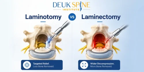
Standard microsurgical discectomy through a posterior approach remains the most commonly performed procedure for upper lumbar disc herniations. The surgeon removes a small window of the lamina (laminotomy) and excises the herniated disc fragment to decompress the nerve root. ⁸
However, surgical outcomes for upper lumbar discectomy have historically been less predictable than those for lower lumbar levels. One multi-center study reported that preoperative signs and symptoms were “highly variable and potentially misleading” in suggesting the level of herniation, and that clinical outcomes were less reliably satisfactory. ⁸
Decompression with fusion
In some cases, particularly when there is concurrent spinal instability or significant facet joint degeneration, surgeons may recommend decompression combined with interbody fusion (such as transforaminal lumbar interbody fusion, or TLIF). A comparative study of decompression alone versus fusion for upper lumbar herniations found more reliable satisfactory rates and better functional scores at three months in the fusion group, though long-term differences narrowed. ¹
Endoscopic and minimally invasive approaches
Full-endoscopic transforaminal discectomy has proven to be a promising treatment for upper lumbar disc herniation cases. These techniques employ minimal incisions, tubular retractors, and high-definition endoscopic imaging to eliminate the herniated discs either under local or general anesthesia. ¹⁹ ²⁰
In a group of 28 patients with upwardly migrated upper lumbar disc herniation treated by full-endoscopic transforaminal discectomy, there was significant improvement in the pain score and disability index of the patients, and 26 out of 28 migrated disc fragments were successfully removed.
No significant complications were reported. ²⁰
Minimally invasive transforaminal lumbar interbody fusion (MIS-TLIF) has also demonstrated advantages over open TLIF for upper lumbar herniations, including reduced intraoperative bleeding, shorter hospital stays, and faster return to work, with comparable long-term outcomes. ⁷
Deuk Laser Disc Repair®
Among endoscopic approaches, Deuk Laser Disc Repair® (DLDR) stands out as a time-tested solution for lumbar disc herniations, including those at the L2-L3 level. Developed by Dr. Ara Deukmedjian, a board-certified neuro-spine surgeon, DLDR is a form of endoscopic laser spine surgery performed under sedation in an outpatient surgery center. The procedure uses a laser to precisely vaporize the herniated disc material and seal the annular tear. The source of nerve compression and pain through a 4-7mm sized incision. ²¹
Unlike traditional open discectomy, laminectomy, or spinal fusion, DLDR does not remove bone, cut muscle, or compromise the structural integrity of the spine. The procedure preserves the full range of motion at the treated segment, which is a critical advantage at the L2-L3 level where maintaining upper lumbar mobility contributes to normal gait mechanics and hip flexion. ²¹
Over 20 years and more than 2,750 patients treated, Lumbar DLDR has achieved a 99.6% success rate with no reported complications. ²¹ Patients are discharged the same day, typically resume walking within one hour of surgery, and return to driving and daily activities within 24 hours. For patients with L2-L3 herniations who have failed conservative management or who wish to avoid the risks and prolonged recovery associated with fusion surgery, DLDR offers a proven, motion-preserving alternative.
Treat the L2-L3 disc without fusion or spinal-cord risk.
L2-L3 herniations account for barely 1% of lumbar disc surgery, which is exactly why they get misdiagnosed as hip pathology or femoral neuropathy for months. The narrow canal and proximity to the conus medullaris make open surgery here higher-risk than at lower levels. Send your MRI for a free review by Dr. Deukmedjian and learn whether Deuk Laser Disc Repair® can decompress the L2 nerve through a 7 mm incision — no fusion, no hardware, same-day discharge.
- 99.6%
- Success rate
- 0.01%
- Complication rate
- 7mm
- Incision, outpatient
FAQs
Is a L2-L3 herniated disc serious?
It depends on what the herniation compresses. A small posterolateral herniation affecting only the L2 nerve root produces pain, numbness, and hip flexor weakness, which is uncomfortable and functionally limiting but typically manageable with conservative care. A large central herniation at L2-L3 is potentially serious because it can compress multiple nerve roots or the conus medullaris, causing bilateral leg weakness, bowel or bladder dysfunction, and cauda equina or conus medullaris syndrome, both of which are surgical emergencies. ⁵ ⁶
Can a L2-L3 herniated disc heal on its own?
Many disc herniations decrease in size over time through a process of resorption, and the majority of patients with upper lumbar disc herniations improve with conservative treatment. ¹² However, “healing” in the sense of complete anatomical restoration is uncommon. What typically occurs is a reduction in inflammation, partial resorption of extruded disc material, and adaptation of the neural structures, leading to symptom improvement even though some degree of disc abnormality persists on imaging.
Why does a L2-L3 herniated disc cause thigh pain instead of leg pain?
The L2 nerve root supplies the anterior thigh through the femoral nerve. Unlike the L5 and S1 nerve roots. Which travel through the sciatic nerve and produce pain down the posterior leg and into the foot. The L2 root produces pain in the groin, anterior thigh, and sometimes medial knee. ³ ⁴ This is why L2-L3 disc herniations are frequently confused with hip problems.
Can a L2-L3 disc herniation cause foot drop?
Rarely, yes. While foot drop is classically associated with L5 nerve root compression from an L4-L5 disc herniation, documented cases exist in which a large L2-L3 herniation compressed the L5 nerve root as it traveled within the thecal sac, causing isolated foot drop without L2 or L3 symptoms. ¹³ This is an uncommon but well-documented phenomenon.
What is the difference between conus medullaris syndrome and cauda equina syndrome at this level?
Conus medullaris syndrome results from compression of the terminal spinal cord (which ends near L1 in most adults), producing upper motor neuron findings such as brisk reflexes and Babinski sign, along with bowel/bladder dysfunction. Cauda equina syndrome results from compression of the nerve roots below the conus, producing lower motor neuron findings such as absent reflexes and flaccid weakness. ⁵ ⁶ At L2-L3, the distinction is often blurred because both the conus and the upper cauda equina may be affected simultaneously, producing a mixed clinical picture.
What physical examination test is used for L2-L3 disc herniations?
The femoral nerve stretch test (also called the reverse straight leg raise or prone knee bending test) is the appropriate provocative maneuver for upper lumbar nerve root compression. ⁸ ¹² It is performed with the patient lying prone while the examiner flexes the knee and extends the hip. Reproduction of anterior thigh pain indicates irritation of the L2, L3, or L4 nerve root.
When should I get a second opinion?
If you have anterior thigh or groin pain with weakness that has been attributed to hip pathology, femoral neuropathy, or “nonspecific back pain” but has not responded to treatment, consider requesting a lumbar MRI that explicitly includes the upper lumbar levels and a consultation with a spine specialist experienced in upper lumbar disc disease. If open surgery with fusion has been recommended, ask whether a minimally invasive or endoscopic approach may be appropriate for your specific herniation pattern.
Sources
View Sources
- Chang HS, et al. Surgical outcomes for upper lumbar disc herniation. BioMed Res Int. 2019.
- Cimatti M, et al. Upper lumbar disc herniation L1-L2 and L2-L3. Eur Spine J. 2007.
- Ozer E, et al. Disc herniation mimicking cervical radiculopathy. Int J Spine Surg. 2017;11(4):30.
- Pulsenotes. Radiculopathies: dermatomal and myotomal distributions.
- StatPearls. Cauda equina and conus medullaris syndromes.
- MSD Manual. Cauda equina syndrome. 2025.
- Li Q, et al. MIS-TLIF vs TLIF for upper lumbar disc herniation. BMC Surgery. 2024;24:153.
- Sanderson SP, Houten JK. Upper lumbar disc herniations. Spine. 1993;18(16):2507-2512.
- Physiopedia. Lumbar radiculopathy.
- Varun S, et al. Asymptomatic lumbar disc herniation prevalence on MRI. PARIPEX. 2025.
- Kanayama M, et al. Lumbar disc degeneration in 200 healthy individuals. J Neurosurg Spine. 2009;11(4):501-507.
- Physiopedia. Lumbar radiculopathy: conservative management and surgical indications.
- Kowalski JM, et al. Isolated L5 radiculopathy from L2-3 disc herniation. Cureus. 2018;10(10):e3472.
- Patel R, et al. Symptomatic upper lumbar disc herniation in the Indian population. Cureus. 2025.
- Rai A, et al. Cauda equina syndrome from L2-L3 disc herniation: case report. JNMA. 2023;61(262):515-517.
- Battié MC, et al. The Twin Spine Study. Spine J. 2009;9(1):47-59.
- Sambrook PN, et al. Genetic influences on disc degeneration in twins. Arthritis Rheum. 1999;42(2):366-372.
- Jensen MC, et al. MRI of the lumbar spine in people without back pain. N Engl J Med. 1994;331(2):69-73.
- Li Y, et al. Endoscopic discectomy for migrated upper lumbar herniations. BioMed Res Int. 2017.
- Kim HS, et al. Endoscopic discectomy for upper lumbar disc herniation. J Clin Med. 2020;9(7):2107.
- Deuk Spine Institute. Lumbar Deuk Laser Disc Repair® benefits.