
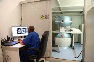

So your insurance has finally authorized your spine MRI after months of trying to prove that it is medically necessary. The big day finally arrives, and after it is finished you’re sent home with the radiologist’s report and told to follow up with your physician next week.
It may be tempting to read the report and try to interpret meaning from it that will help you to understand what is going on with your back. Curiosity is a natural Human instinct after all. But the language doctors use to communicate with each other may end up leaving you confused and more afraid. So to help you interpret your report, we will be covering some terms that you might see on it.
NOTE: This article is an educational tool only, and not a diagnostic tool. MRIs can never identify the source of pain. They only show structural changes in your anatomy. Pain is a physiological condition and the proper interpretation of symptoms can only be performed through physical examination and professional symptom analysis. The Deuk Spine Institute offers telemedicine examinations via mri.deukspine.com if you have any further questions about your current diagnosis or recommendation.
Find Your Term
Find the word on your report below and jump straight to what it means.
- Spinal stenosis: foraminal stenosis, lateral recess stenosis, central stenosis
- Disc protrusion: disc protrusion, annulus fibrosus, nucleus pulposus
- Vertebral body and endplate findings: Schmorl’s nodes, Modic changes
- Spondylolisthesis: retrolisthesis, anterolisthesis, lateral listhesis
- Joint hypertrophy: facet hypertrophy, uncovertebral hypertrophy, synovial and facet cysts
- Spinal cord and nerve damage: myelomalacia, thecal sac effacement, Tarlov cysts, myelitis
- Report language: what words like mild or minimal actually mean
Spinal Stenosis
Also known as spinal encroachment or spinal narrowing, spinal stenosis is a constriction of the space within the spine. Narrowing is the key word to remember here. There are several kinds of stenosis, each with different implications for treatment, and almost all of them can be treated with the correct surgery.
Foraminal Stenosis
Foraminal stenosis is the second most common type of spinal narrowing. It involves narrowing of the neuroforamen, the pair of openings to either side of the vertebrae where the nerve roots exit the spinal canal and travel down the arm or leg. It can occur due to several different conditions, such as a degenerated disc, a herniated disc, facet hypertrophy, facet arthropathy, ligament thickening, short pedicles, and bone spurs.
On the report, this signifies a narrowing or compression of the space between the neuroforamen.
Lateral Recess Stenosis
This is the most common type of spinal stenosis in the lower back. Narrowing occurs medial to the pedicles and is due to facet joint hypertrophy, short pedicles, and disc herniations.
Central Stenosis
Also known as central canal stenosis, this is the least common type of spinal stenosis. When it occurs in the neck it can cause damage to the spinal cord, which is called myelopathy.
When stenosis is identified on your MRI report, it may lead your physician to look into the possibility of nerve impingement, known as radiculopathy. This dysfunction occurs when nerves are compressed, and the signals they typically transmit to other parts of your body become limited by the injury. Whether it is central, foraminal, or lateral recess stenosis, you may experience nerve symptoms with any of them.
Disc Protrusion
Also known as disc herniation, disc bulge, ruptured disc, slipped disc, herniated nucleus pulposus, or prolapsed disc among others, these terms are all related to the same condition, a damaged spinal disc. When it is identified on your report, it means that at the level specified you have a tear in the tough outer layer of your vertebral disc (annulus fibrosus), and through that tear some of the soft material from inside of the disc (nucleus pulposus) has been squeezed out, almost like poking a hole in a balloon full of playdough.
Annulus Fibrosus
This is the tough outer layer of your disc. It is a ring of collagen that surrounds the soft shock-absorbing interior. When your disc herniates, the nucleus pulposus forces its way out through a tear in this ring of tissue that resulted from a prior injury to the annulus fibrosus.
Common causes of injury include motor vehicle accidents, work injuries, sports injuries, slip and fall, and repetitive motion injuries.
Nucleus Pulposus
This is the soft gelatinous interior of your disc. It absorbs the shock of bodily motion and movement down through your spine. It remains in the center of your disc unless there is a tear present in the annulus, where it takes the path of least resistance and pushes its way out into the surrounding spaces.
Vertebral Body and Endplate Changes
Two more findings show up constantly on lumbar and cervical MRI reports and are worth knowing on sight.
Schmorl’s Nodes
These are small herniations of disc material through a weak point in the endplate, the thin layer of bone and cartilage that caps each vertebra, pushing into the vertebral body itself instead of into the spinal canal. Many are old and produce no symptoms at all, though a newer one can appear alongside disc height loss or a compression injury.
Modic Changes
These describe how the bone marrow inside a vertebral body responds, on MRI, to a degenerating disc next to it. Type 1 changes usually reflect active inflammation and can correlate with pain, while Type 2 and Type 3 changes reflect longer-standing fatty or bony conversion.
Spondylolisthesis
This term refers to the relative position of your spinal vertebrae. The word spondy relates to the spine and the word listhesis refers to slippage. There are several types of spondylolisthesis and different grades for each.
Retrolisthesis
This is a posterior, or backwards, slippage, where one vertebral body has slid backwards and is now resting posteriorly relative to the vertebrae below it.
Anterolisthesis
This is an anterior, or forward, slippage, where one vertebral body has slid forward and is now resting anteriorly relative to the vertebrae below it.
Lateral Listhesis
The bones slip sideways relative to each other, resulting in scoliosis.
The grades of a spondylolisthesis go from 1 to 5. A grade 1 is only 25% slippage, up to 50% for grade 2, 75% for grade 3, 100% for grade 4, and then grade 5, which means your vertebral body has completely fallen out of your spine (called spondyloptosis). Grade 5 is one of the most uncommon, and typically any grade below a 3 will not need urgent treatment to correct spinal deformity. However, grade 3 to 4 slippage may need to be corrected with surgery if it is resulting in herniations and severe destabilization of the spine.
Joint Hypertrophy
When hypertrophy is indicated on the report in any capacity, it refers to a structure that has enlarged in size. There are different types of hypertrophy, just like there are different types of stenosis in the spine.
Facet Hypertrophy
This indicates an enlargement of the facet joints, or a swelling of the joints, which may be indicative of chronic inflammation and underlying diseases like arthritis.
Uncovertebral Hypertrophy
These are joints located in your cervical spine adjacent to each vertebra. Hypertrophy in these joints may be indicative of bone spurs (known as disc osteophytes or a disc osteophyte complex) and can cause similar issues with neck and arm pain.
Synovial and Facet Cysts
A synovial cyst is a fluid-filled sac that forms off an arthritic facet joint, most often in the lower back, and it can narrow the same space a herniated disc or facet hypertrophy would narrow. Not every cyst causes symptoms, but a larger one pressing on a nerve root can produce leg pain similar to a herniated disc.
Spinal Cord and Nerve Damage
There are many disorders of the spinal cord and nerves that may be identified on a radiologist’s report. They can range from an indication of swelling, spinal fluid leaks, or abnormal artifacts and growths that may need additional diagnostic testing to identify. Some of the terms you may see include the following.
Myelomalacia
A pathological softening of the spinal cord, typically caused by trauma to the cervical spine such as a motor vehicle accident, a sports injury, or compression from a large extruded disc.
Thecal Sac Effacement
The thecal sac, or dural sac, is the membranous sheath (theca) or tube of dura mater that surrounds the spinal cord and the cauda equina. It contains the cerebrospinal fluid that provides nutrients and buoyancy to the spinal cord. When effacement or impingement is identified on the report, it means that the sheath surrounding your spinal cord and nerves is being pressed on.
Tarlov Cysts
Tarlov cysts are fluid-filled sacs that affect the nerve roots of the spine, especially near the base of the spine (sacral region). Not all individuals will experience symptoms from a Tarlov cyst, and for those who do, there are few surgeries that can fully remove them or prevent them from growing back.
Myelitis
A pathological inflammation of the spinal cord, typically shown on MRI as white matter within the grey material of the spinal cord. This inflammation is easy to identify and typically relates to some sort of traumatic spinal cord injury.
What Descriptive Words Like Mild or Minimal Actually Mean
Adjectives used in an MRI report such as mild, minimal, small, insignificant, or nominal are all subjective and based on size and appearance alone. A radiologist’s terms have no correlation to the symptoms you may be experiencing from the specified area. We strongly recommend you disregard these descriptive terms and get an opinion from an experienced clinician to understand what the abnormality actually means for you.
Deuk Spine Institute offers free evaluation and teleconferences to discuss findings in depth and help you understand the source of your symptoms, which may not be apparent from the MRI report alone.
Now that we’ve covered some of the most common terminology that may appear on your MRI report, let’s go over an example and extrapolate the information from it.
If your MRI Report identifies the findings as the following;
“There is left uncovertebral joint hypertrophy at C4-C5 causing mild left neural foraminal stenosis. At C5-C6 there is a mild broad-based central disc protrusion. The central canal and neural foramina are within normal limits.”
Broken down, the radiologist is reporting that the patient has NARROWING (stenosis) at the LEFT nerve root of C4-C5 due to HYPERTROPHY (swelling) of the joint tissue. This may correlate with pain or numbness in the LEFT arm or hand. They are also reporting a mild PROTRUSION (blown-out disc) at C5-C6, but no notable narrowing, which means this disc may not be the source of any pain or nerve symptoms.
This is just one example, and some radiologists use different language. But this key should be helpful while trying to read and understand the report that was given to you.

For further assistance understanding your diagnosis, visit here where the Deuk Spine Foundation provides free telemedicine consultations and MRI reviews. Our team of professional physicians can help you interpret the information in your report and give you a proper medical opinion.
And take a look at our in depth reviews of institutes across the country, like the Total Spine and Orthopedics Wellness review.
DISCLAIMER: This article is for educational purposes only and is not a substitute for professional medical advice, diagnosis, or treatment. Always consult a qualified physician regarding any questions about a medical condition or an MRI finding.