
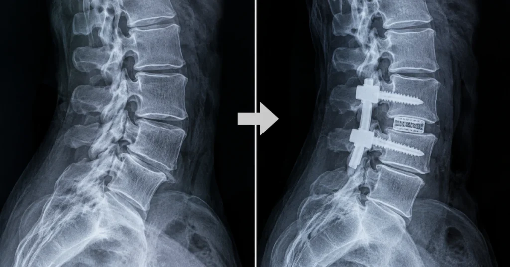
Before and after spinal fusion, the biggest change is that two or more vertebrae that used to move separately become one solid segment of bone. On an X-ray, that shows up first as screws, rods, and often a spacer cage, then as new bone bridging the level over the following months. Many patients have less pain and a more stable spine afterward, but motion at the fused level is gone for good.
That is only the headline, though. Whether the bone actually fuses, how much stiffness you notice, and how much extra load lands on the discs above and below all depend on the level, the number of segments, and the diagnosis behind the surgery. Most of the dramatic before and after spinal fusion surgery photos online also show scoliosis corrections, which look nothing like a one-level fusion for disc pain.
If you have been told you need a fusion and have not had it yet, a free review of your MRI gives you a second look at your imaging while every option is still open. Neurosurgeon Ara Deukmedjian, MD, and his team use it to identify the structure most likely causing your pain and to check whether a motion-preserving procedure such as Deuk Laser Disc Repair® could treat it. Here is what the full before and after picture of a fusion includes, starting with what the surgery changes.
Medical Disclaimer: This article is educational and is not a diagnosis or treatment recommendation. Imaging findings and surgical outcomes vary from person to person. Talk with a qualified spine specialist about your own condition and treatment options.
No cost · No obligation
Live Pain Free
Upload your MRI for a free expert review by Dr. Ara Deukmedjian, M.D. — board-certified neurosurgeon. Ten minutes can change your life.
What Does a Spinal Fusion Change?
A spinal fusion permanently joins two or more vertebrae so they heal into a single, immovable piece of bone. The surgeon removes or prepares the disc or joint surfaces, packs the space with bone graft, and holds everything still with hardware while the bone grows across.
The goal is to stop painful motion or instability at that level, or to hold the spine in a corrected position. Unlike most spine operations, the change is permanent by design. Fusion is used for conditions such as spondylolisthesis, instability after a decompression, fractures, spinal deformity, and some cases of severe disc degeneration.
What Does Spinal Fusion Look Like Before and After on an X-Ray?
Before and after spinal fusion, an X-ray goes from showing a narrowed or shifted disc space to showing metal hardware and, over months, a continuous column of bone across that level.
Before Surgery: What Your Imaging Usually Shows
Before a fusion, the X-ray shows the bones and their alignment, while the MRI shows the discs, nerves, and soft tissue. Common findings include a collapsed disc space, bone spurs along the edges of the vertebrae, one vertebra slipped forward on another, or a curve.
These are the findings a surgeon uses to recommend fusion. They are also the findings that deserve a second look, because a worn disc on a scan is not always the disc that is causing the pain.
Right After Surgery: Screws, Rods, and Cages
Right after surgery, the hardware is the most visible part of the image. Metal appears bright white on an X-ray, so the construct stands out clearly against the gray of the bone.
- Pedicle screws: Angled into the back of each vertebra in the lower or mid back, usually placed in pairs.
- Rods: Vertical bars that connect the screws on each side and lock the level in place.
- Plates: In a neck fusion done from the front, such as an anterior cervical discectomy and fusion (ACDF), a small plate with screws usually spans the disc space.
- Interbody cages: Spacers placed where the disc used to be. Titanium cages show up bright white, while PEEK plastic cages are nearly invisible except for small metal marker dots.
At this stage the bone graft is still loose material, so it looks faint or patchy. The hardware is doing all of the holding.
Months Later: Bone Bridging Across the Level
Over the following months, the graft should turn into living bone that grows from one vertebra to the next. On imaging, this appears as a continuous bridge of bone with a visible internal texture, called trabecular bone, crossing the disc space or the area along the sides of the spine.
A successful fusion also looks quiet around the hardware. There should be no dark rim around the screws and no change in their position compared with earlier films.
Why Before and After Photos Online Can Be Misleading
Many of the most shared before and after images on social media show scoliosis surgery, where long rods straighten a curve across 10 or more vertebrae. Those images are striking, but they do not represent what a one or two level fusion for back or neck pain looks like.
A typical degenerative fusion changes little about how your back looks from the outside. The meaningful changes are internal.
How Does a Surgeon Confirm the Fusion Took?
A surgeon confirms a fusion by looking for continuous bone bridging across the level and no movement between the fused vertebrae.
Flexion and extension X-rays, taken while you bend forward and lean back, show whether the fused vertebrae still shift.
A CT scan is the most detailed way to see bone, and it can catch problems that standard X-rays miss. In one study of lumbar interbody fusion, CT taken in extension detected more nonunions than bending X-rays at an average of about 15 months after surgery.
| What the Surgeon Checks | Solid Fusion | Fusion That Has Not Taken (Nonunion) |
|---|---|---|
| Bone across the level | Continuous bridging bone | Gaps, clefts, or graft that has thinned or disappeared |
| Motion on bending X-rays | None or minimal | Measurable shifting between the vertebrae |
| Bone around the screws | Tight contact with no dark rim | A dark halo that suggests loosening |
| Hardware position | Unchanged from earlier films | Broken rods or screws, or a cage sinking into the bone |
| Symptoms | Often improved | Pain may persist or return, though some nonunions cause no symptoms |
A fusion that does not heal is called a pseudarthrosis, or nonunion. Smoking, diabetes, osteoporosis, and multi-level surgery all raise the risk, and some patients with a nonunion need another operation.
Published estimates of spinal fusion success commonly fall between about 70 and 90%. That range depends on the condition treated, the technique, and whether success means solid bone on imaging or relief of pain, and it is usually judged one to two years after surgery.
What Changes in Movement and Daily Life After Spinal Fusion?
After spinal fusion, the fused level no longer bends or rotates, and the rest of your spine takes over that work. For many people with a one-level fusion, that loss is hard to notice in everyday life, and a lot of patients do well, with less pain and more confidence in their back.
The more levels that are fused, the more noticeable the change. A long lumbar fusion can make it harder to bend down to tie your shoes, and a multi-level neck fusion can limit turning your head while driving.
- Less of the pain that came from the unstable or degenerated level, for many patients
- A stiffer feel at the fused area, especially when bending or twisting
- Bending more from the hips and knees to make up for the lost motion
The loss of motion is permanent,, and patients are often not walked through it before surgery. If staying flexible matters to you, it helps to understand how different spine surgeries affect your range of motion before you decide.
What Does Not Improve After Spinal Fusion?
A spinal fusion fixes motion at one level, but it does not repair the rest of the spine or reverse long-standing nerve damage.
- Degeneration elsewhere: Discs and joints at other levels keep aging at their own pace.
- Long-standing nerve symptoms: Numbness or weakness from a nerve that was compressed for a long time may only partly recover.
- Pain from a different source: If the fused level was not the true pain generator, the pain can remain after a technically successful fusion.
That last point is why diagnosis matters so much. A fusion can heal perfectly on an X-ray and still leave a patient in pain if the imaging finding and the pain source were not the same thing.
What Happens at the Levels Above and Below a Fusion?
The levels directly above and below a fusion take on extra motion and load, because the fused segment can no longer share the work. Over time, that added stress can speed up wear on those neighboring discs and joints.
When that wear causes symptoms, it is called adjacent segment disease. It does not happen to everyone, and it often appears years later, which is why it rarely shows up in early after photos.
How often it develops varies by spinal level and follow-up period, and the research on adjacent segment disease after fusion breaks those rates down for the neck and lower back.
How Long Before You See the Final Result?
The final before and after result of a spinal fusion is usually clear between 6 and 12 months, though bone can keep maturing for up to two years.
- First few weeks: Hardware is in place, and the graft has not yet become bone.
- Around 3 to 6 months: Early bridging may start to appear on imaging.
- Around 6 to 12 months: Many surgeons assess whether the fusion is solid.
- 12 to 24 months: Bone continues to remodel, and a CT scan may be used if the fusion still looks incomplete.
How you feel follows a separate track from what the X-ray shows, and the day-to-day side of healing follows its own week-by-week recovery timeline.
If You Have Not Had Spinal Fusion Yet, What Should You Ask?
If you have not had the surgery yet, the most useful step is confirming that the level being fused is the actual source of your pain. Fusion is an established operation with a clear role, and the most important variable is how precisely the pain source has been identified.
Questions worth asking:
- Which structure on my imaging is causing my pain, and how was that confirmed?
- Is the level unstable, or is the main problem a damaged disc or a pinched nerve?
- How many levels will be fused, and what will that mean for my movement?
- Is there a motion-preserving option for my specific diagnosis?
Some conditions, such as significant instability or deformity, are commonly treated with fusion. In other cases, the pain comes from a specific disc injury that can be treated without joining vertebrae, which is the problem Deuk Laser Disc Repair® was developed to address.
Before committing to a permanent change, it is worth reading how spinal fusion works as a treatment option, including when it is used and what it involves.
Bottom Line
Before and after spinal fusion surgery, the core change is simple: a moving level becomes solid bone, held by hardware until the fusion heals. Many patients feel better afterward, but the stiffness at that level is permanent and the neighboring levels carry more load for life.
If fusion has been recommended, make sure the diagnosis is precise before the change becomes permanent. Submit your MRI for a free expert review with no obligation, or contact our team to find out whether your pain source could be treated while keeping your spine’s natural motion.

Diagnosis. Answers. Relief.
Submit your MRI for a free expert review by Dr. Ara Deukmedjian, M.D. —
board-certified neurosurgeon. No obligation. Real answers.
Frequently Asked Questions
What Does a Fused Spine Look Like on an X-Ray?
A fused spine on an X-ray looks like two or more vertebrae joined by a continuous bridge of bone, usually with screws, rods, a plate, or a cage visible as bright white metal. Over 6 to 12 months, the graft between the vertebrae should fill in as solid bone.
Can an X-Ray Show If a Spinal Fusion Failed?
An X-ray can often show if a spinal fusion failed, through signs such as movement between vertebrae on bending films, a dark halo around screws, or broken hardware. A standard X-ray can also miss a nonunion, so surgeons often order a CT scan when symptoms or images raise doubt.
Do You Lose Flexibility After Spinal Fusion?
You do lose flexibility after spinal fusion at the levels that are fused, and that loss is permanent. With a single level, many people notice little difference in daily life, while fusions across several levels can noticeably limit bending or turning.
Will the Screws and Rods Stay in Forever?
The screws and rods usually stay in forever after a spinal fusion, because removing them is a separate surgery with its own risks. Surgeons sometimes take hardware out if it loosens, breaks, or causes irritation, but the fused bone stays fused.
Does Spinal Fusion Change Your Posture or Height?
Spinal fusion can change your posture or height when it corrects a curve or a slipped vertebra, as in scoliosis surgery. For a typical one or two level fusion for disc problems, the outward change is usually small.
Can a Spinal Fusion Be Reversed?
A spinal fusion cannot be reversed once the bone has healed, because the vertebrae have become a single piece of bone. That permanence is the main reason to confirm your diagnosis and review motion-preserving options before agreeing to surgery.
Medical Disclaimer: This content is for educational purposes only and does not constitute medical advice. Individual results vary. Always consult your healthcare provider about your specific condition and treatment options.