Transforaminal Lumbar Interbody Fusion (TLIF): Potential Complications And Occurrence Rates
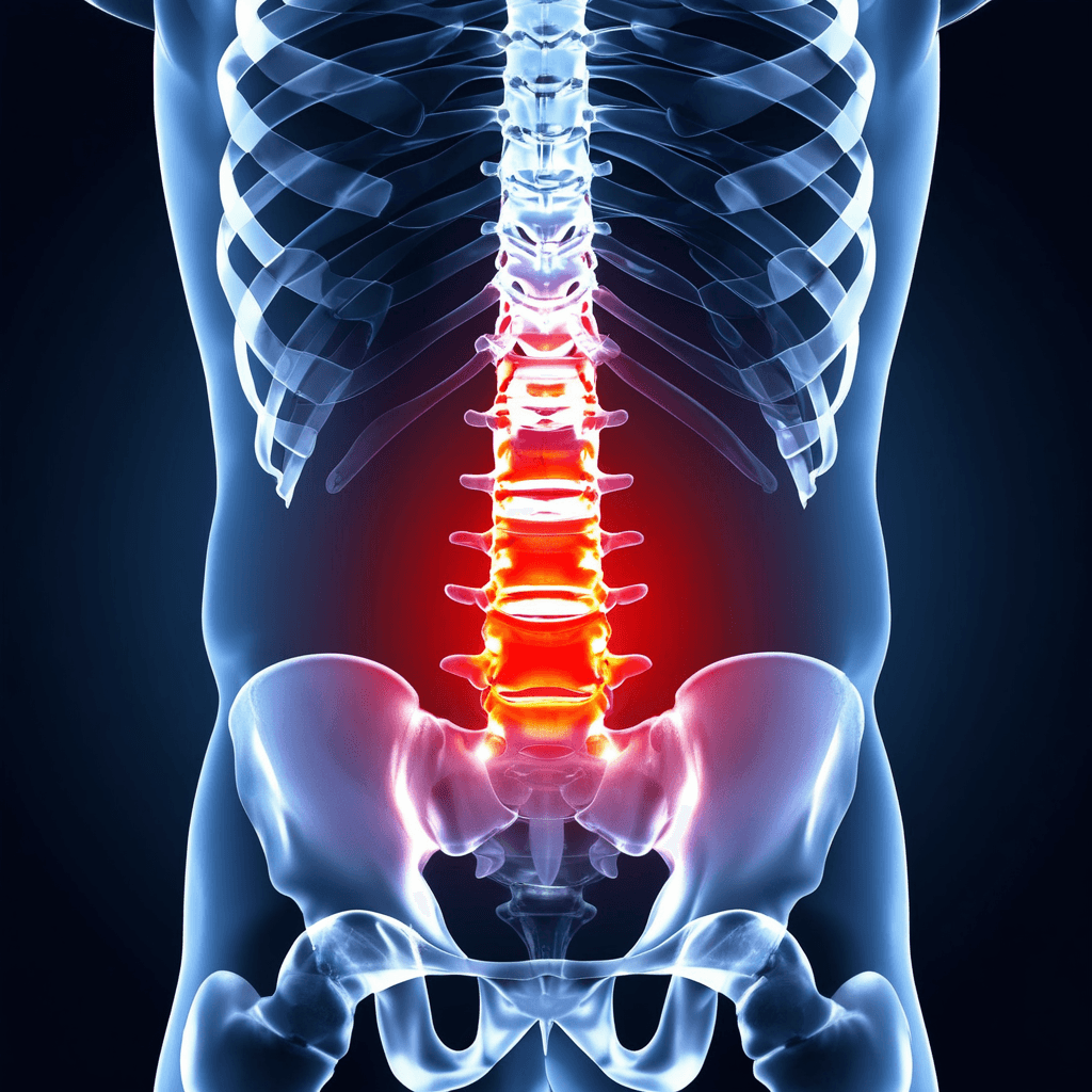
Transforaminal Lumbar Interbody Fusion (TLIF) is a surgical procedure used to fuse or join two or more vertebrae in the lumbar (lower) spine. As a type of spinal fusion, its primary goal is to stabilize the spinal vertebrae and reduce pressure on spinal nerves. The most common indications for TLIF are degenerative disc disease, spondylolisthesis, spinal stenosis, foraminal stenosis, and recurrent herniated disc. This article reviews this procedure for this invasive surgical technique and its potential complications.
TLIF: An Invasive Approach
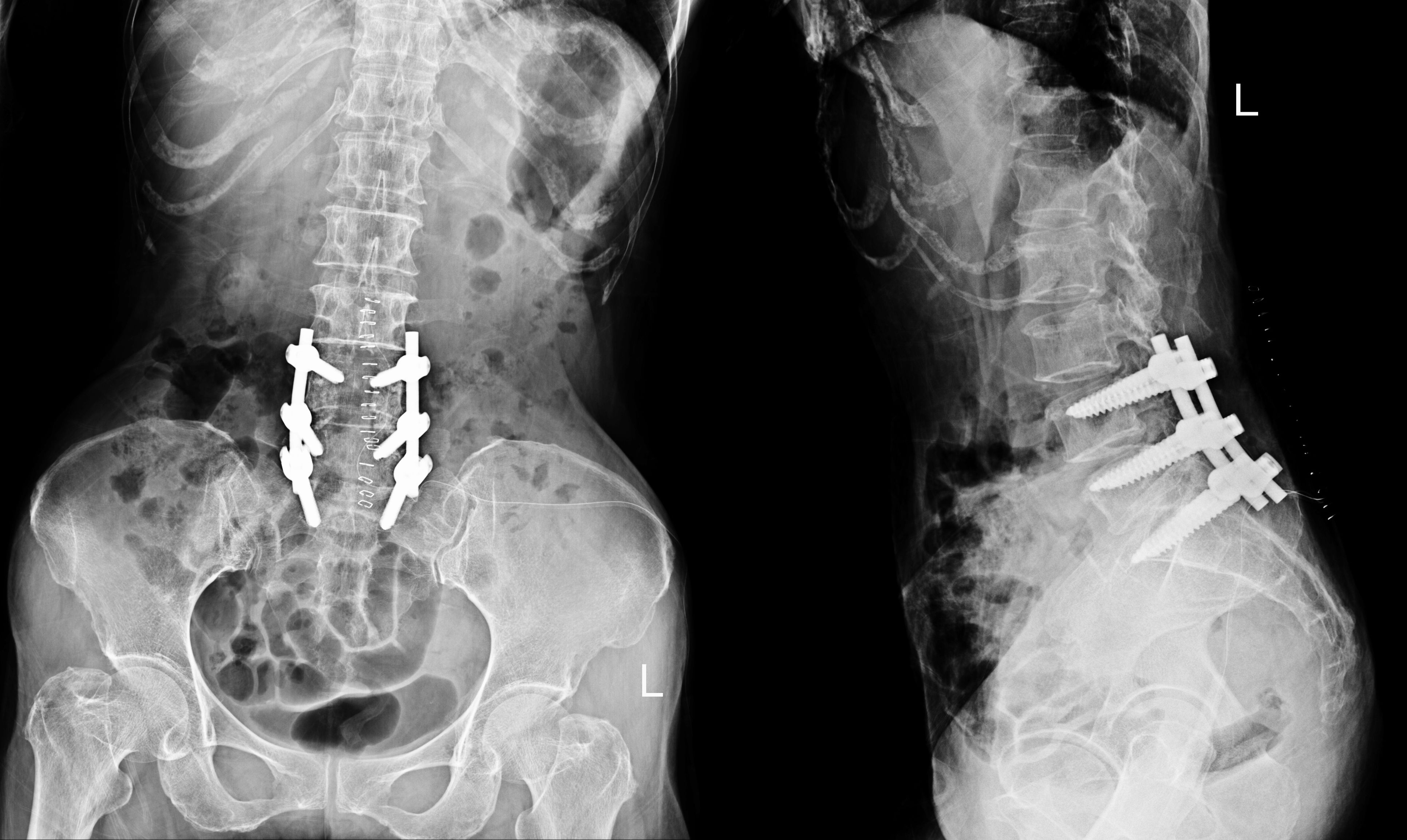
The main goal of TLIF is to remove a damaged intervertebral disc—the cushion between the vertebrae—and replace it with a bone graft and a structural spacer, usually a cage made of titanium or a high-performance polymer like PEEK. This graft and cage restore the correct height between the vertebrae and help the fusion process. Over time, the bone graft grows and fuses the adjacent vertebrae into a single, solid bone, eliminating movement at that segment. To provide immediate stability while the fusion matures, the surgeon also installs hardware, such as pedicle screws and rods.
TLIF requires the removal of part of the facet joint and lamina to access the intervertebral disc space. Muscle retraction and some bone removal are necessary, but the unilateral approach (one side of the spine) reduces overall tissue damage.
TLIF: A Step-By-Step Overview
The TLIF procedure is performed under general anesthesia. While specific techniques can vary, the general steps are as follows:
- Incision and Exposure: The surgeon makes an incision on the patient's back over the affected spinal level. The muscles are retracted to expose the lamina, facet joints, and transverse processes of the vertebrae.
- Decompression and Facetectomy: To access the intervertebral disc and foramen, the surgeon performs a laminectomy(removal of a portion of the lamina) and a facetectomy (removal of a facet joint) on one side. This action immediately relieves pressure on the nerve roots.
- Discectomy: Working through the newly created path, the surgeon removes the damaged intervertebral disc. The surfaces of the endplates (the top and bottom of the vertebrae) are then prepared to receive the bone graft.
- Interbody Spacer and Graft Placement: The surgeon inserts an interbody cage filled with bone graft material into the empty disc space. The cage acts as a structural support to maintain disc height, while the bone graft promotes fusion.
- Pedicle Screw Fixation: To provide rigid stabilization, the surgeon places pedicle screws into the vertebrae above and below the fused level. These screws are then connected with rods, creating a solid internal construct that holds the vertebrae in place while the bone fuses.
- Closure: The muscles are returned to their normal position, and the incision is closed with sutures or staples.
TLIF: The Complications
TLIF’s unique surgical approach allows for both decompression of nerves and stable fusion through a single incision. However, it is an invasive surgery with a distinct set of potential perioperative complications.
Complications can be categorized as intraoperative (occurring during surgery), early postoperative (within days or weeks), and late postoperative (months to years later). The surgery can also cause long-term side affects that may affect patients for a lifetime.
Infection: Infections can be superficial (in the incision) or deep (around the hardware and spine). Deep wound infectionsare more serious and may require additional surgery to clean the area and a long course of antibiotics.
Nerve Damage (Neurological Deficit): Because the surgery is performed near critical nerve structures, there is a risk of nerve root injury. This can lead to new or worsening weakness, numbness, or pain in the legs. Permanent neurological injury is rare, but temporary nerve irritation is more common.
Blood Clots (Deep Vein Thrombosis): After any major spinal surgery, there is a risk of developing blood clots in the legs, which could travel to the lungs and cause a pulmonary embolism. Proactive measures, such as early mobilization and the use of blood thinners, are standard protocols for all these surgeries to reduce this risk.
Dural Tear (Cerebrospinal Fluid Leak): The dura mater is the membrane surrounding the spinal cord and nerves. It can be accidentally torn during surgery, causing a cerebrospinal fluid (CSF) leak. Most dural tears are identified and repaired during the procedure. If a leak persists, it can cause headaches and increase the risk of infection.
Hardware Failure or Malposition: The screws and rods provide the initial stability for the fusion. Hardware can break, loosen, or be misplaced. Screw malposition can irritate nerves or compromise the stability of the construct. Hardware failure may require revision surgery..
Non-union (Pseudarthrosis): Fusion is not guaranteed. Pseudarthrosis occurs when the vertebrae fail to fuse into a single, solid bone. This can lead to persistent pain and may necessitate revision surgery. Risk factors include smoking, obesity, osteoporosis, and multi-level fusion. The rate of pseudarthrosis after
Cage Migration or Subsidence: The interbody cage can shift from its intended position (migration) or sink into the adjacent vertebral bone (subsidence). Significant subsidence can lead to a loss of disc height and potential re-stenosis of the foramen. Clinically significant cage migration is uncommon, but subsidence rates can be higher, though often asymptomatic.
Adjacent Segment Disease (ASD): Fusing a spinal segment transfers stress to the vertebrae above and below the fusion. Over time, this increased stress can accelerate degeneration at these adjacent levels, potentially leading to new symptoms that require further treatment. The risk of developing symptomatic ASD is estimated to be around 2% to 4% per year.
Nerve Root Injury: TLIF is performed by accessing the spine from the back and slightly off to one side, bringing the surgeon close to the exiting nerve roots. Direct nerve root manipulation raises the risk of injury, leading to leg pain or weakness.
Increased Muscle Dissection: The posterior approach requires moving or dissecting back muscles, which can cause more postoperative pain or, on occasion, longer recovery due to muscle trauma.
Scar Tissue Formation: The posterior route may lead to more scar tissue around nerve roots, which could contribute to recurrent symptoms in the long term.
DEUK LASER DISC REPAIR (DLDR): No Complications
Deuk Spine Institute’s proprietary DLDR is performed without cutting or damaging healthy muscle, fascia, or bone, without using risky hardware, and without post-operative pain. Unlike TLIF, there are no complications. None. Best of all, the DLDR procedure takes less than an hour, and recovery takes less than three days.
Our success rate is 99.5%, and we have treated thousands of patients who are now living pain-free.
Make Your First Pain-Free Move
If you’re seeking relief from lumbar pain, have been recommended for a TLIF spinal fusion, or are still dealing with chronic neck pain after a failed fusion surgery, we can help improve your quality of life and enable you to live pain-free.
Upload your latest MRI for a free review and a personal consultation with acclaimed spinal surgeon Dr. Ara Deukmedjian, M.D., founder of Deuk Spine Institute and creator of the Deuk Laser Disc Repair® procedure.