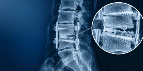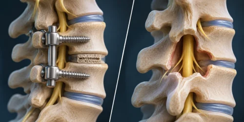


You had the fusion. The radicular pain that sent you to surgery may even be gone. Then a follow-up MRI report uses a phrase you were never warned about, cord signal change or myelomalacia, and no one on your care team has explained what it means for the rest of your life.
Here is the direct answer. A T2 signal change on your cord means the spinal cord tissue itself was compressed long enough to change structurally, not just to hurt. Decompression and fusion stop new injury from happening, but they do not reliably reverse damage the cord already sustained, and no honest surgeon can promise that they will.
That does not mean nothing else matters. Whether you improve, plateau, or stay the same depends on how long the compression lasted before surgery, how much of the cord shows the change, and where you started functionally. Some patients regain real function over the following year, while others reach a plateau and stay there.
The rest of this article walks through what drives that difference, what symptoms need care today rather than at your next appointment, and what a realistic timeline looks like, including well past the 12-month mark most patients are told to expect.
What Cord Signal Change on a Post-Op MRI Means
On a T2-weighted MRI, the spinal cord normally shows a uniform, dark signal. A bright, high-intensity area inside the cord on that sequence is what radiologists call increased signal intensity, and it is the finding behind terms like cord signal change, T2 hyperintensity, or myelomalacia. It shows up at the exact level where the cord was compressed, most often at C5-C6 or C6-C7, the two levels that carry the most mechanical load in the neck.
The finding is not a measurement of your pain or your function, it is a picture of tissue change. Two patients with an identical-looking bright spot on MRI can have very different exam findings, which is exactly why the report alone cannot tell you your prognosis. Correlating the image with a physical exam, your symptom history, and how the finding has changed between scans is what actually predicts where you are headed.
Myelomalacia in Plain Language
Myelomalacia literally means softening of the spinal cord. It describes a more advanced, usually longer-standing stage of the same process that starts as a T2 signal change. Where an early signal change can reflect swelling and inflammation that has real potential to settle down, myelomalacia usually reflects tissue that has undergone lasting structural change, sometimes including small areas of cavitation inside the cord.
Radiologists do not always use the two terms consistently. Some use “myelomalacia” for any T2 bright signal, while others reserve it for cases with visible cord atrophy or cavitation on a follow-up scan. If your report uses the word, ask your surgeon or a specialist which meaning applies to your images, because the two versions carry different weight for what comes next.
Red Flags That Mean Urgent Care Now
A handful of symptoms mean you should not wait for your next scheduled visit. Get evaluated the same day, or go to an emergency department, if you notice any of these signs.
- Progressive weakness in an arm, hand, or leg that is getting worse day to day, not just present.
- Worsening gait or balance, including new stumbling, a wider stance to stay steady, or trouble on stairs you managed fine last month.
- New bowel or bladder change, including urgency, retention, or loss of sensation, which can signal a change severe enough to need immediate imaging.
- New or worsening hand clumsiness, such as dropping objects or trouble with buttons and zippers.
- A sudden change in symptoms following a fall, a car accident, or any new neck trauma after your fusion.
None of these findings mean your fusion failed. They mean the cord is under active stress that a follow-up scan needs to catch quickly, and waiting costs recovery potential that timing can protect.
Does Cord Damage Recover? The Honest Answer
Established myelomalacia, meaning cord tissue that has undergone lasting structural change, can be irreversible. That is the honest limit of what surgery and medicine can currently offer, and no procedure, including any performed at Deuk Spine Institute, reverses that specific tissue change once it has set in.
What decompression and fusion accomplish is removing the ongoing mechanical pressure that was driving the injury forward. Stopping the compression protects whatever cord function remains and gives the tissue its best chance to recover on its own. For background on what a fusion is designed to accomplish and where its limits are, see our overview of spinal fusion.
The factors that most influence the outcome are how long the cord was compressed before surgery, how many spinal levels show signal change, and the patient’s neurological exam before the operation. A study following patients with cervical myelopathy for five years after decompression surgery found that improvement in the T2 signal at the one-year mark predicted a better midterm outcome (postoperative cord signal recovery study), which is one reason your surgeon may recommend a follow-up scan at that stage rather than relying on symptoms alone.
What the Timeline Looks Like, Including Past 12 Months
Most of the recovery patients experience happens in the first 3 to 6 months after decompression, as swelling resolves and the cord adjusts to reduced pressure. A second, slower phase of improvement can continue for up to a year, which is why many surgeons wait until the 12-month mark before calling a result final. For a general recovery timeline after cervical decompression and fusion, see our guide to neck surgery recovery time at C5-C6-C7.
Patients past that window often ask whether anything can still change at 13 months, 18 months, or later. The honest answer is that further meaningful neurological improvement past the one-year mark is less common, but it is not impossible, and small gains in strength, sensation, or hand function have been documented well beyond a year in patients whose compression was addressed. What rarely happens is a return to fully normal function once myelomalacia has become established on imaging.
If you are past 12 months and still seeing changes, whether better or worse, that is worth documenting with your surgeon rather than assuming the window has simply closed.
What “Cord Thinning” Means on a Report
Cord thinning, sometimes written as cord atrophy, describes a measurable decrease in the width or cross-sectional area of the spinal cord on a follow-up MRI compared with an earlier scan. It reflects a later stage than a simple T2 signal change and generally indicates that some of the cord’s internal structure has been lost rather than just altered.
Thinning does not automatically mean your symptoms will worsen, and some patients with visible atrophy have stable, functional exams for years. What it does mean is that the finding deserves a conversation with a spine specialist about what your specific exam shows now, not just what the image shows.
When a Second Opinion Is Worth Getting
A second opinion makes sense when your symptoms and your MRI report do not seem to match, when a new symptom has appeared since your last visit, or when you were told to “watch and wait” and you want a second read on whether that is still the right call. It is also reasonable if you simply want your images reviewed by someone outside the surgical team that performed your original procedure.
To be clear about what this can and cannot offer. A second review of your post-fusion MRI can confirm what the signal change or thinning represents today, and it can flag whether an unrelated pain source, such as facet joint irritation or a problem at the level above or below your fusion, is contributing to symptoms the myelomalacia itself does not explain. It cannot reverse established cord damage, and any specialist who implies otherwise should be questioned closely.
Pain that returns after a technically successful surgery is common enough to have its own name, and our guide to failed back surgery syndrome covers how that diagnosis works.
If you want that kind of MRI review, send your images for a free MRI review and Dr. Deukmedjian’s team will tell you plainly what the findings show and whether anything else may be contributing to your symptoms.
What to Track Between Scans
Between follow-up MRIs, the most useful thing you can do is track function, not just pain. A simple log covering the items below gives your surgeon real data at your next visit instead of a general impression.
- Grip strength and fine motor tasks, like buttoning a shirt or opening a jar, noted as easier, the same, or harder.
- Walking distance and balance, including any new use of a wall, railing, or cane.
- Sensation changes in the hands or feet, described by location and whether they are spreading.
- Bowel or bladder function, tracked honestly even if it feels like an awkward detail to raise.
- Any fall or near-fall, with the date and what was different about your footing or balance that day.
Bottom Line
If your follow-up MRI shows a cord signal change or myelomalacia, the finding tells you the cord was compressed long enough to change, and your surgery’s job was to stop that compression from continuing. Recovery beyond that point depends on how long the damage had been building and how much of the cord it affected, not on anything a second procedure can undo. If your symptoms and your imaging do not line up, or if a new red flag shows up, contact our team or submit a free MRI review to get a direct answer.

Diagnosis. Answers. Relief.
Submit your MRI for a free expert review by Dr. Ara Deukmedjian, M.D. —
board-certified neurosurgeon. No obligation. Real answers.
Frequently Asked Questions
Does myelomalacia always get worse over time?
No, many patients with an established finding on imaging have a stable exam for years once the compression causing it has been addressed. Worsening after successful decompression is the exception, not the rule, and it is one reason follow-up imaging and a documented functional exam matter more than the report label alone.
Can Deuk Laser Disc Repair® treat myelomalacia?
No, Deuk Laser Disc Repair® treats disc-sourced pain from a torn or herniated disc, and it does not reverse established cord tissue change. If a follow-up scan shows a myelomalacia finding, that is a spinal cord issue best managed by the surgeon who performed your decompression or by a specialist reviewing the full picture, not a candidacy question for a disc procedure.
How long after neck surgery should I expect a follow-up MRI?
Practice varies, but many surgeons order a follow-up scan around the 12-month mark specifically to check whether a T2 signal change has improved, since that early change has been linked to how a patient does over the next several years.
What if my pain came back even though the myelomalacia looks stable?
Returning pain with a stable cord finding usually points to a separate pain generator rather than a cord problem, and it deserves its own workup instead of being attributed automatically to the original finding. This pattern is common enough after spine surgery to have its own clinical name, and it is worth raising directly with your surgeon rather than assuming nothing more can be done.
Is fusion still the right call if imaging shows cord changes before surgery?
That decision depends on your specific exam, the extent of compression, and how long symptoms have been present, and it is not something imaging alone can answer. A specialist reviewing your case can walk you through what the procedure is designed to accomplish and where its limits are.
Medical Disclaimer. This article was reviewed for medical accuracy but is not a substitute for individualized care from your treating physician. Every patient’s imaging and exam findings are different, and only a specialist examining you directly can give you a prognosis specific to your case.