Treatment Not Recommended

Ultrasonic Spine Treatment

Risks & Better Alternatives

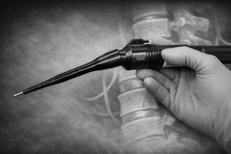
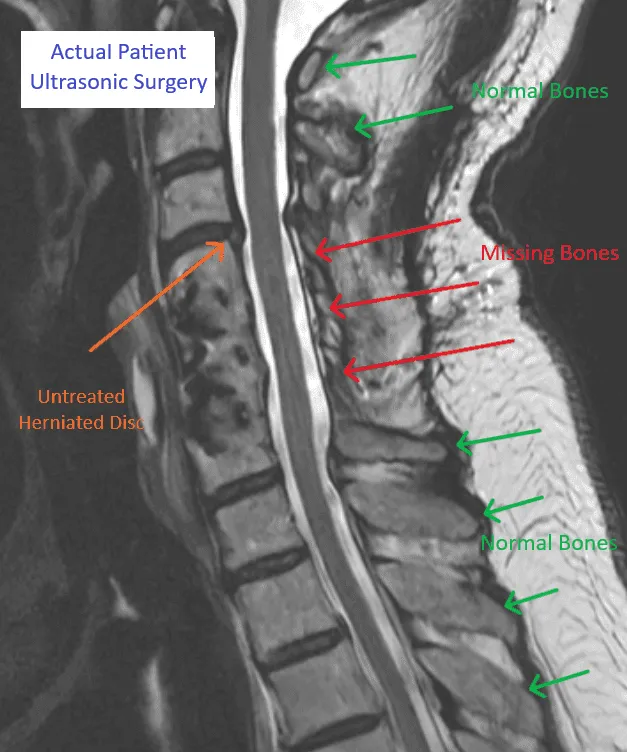
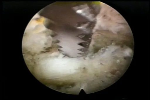
Ultrasonic Spine Treatment destroys normal bone and bone marrow from the spine using an ultrasonic probe, does not treat the actual source of pain — disc injuries — and may lead to spinal instability, excessive bleeding, and the need for additional surgery. Discover safer, minimally invasive alternatives.
A gloved hand holding a medical tool in front of a spine X-ray.

⚠️ While Deuk Spine Institute can perform Ultrasonic Spine Surgery, we do not recommend it.
This page explains why — learn the risks before considering this procedure.

THE PROCEDURE

How Is Ultrasonic Spine Treatment Performed?

Graphic Surgical Content

The images below contain surgical illustrations. Viewer discretion is advised.

A gloved hand performing surgical incision on a patient with blood visible.

Step 1: Cut Skin Behind Spine

A large incision is made in the skin over the spine to access the vertebral column.

A gloved hand holding a medical tool in front of a spine X-ray.

Step 2: Ultrasonic Probe Removes Normal Bone Along With Bone Spurs

An ultrasonic device (CUSA) is used to break apart and remove both normal bone and pathological bone spurs.

Surgical setup with tools surrounding an incision site covered in iodine and blue drapes.

Step 3: Create a Channel in the Bone of the Vertebrae

The probe creates a channel through the vertebral bone, destroying normal bone and bone marrow in the process.

CRITICAL RISKS

Why Should Ultrasonic Spine Treatment Be Avoided?

11 documented reasons why this procedure fails to deliver lasting results.

Graphic Medical Content

Images below contain real surgical procedures and medical imagery.

MRI of the spine showing missing vertebrae, marked by red arrows.

Highly Invasive

Surgeons operating with instruments and a red tube during a surgical procedure.

Excessive Bleeding

MRI image of a spine showing areas with normal and missing bones labeled in green and red.

Destruction of Stabilizing Bones

MRI showing spinal structure with labeled scar tissue from muscle damage.

Muscle Damage

Close-up of lung tissue showing bronchioles in pink and red hues.

Scar Tissue Formation

Medical scan showing normal and missing spinal bones with herniated disc, labeled for ultrasonic surgery.

Pain Source NOT Treated

MRI image of a spine showing untreated herniations marked with arrows.

Herniated Discs Not Treated

Close-up of arthroscopic knee surgery showing a surgical tool inside a joint.

Bone Necrosis

Person choosing pills from assorted medication on a table.

Excessive Pain & Opioid Dependency

X-ray images showing spinal fusion with metal screws in the lower back.

Back Pain Becomes Worse

X-ray showing spinal hardware in the lower back from front and side views.

Additional Surgery Necessary

DOCUMENTED COMPLICATIONS

What Can Go Wrong?

Graphic Medical Content

Images below show real surgical complications. Viewer discretion is advised.

A person's lower back with a large blister and several scars visible.

Nerve Root Damage

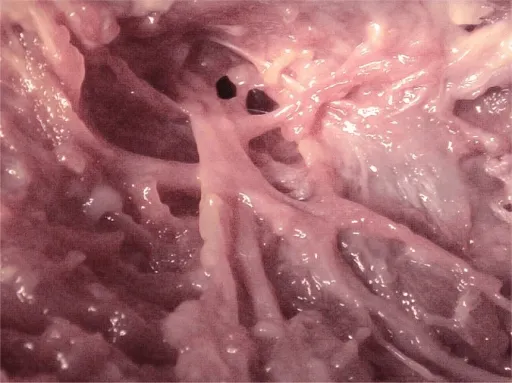
Ultrasonic probes can damage delicate nerve roots during bone removal.
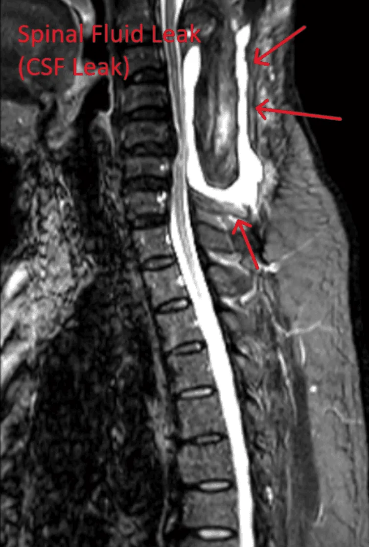
MRI showing a spinal fluid leak with arrows indicating the affected area.

Spinal Fluid Leak

Dural tears can occur, causing cerebrospinal fluid leakage.

Illustration of a herniated disc pressing against spinal nerves.

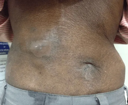
Infection of Bone or Spine

Bone destruction creates an environment susceptible to infection.
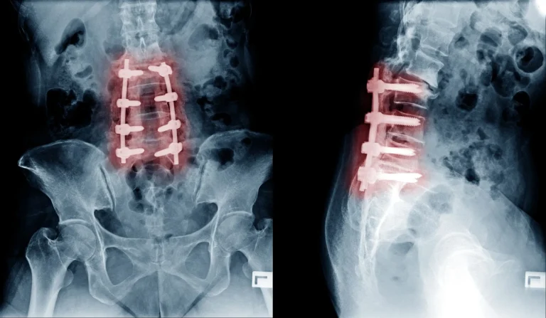
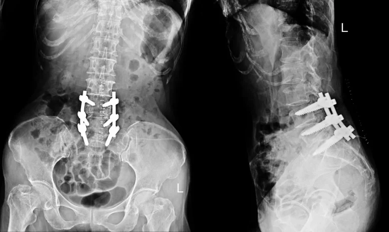
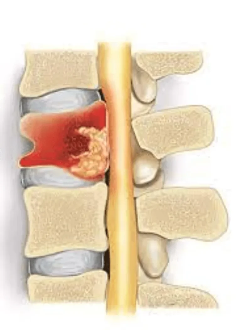
CT scans showing a spine fracture in sagittal (a) and axial (b) views.

Spinal Instability

Removal of stabilizing bone can lead to spinal instability requiring fusion.

Illustration of a spinal vertebra with a visible fracture and surrounding inflammation.

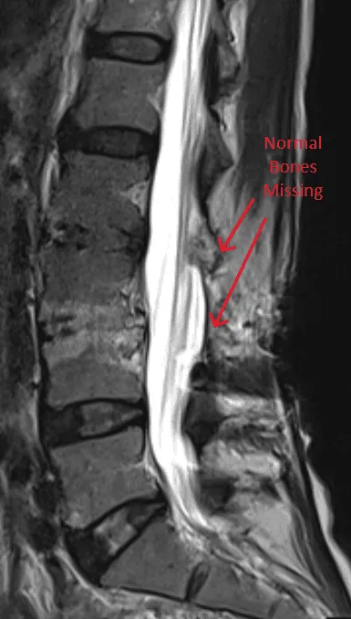
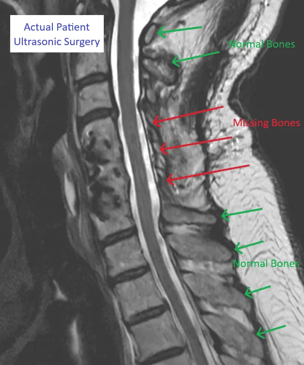
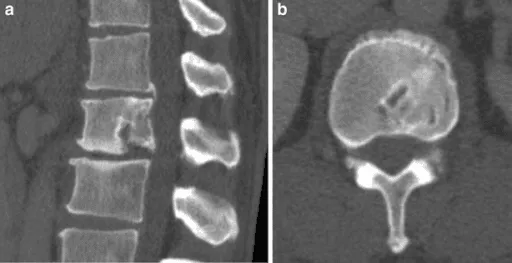
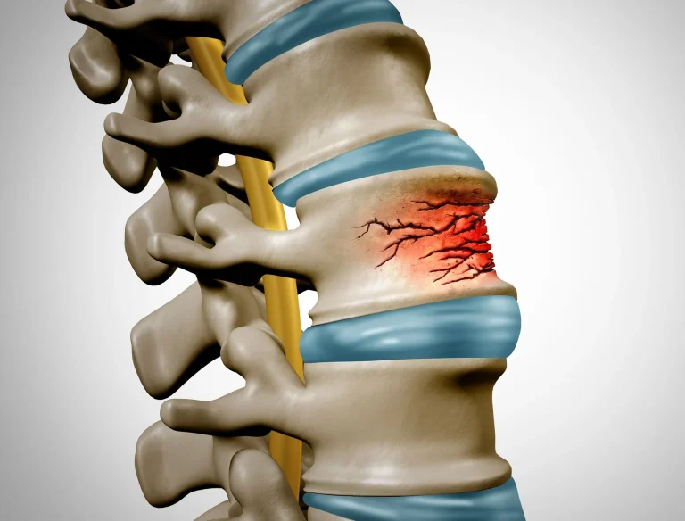
Fractured Bones in Spine

Ultrasonic energy can weaken and fracture remaining vertebral bone.

Man holding lower back with highlighted spine in pain.

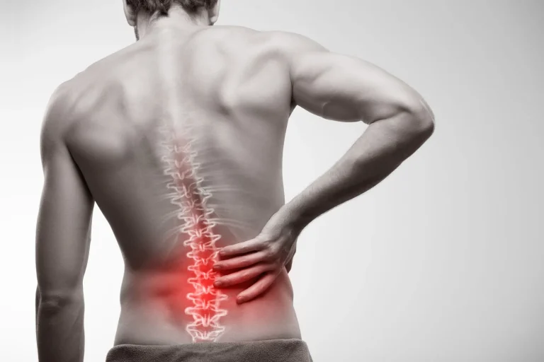
Failure to Eliminate Pain

The actual source of pain is often untreated, leaving patients in chronic pain.

WHAT WE RECOMMEND INSTEAD

Safer Alternatives to Ultrasonic Spine Treatment

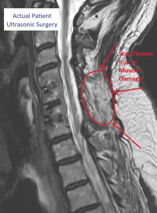
Instead of destroying bone and muscles with ultrasonic probes, these minimally invasive procedures treat the actual source of pain with precision and safety.

Deuk Laser Disc Repair

Minimally invasive laser treatment that repairs the disc without destroying bone or muscle tissue.

Deuk Piriformis Muscle Release

Endoscopic release for piriformis syndrome — targeted relief without invasive bone surgery.

Deuk Plasma Rhizotomy

Plasma technology for facet joint pain — precise nerve treatment without bone destruction.

A BETTER ALTERNATIVE

Deuk Laser Disc Repair®

Minimally invasive, outpatient procedure with a 0.01% complication rate and 95% patient satisfaction. No bone destruction. No muscle damage. Same-day recovery.

95%

Patient Satisfaction

0.01%

Complication Rate

1 Hour

Procedure Time