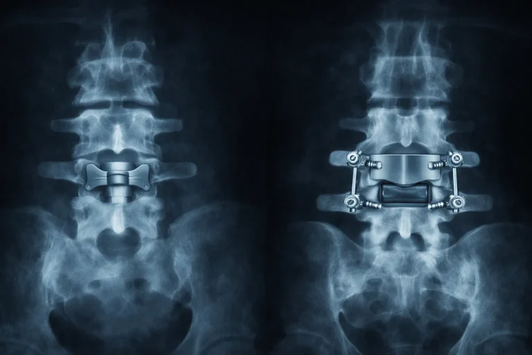
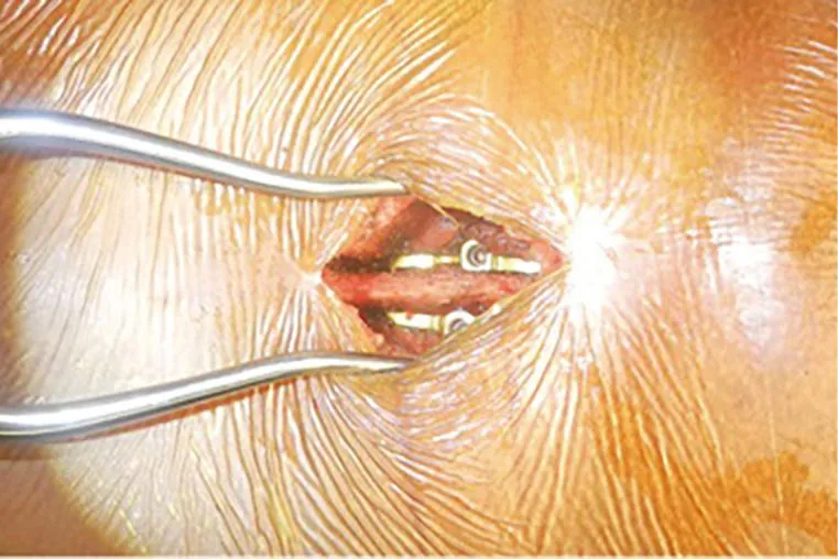
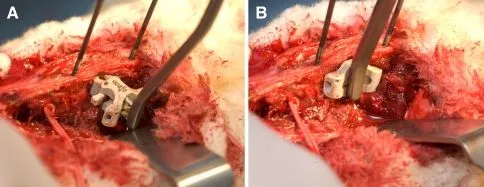
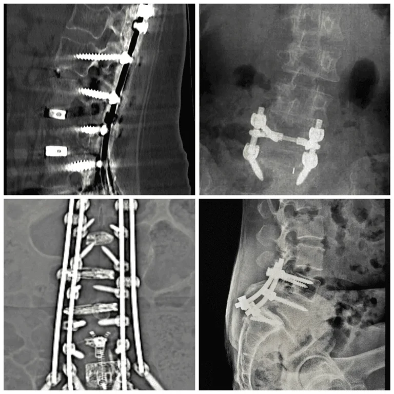
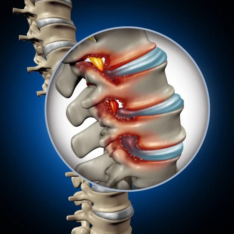
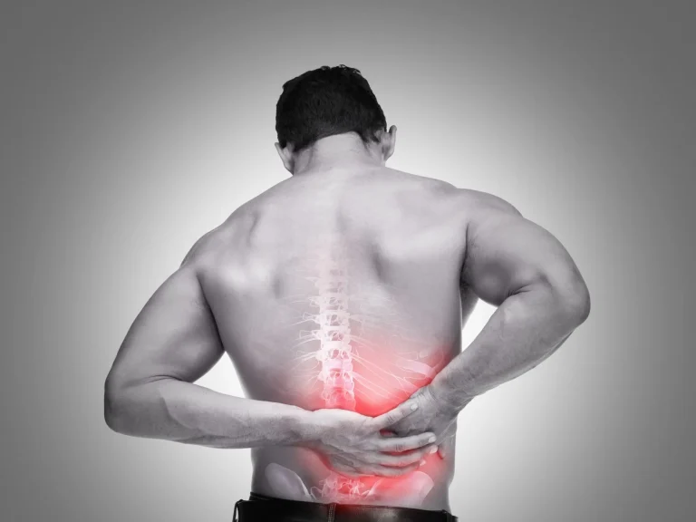
Interspinous and interlaminar devices are metal implants attached between vertebrae that destroy stabilizing spinal structures during placement, frequently fail due to migration and hardware breakdown, and often leave patients worse off than before surgery.
⚠️ While Deuk Spine Institute can perform Interspinous & Interlaminar Device implantation, we do not recommend it. This page explains why — learn the risks before consenting to this procedure.
Why Do Some Surgeons Recommend Interspinous & Interlaminar Devices?
While there are legitimate medical reasons, financial incentives from expensive hardware often influence surgical recommendations.
Medical Reasons Cited
Radiculopathy
Nerve root dysfunction from compressed or pinched nerves. However, these devices rarely address the actual source of nerve inflammation.
Sciatica
Severe leg pain from compressed nerves. The device placement destroys stabilizing structures while leaving the herniated disc untreated.
Cauda Equina Syndrome
Poly radiculopathy from compressed nerves. A rare emergency often used to justify device implantation for far less severe conditions.
Spinal Stenosis
Canal narrowing causing cord compression. Surgeons may overdiagnose this on MRI to justify device implantation for mild cases.
Recurrent Disc Herniation
Repeat disc bulging or rupture. Rather than treating the disc itself, surgeons attach hardware that fails to address the root cause.
Financial Incentives Behind It
Device Costs $5K–15K Per Implant
Each interspinous or interlaminar device generates massive revenue for the manufacturer and hospital, creating a strong incentive to implant.
Surgeon Implant Bonuses
Surgeons receive premium reimbursement for device implantation procedures, creating powerful financial incentive even when less invasive options exist.
Hospital Revenue Per Case
Extended operating time, overnight stays, device costs, and complex post-surgical care drive massive hospital revenue per procedure.
Repeat Procedures When Devices Fail
Device migration, separation, and hardware failure are common, guaranteeing additional lucrative revision surgeries and replacements.
Pain Management Pipeline
Failed device implantations create patients needing ongoing injections, ablations, stimulators, and opioids for life — generating continuous revenue.
Volume Over Outcomes
Surgeons receive the same compensation whether pain resolves or worsens. The system rewards volume of device implantations, not patient results.
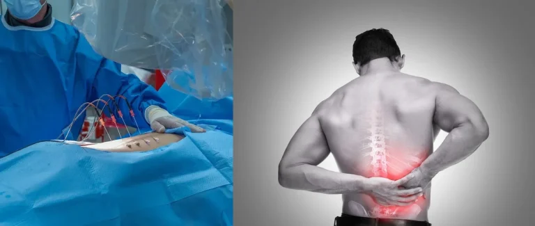
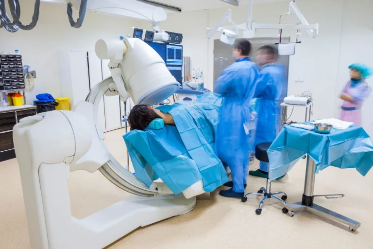
THE SURGICAL PROCESS
How Are Interspinous & Interlaminar Devices Implanted?
Graphic Surgical Content
The videos below contain real surgical footage. Viewer discretion is advised.
Permanent nerve damage causing weakness, numbness, or paralysis in extremities.
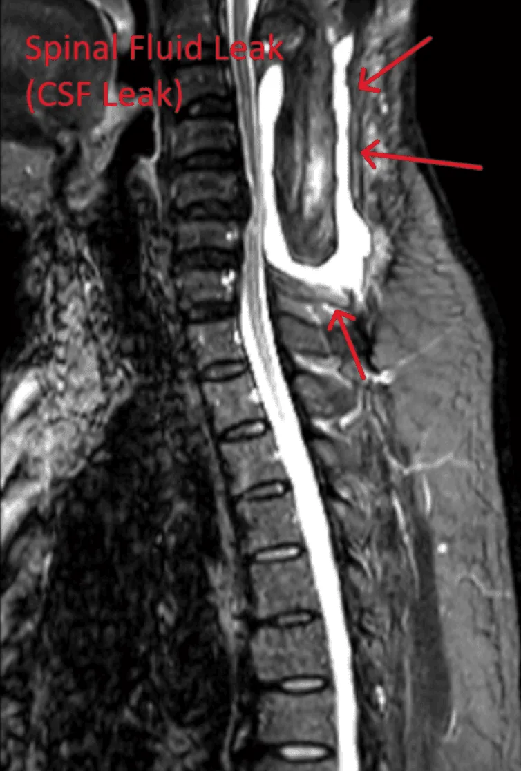
Spinal Fluid Leak
Dural tears leading to cerebrospinal fluid leaks, requiring additional repair surgery.
Infection
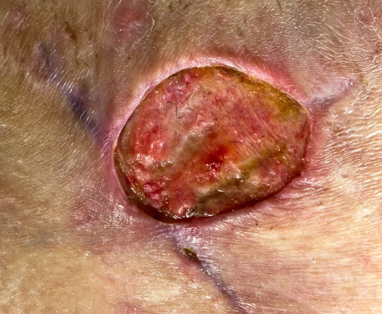
Discitis, osteomyelitis, and infections of blood, spinal fluid, lungs, bladder, and kidneys.
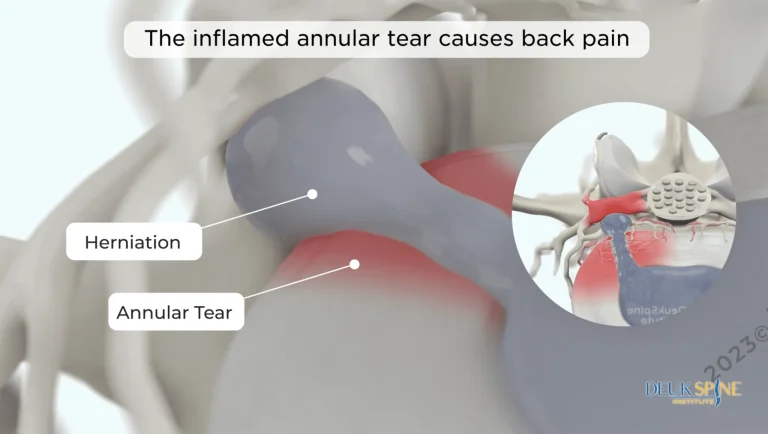
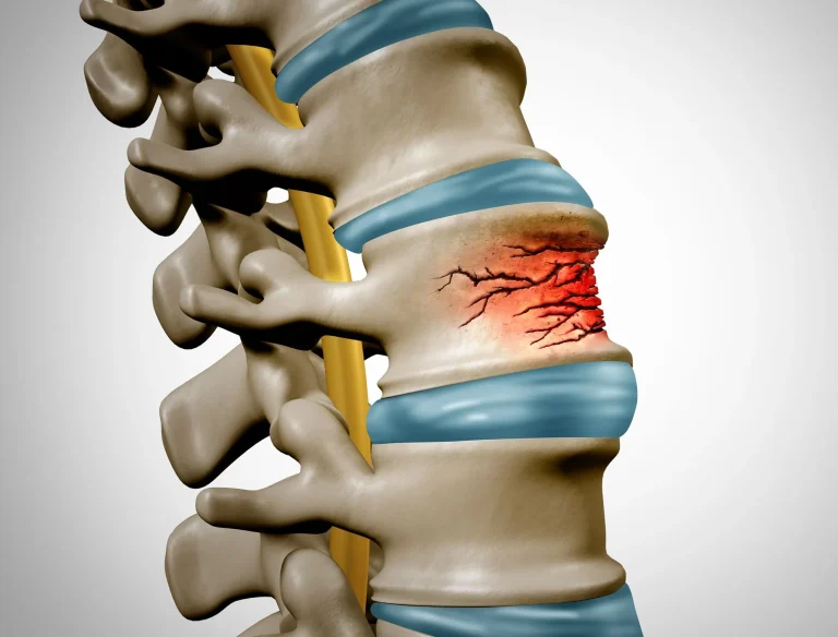
Recurrent Disc Herniation
Disc herniation returns because the device does not treat the underlying disc injury.
Residual Stenosis
Stenosis persists or reoccurs after device placement, requiring additional surgery.
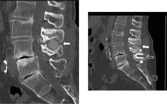
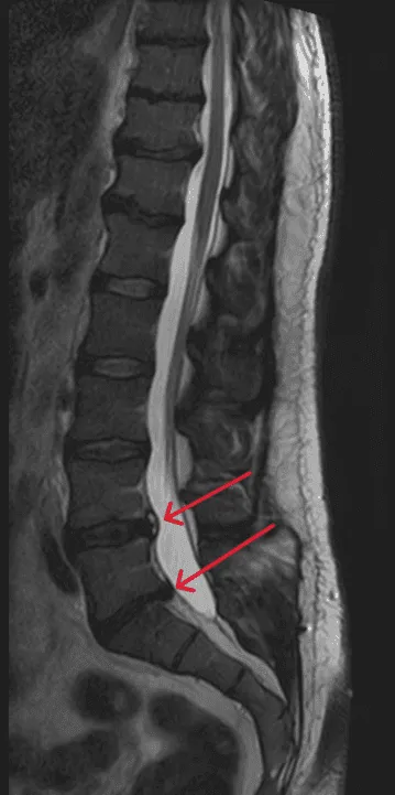
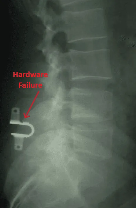
Hardware Failure
Implant migration or separation requiring emergency revision surgery.
Proximal Junction Kyphosis
Proximal junction kyphosis and failure causing abnormal spinal curvature above the device.
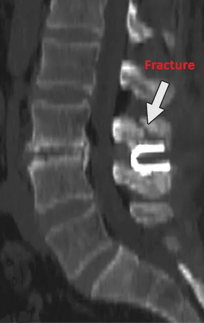
Vertebral Fracture & Subsidence
Vertebral fracture, subsidence, and telescoping of the implant into weakened bone.
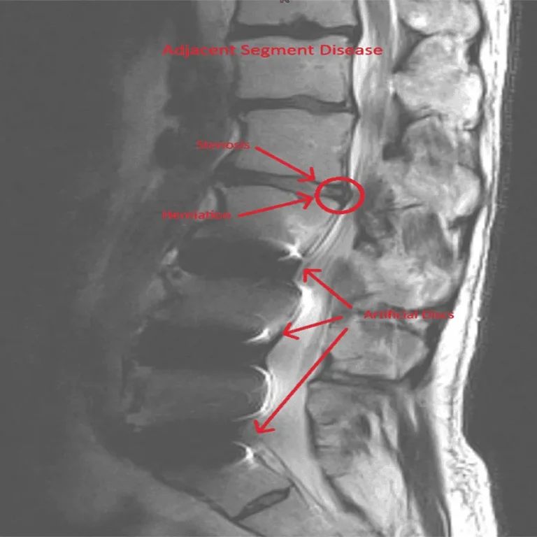
Adjacent Segment Disease
Adjacent segment disease and spinal instability caused by altered biomechanics.
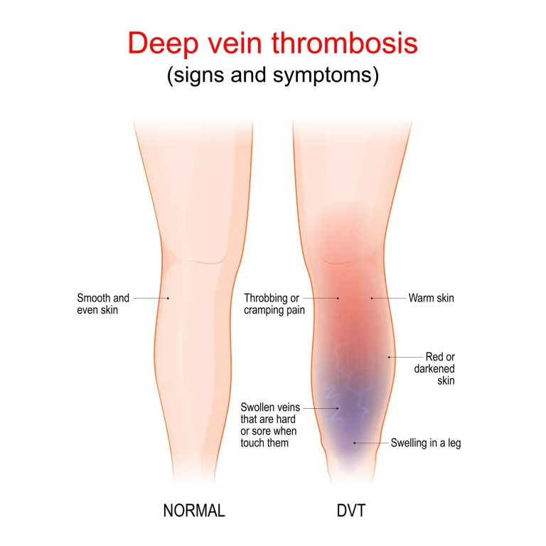
DVT, Embolism & Heart Attack
Deep venous thrombosis, pulmonary embolism, heart attack, and pneumothorax.
Failed Back Surgery & Chronic Pain
Permanent failed back surgery syndrome with unrelenting chronic pain.
Sexual Dysfunction
Sexual dysfunction and retrograde ejaculation from surgical nerve damage.
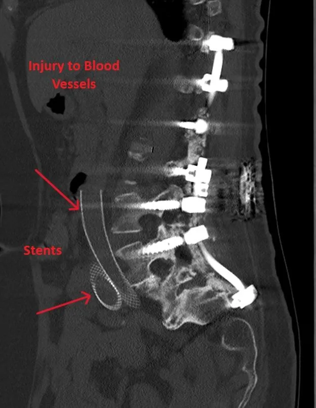
Vascular & Organ Injury
Injury to blood vessels, bladder, or ureters during surgical approach.
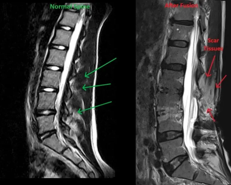
Excessive Scar Tissue
Excessive scar tissue formation around the spine and nerves post-surgery.
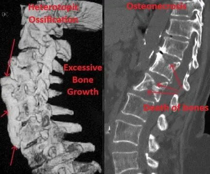
Heterotopic Ossification
Heterotopic ossification and osteonecrosis — abnormal bone growth and bone death.
Death
Fatal complications from surgery, anesthesia, blood clots, or post-operative events.
Excessive Radiation Exposure
Excessive radiation exposure from fluoroscopy during device placement and follow-up imaging.
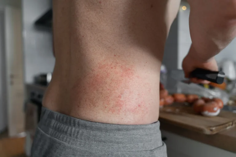
Allergic Reactions
Allergic reactions to metal implant materials causing inflammation and tissue damage.
WHAT WE RECOMMEND INSTEAD
Deuk Laser Disc Repair®: A Safer, Proven Alternative
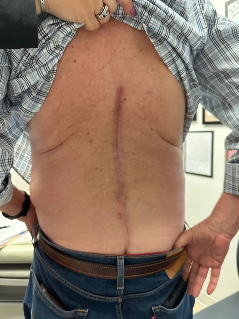
Instead of attaching metal hardware to your spine, Deuk Laser Disc Repair® uses endoscopic technology and laser precision to treat the actual source of pain — the damaged disc — through an incision smaller than a fingernail.
No Bone Removal
Your spine's stabilizing structures remain completely intact. No lamina, spinous process, or ligament destruction. No metal hardware attached.
Treats the Root Cause
Laser technology directly repairs the damaged disc — the actual source of pain that interspinous and interlaminar devices completely ignore.
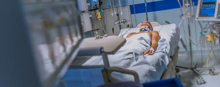
Same-Day Recovery
Outpatient procedure under light sedation. Walk out the same day — no hospital stay, no intubation, no opioids required.
Proven Results
95% patient satisfaction, 0.01% complication rate, and over 1,300 successful procedures performed by Dr. Deukmedjian.
Feature
Interspinous/Interlaminar Devices
Deuk Spine Procedure
Procedure Type
❌ Invasive, hardware implantation
✅ Minimally invasive, endoscopic
Incision Size
❌ 2–4 inches
✅ Less than 1/4 inch
Anesthesia
❌ General (intubated)
✅ Light IV sedation
Hardware
❌ Metal implant attached to spine
✅ None
Treats Disc Injury
❌ No — disc left untreated
✅ Yes — laser repairs the disc
Hospital Stay
❌ 1–3 days inpatient
✅ Outpatient — go home same day
Recovery Time
❌ 4–8 weeks
✅ Days
Complication Rate
❌ High — device failure common
✅ 0.01%
Success Rate
❌ Variable — many end in pain management
✅ 95% patient satisfaction
Repeat Surgery
❌ Common — device migration, failure
✅ Rarely needed
A BETTER ALTERNATIVE
Deuk Laser Disc Repair®
Minimally invasive, outpatient procedure with a 0.01% complication rate and 95% patient satisfaction. No bone removal. No metal hardware. Same-day recovery.