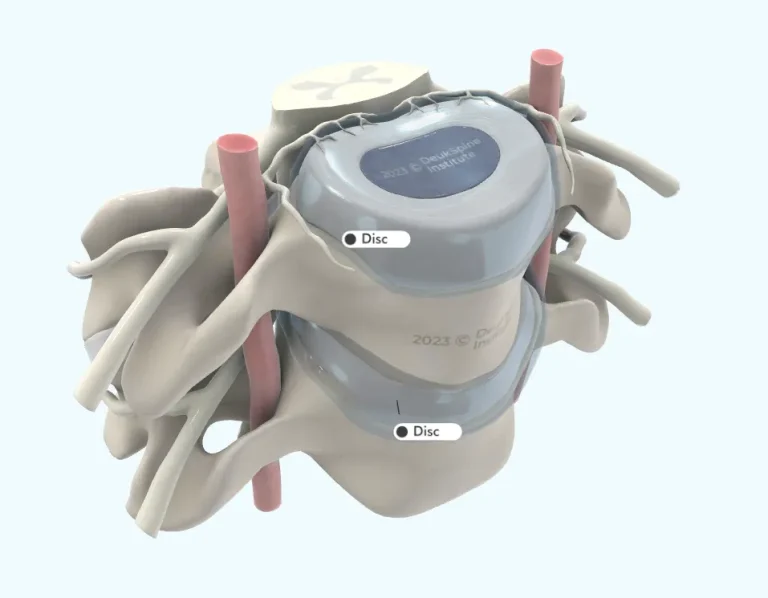
Spacer located between two adjacent vertebral bodies. The disc has a soft, compressible core called the "nucleus pulposus" surrounded by a firm, elastic outer ring-like wall called the "annulus fibrosus". The nucleus pulposus is normally an avascular, water-rich compressible semi-solid (think raw shrimp meat) that acts as hydraulic fluid distributing spinal compression forces outward towards the annulus fibrosus. In an injured disc, the herniated nucleus pulposus causes a severe inflammatory response within the outer annulus fibrosus. Herniated discs are the most common cause of neck pain and can be treated best with Deuk Laser Disc Repair.
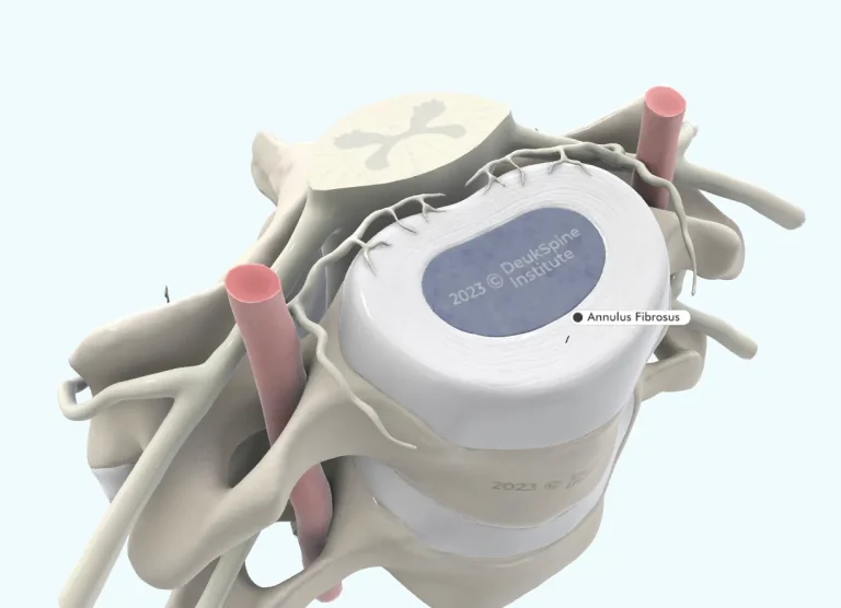
Outer ring of dense, circular connective tissue making up the spinal disc. The annulus fibrosus is composed of 20 concentric layers of collagen fibers wrapped around a soft core, the nucleus pulposus. It is important to note the inner half of annulus fibrosus is normally without blood vessels or nerves. On the other hand, the outer half of annulus fibrosus has blood vessels capable of inflammation and innervation from the Sinuvertebral nerve capable of transmitting pain.
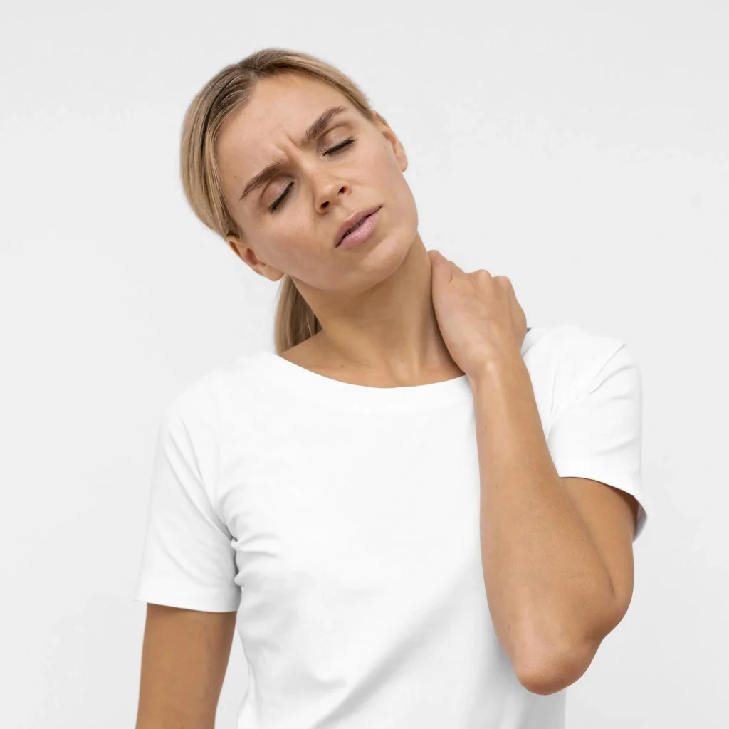
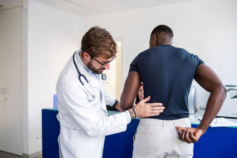
The most common cause of neck pain is an inflamed posterior annular tear of the spinal disc. This occurs when a traumatic injury tears the back wall of the disc (posterior annular tear) thus allowing pieces of nucleus pulposus to escape (herniate) into the tear and cause inflammation within the tear. Inflammation within an injured disc's posterior annular tear is the most common source of neck pain worldwide. The best treatment is Deuk Laser Disc Repair.
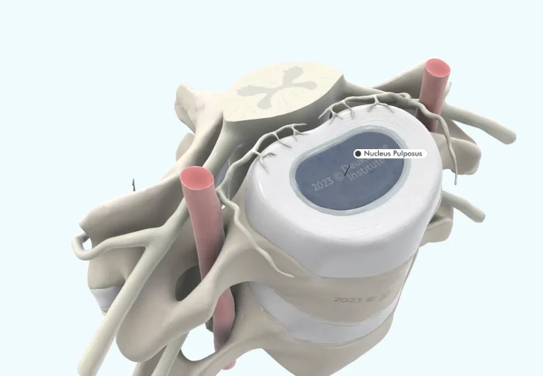
Centrally located hydraulic soft tissue contained within the middle of the spinal disc, embryologically derived from the notochord. The annulus fibrosus forms a circular wall around the nucleus pulposus to hold it in place during normal activities like bending and twisting. The nucleus pulposus is biomechanical "cushion" that, under loading forces, absorbs and redistributes mechanical forces evenly to the inner annulus fibrosus wall. When overloaded, a traumatic annular wall tear is the most common type of disc injury. The annular tear results from excessive force applied to the spine causing a failure of the disc.
Once an annular tear occurs there is now a pathway for pieces of nucleus pulposus to escape the disc and herniate out through the tear in the annular wall. Herniation is the movement of the nucleus pulposus into the annular tear and sometimes out the other side. There are many names given to a herniated disc but they are all part of the same condition at different stages. Small herniations are called disc bulges or protrusions. Larger herniations are called prolapsed, slipped, extruded, migrated and even sequestered discs. The herniated nucleus pulposus can cause inflammation and pain within the annular tear. Bulging discs are the most common cause of neck pain. The best treatment is Deuk Laser Disc Repair.
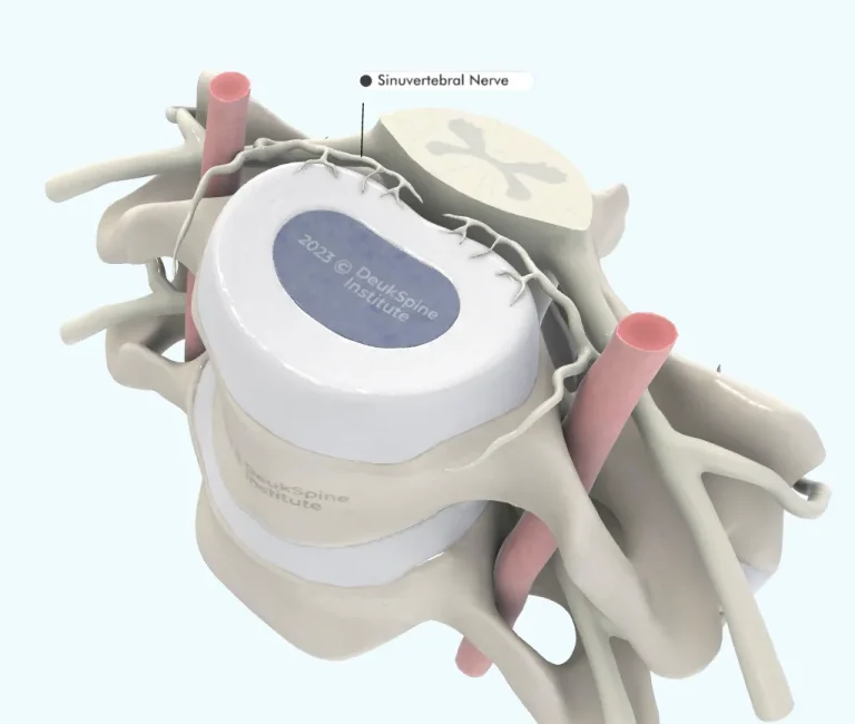
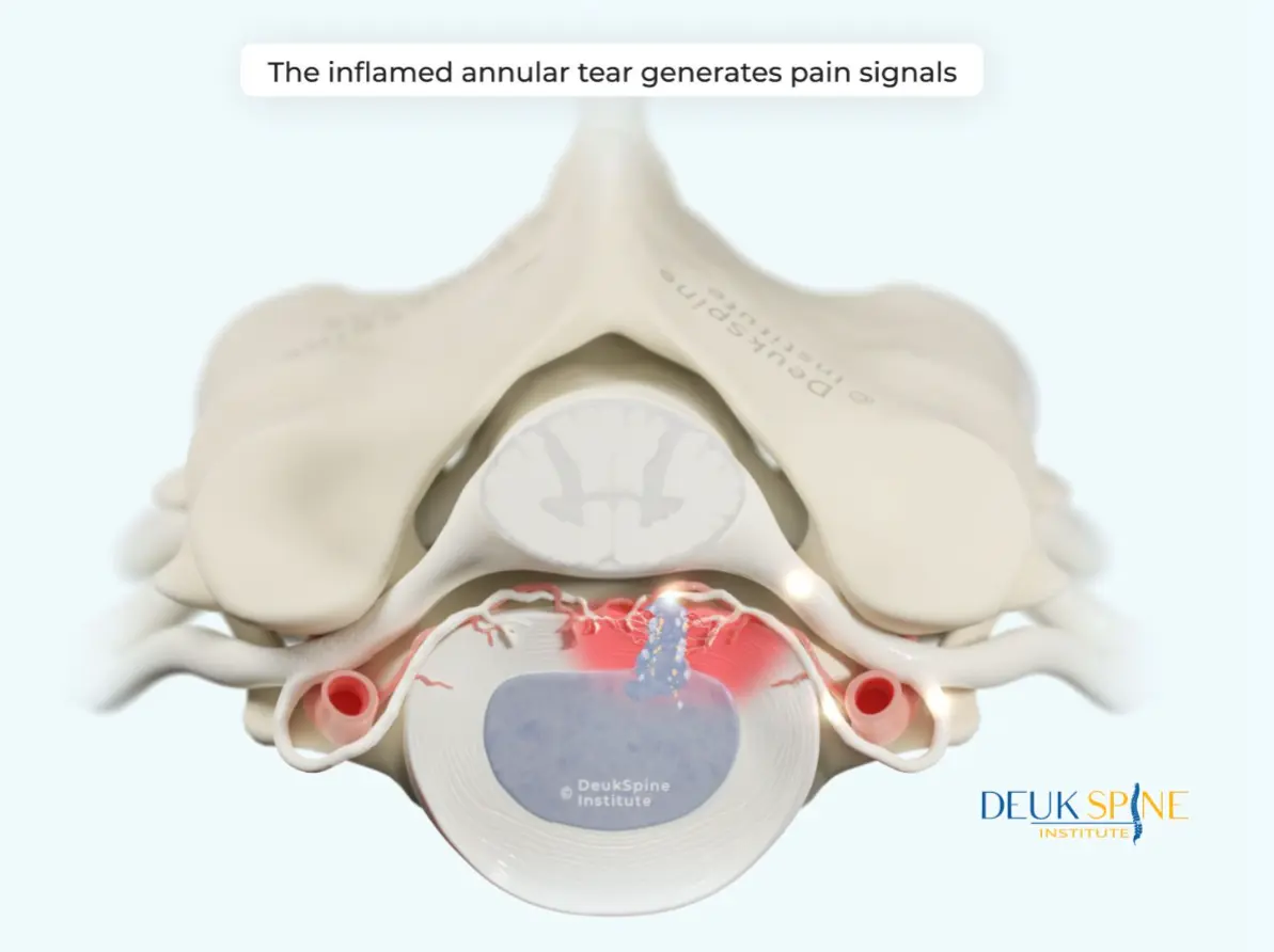
Sensory nerve to the back of the disc (posterior annulus fibrosus) and posterior longitudinal ligament. Somatic afferent nerve fibers transmit painful stimuli from the posterior annulus fibrosus through the dorsal root ganglion via neural pathways to the primary somatosensory cortex in the parietal lobe of the brain. Discogenic pain is only possible because of this nerve carrying pain signals to our consciousness.
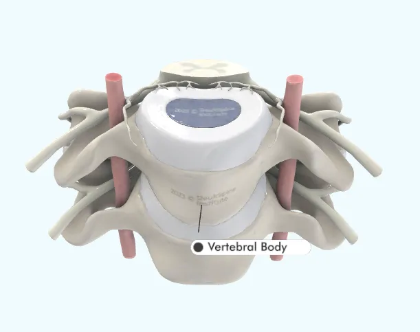
Large weight bearing rectangular bone located in the front of the spine between spinal discs. May fracture with osteoporosis or spinal trauma.
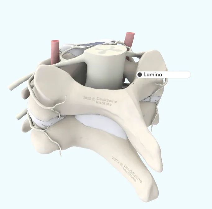
Vertebral flat bony plate-like projection in the back of the spine where paraspinal muscles and stabilizing ligaments attach. Commonly damaged and/or removed during laminectomy, discectomy, microdiscectomy, fusion and foraminotomy open spine surgeries. Deuk Laser Disc Repair does not damage the lamina or any other normal spine structures.
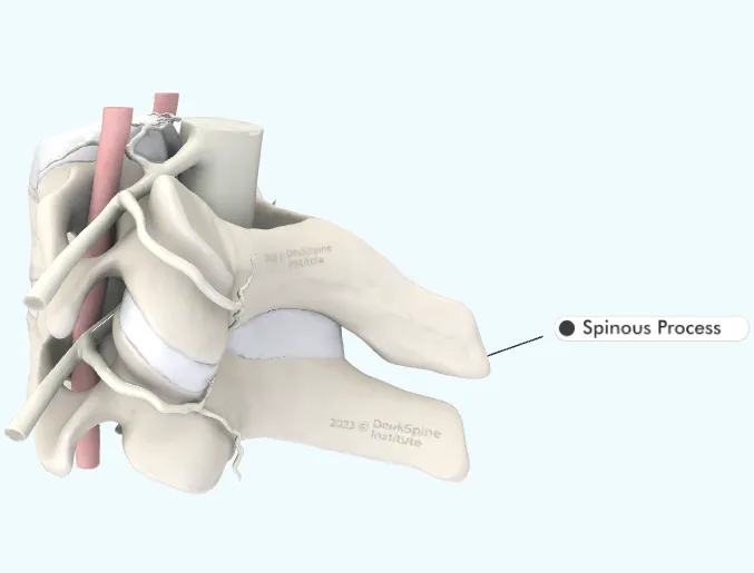
Vertebral bony projection in the back of the spine where paraspinal muscles and ligaments attach for postural movement and spinal stabilization. Commonly damaged during laminectomy, discectomy, microdiscectomy, fusion and foraminotomy open spine surgeries. Deuk Laser Disc Repair does not damage the spinous process or any other normal spine structures.
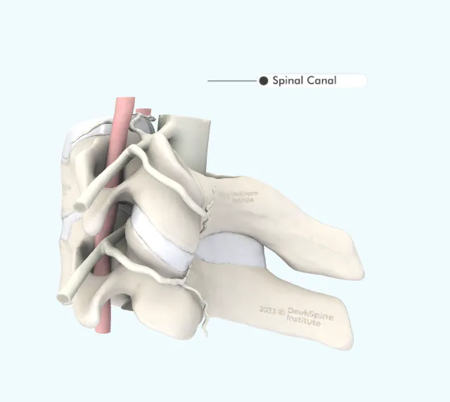
The space in the center of the spine where the spinal cord and nerve roots pass through protected from outside forces as they course to their target locations. Located behind the vertebral body and discs, in front of the lamina and ligaments, the spinal canal is filled with spinal fluid bathing delicate neural structures. Narrowing of the spinal canal is called central stenosis.
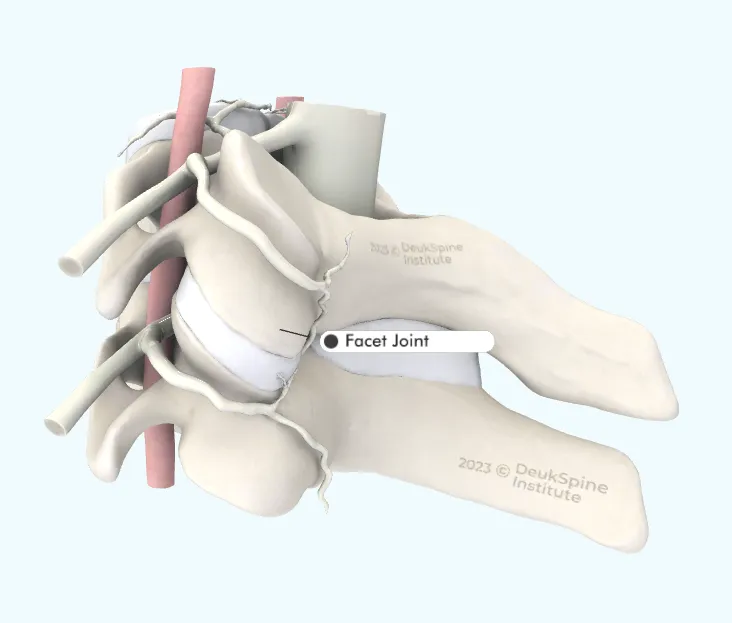
Paired diarthrodial, segmental spinal joints that stabilize the spine's movement with twisting and bending. Each spinal disc has a pair of facet joints located immediately behind the disc on the right and left. Injury to facet joints with resulting inflammation of the joint is the second most common cause of back pain or neck pain and stiffness. These joints are injured with trauma to the spine and during commonly performed open spine surgeries like laminectomy, discectomy, microdiscectomy, fusion and foraminotomy. Deuk Laser Disc Repair does not damage these structures.
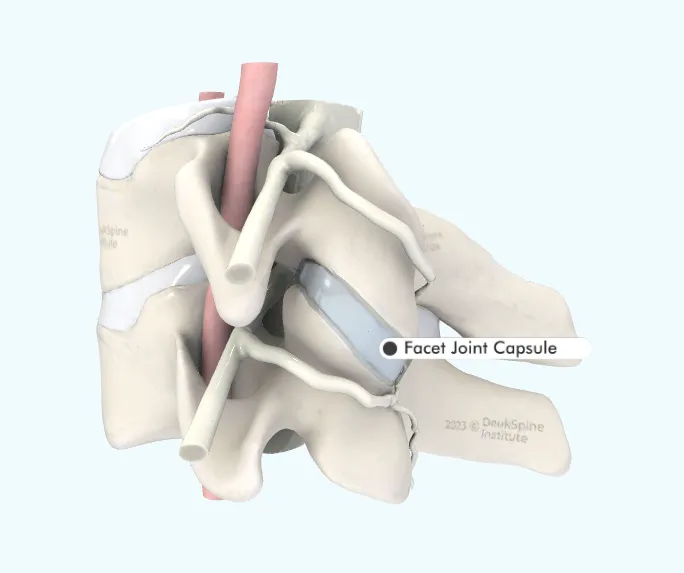
Connective tissue membrane wrapping around the facet joint stabilizing the movement of the bones and bolding the synovial fluid lubricating the facet joint. Damage to the facet capsule allows excessive movement of the facet join t leading to spinal instability and pain. Trauma, such as moto vehicle accidents or falls, is the leading cause of facet joint capsule infury. Surgical trauma during invasive spine surgery (laminectomy, microdiscectomy, MILD procedure, artificial disc and fusion) si another leading cause. Deuk Laser Disc Repair does not damage the facet joint capsule.
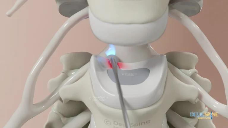
The sensory nerve to the facet joint capsule is the medial branch of the dorsal ramus. This nerve contains somatic afferent sensory fibers that transmit pain signals to the primary somatosensory cortex of the brain allowing conscious localization of the painful facet joint(s). The best treatment for facet joint pain or facet capsule pain is Deuk Plasma Rhizotomy of the painful facet joint(s).
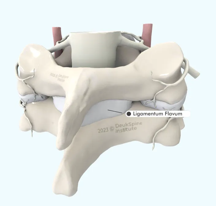
Thick plate of connective tissue ligaments within the posterior spinal canal, attaching adjacent lamina bones. These ligaments stabilize the spine from excessive movement. Injury to the ligamentum flavum causes spinal instability and pain. Ligamentum flavum is either damaged or removed during invasive spine surgery. The most common spine surgeries that damage or remove the ligamentum flavum include laminectomy, microdisecctomy, discectomy, MILD procedure, interspinous devices. Deuk Laser Deisc Repair does not damage the ligamentum flavum.
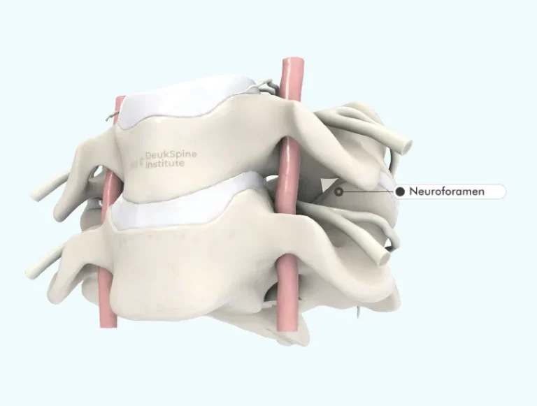
Natural paired, round openings on the sides of the spinal canal where nerve roots pass in and out of the spine on their way to target tissues. Located behind the spinal disc and in front of the facet joints, damage to either structure could result in neuroforaminal stenosis (narrowing) affecting the nearby nerve root. The most common form of spinal stenosis is neuroforaminal stenosis and the most common cause of neuroforaminal stenosis is a herniated disc. The best treatment for a herniated disc is Deuk Laser Disc Repair.
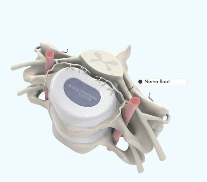
A bundle of nerve fibers responsible for transmitting motor and sensory data between the brain and extremities. Connects brain & spinal cord to muscles for movement and sensors located in skin, joints and muscles. Nerve roots pass behind the spinal discs through natural openings in the spine called neuroforamen. Disc herniations can easily irritate or pinch nerve roots within the neuroforamen. Spinal stenosis typically affects nerve roots with pressure causing arm or leg weakness, numbness, or tingling (radiculopathy). Inflammation of the nerve root typically causes arm pain or leg pain (radiculitis). Nerve root compression or irritation never causes back pain or neck pain.
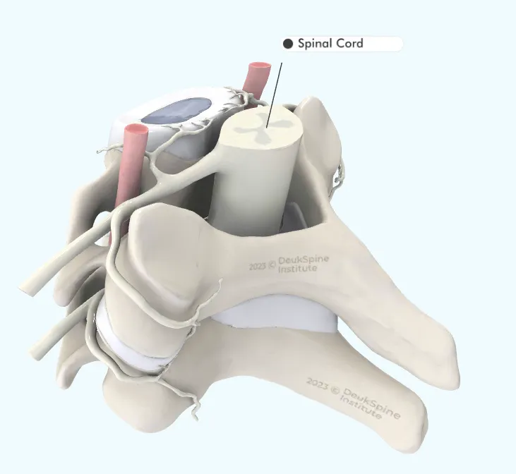
Large bundle of nerves connecting the brain with the rest of the body outside the skull. The spinal cord may become damaged by herniated discs in the cervical spine. Myelopath is spinal cord dysfunction and may manifest as balance difficulty, decreased coordination of the urinary urgence. The spinal cord gives off verve roots that control movement and feeling in the legs.
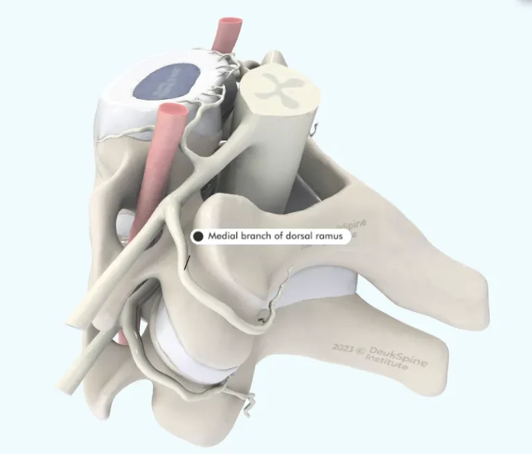
The normal sensory nerve to the facet joint that transmits pain signals from the injured facet joint to the brain. The signals are interpreted as back pain or neck pain depending on which facet joint(s) are the source of pain. This "pain nerve" is the target of the patented Deuk Plasma Rhizotomy procedure first performed at Deuk Spine Institute in 2022 providing the first permanent treatment for facet joint pain in the cervical, thoracic, lumbar, and sacral spine.
Large weight bearing rectangular bone located in the front of the spine between spinal discs. May fracture with osteoporosis or spinal trauma.
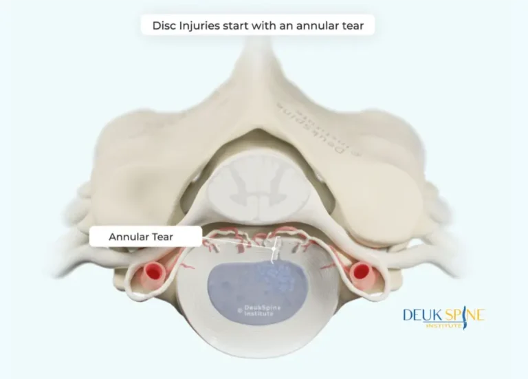
All disc injuries begin with a traumatic tear of the annulus fibrosus ring. An annular tear occurs when traumatic forces on the disc exceed its elastic property limits and cause structural failure of the annulus fibrosus and nucleus pulposus. At the moment of structural failure, linear annular tears occur in the wall of the disc while the adjacent nucleus pulposus is crushed into multiple fragments. Although not all annular tears immediately result in a disc herniation, the tear is a weakened area of the disc wall prone to herniations of the adjacent fragmented nucleus pulposus.
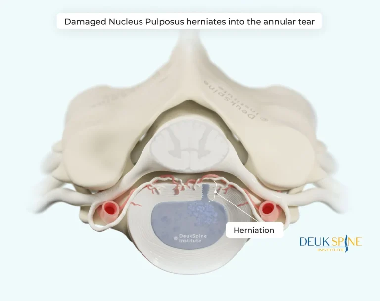
Disc herniations occur when fragments of damaged nucleus pulposus are pushed into the annular tear during physical activities like bending, lifting, twisting, sports, and work. Once herniation of a fragment of nucleus pulposus into the annular tear occurs, the annular tear can no longer heal itself closed. In fact, fragments of nucleus pulposus embedded within the annular tear prevent healing and closure of the annular tear. Over time more pieces of the damaged nucleus pulposus push out into the non-healed annular tear and the herniation gets bigger.
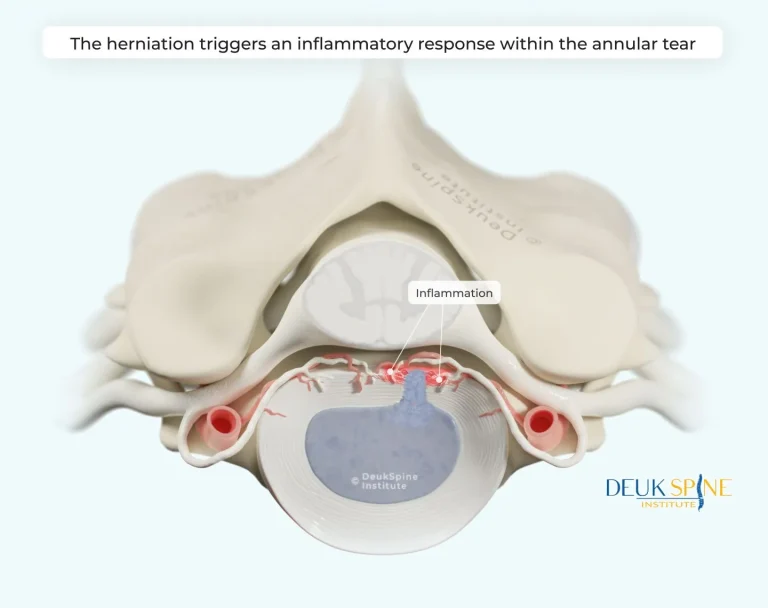
Discogenic neck pain may occur once the herniated fragments of nucleus pulposus reach the outer 1/3 of the posterior annulus fibrosus. The inner 2/3 of the annulus fibrosus normally has no blood vessels, no blood supply and no innervation. Nucleus pulposus present within the outer 1/3 of the annular tear will trigger the body's inflammatory defense system to turn "on" and begin to attack the pieces of herniated nucleus pulposus. The resulting inflammation within the walls of the posterior annular tear of an injured disc is the source of discogenic neck pain (pain generator). When the inflammation spreads to nearby nerve roots it causes sciatica (radiculitis). Inflammation lasting weeks or months within the annular tear further weakens the wall and makes the disc more susceptible to reherniation. Untreated, the annular tear remains open and vulnerable to additional herniation, inflammation and pain occurring in cycles over a lifetime.
Discogenic pain is localized pain that originates from a specific injured spinal disc. The source of discogenic pain is inflammation within the posterior annular tear of the injured disc. Inflammation is a normal part of healing any injury. In this case, the annular tear is the injury that needs to heal. Unfortunately, the fragments of herniated nucleus pulposus stuck within the walls of the posterior annular tear prevent the tear from healing completely. Because the annular tear remains unhealed and "open", more herniations occur and the cycle of inflammation and pain goes on. Discogenic pain can last for decades and is the most common cause of chronic lower neck pain.