


Herniated Disc In Neck MRI
A herniated disc in neck MRI provides the clearest view of spinal disc injuries, making it the most effective tool for diagnosing cervical disc herniations.
This article explores everything you need to know about a herniated disc in the neck as seen on an MRI. We will discuss how an MRI works, what a herniated disc looks like on the scan, and how to differentiate between a normal and herniated disc.
Then we will cover what doctors look for in MRI results and the various treatment options that follow.
MRI For A Herniated Disc In The Neck
MRI, CT scans, and X-rays all provide different insights when diagnosing a herniated disc, with MRI being the superior imaging modality.
MRI scans reveal detailed images of soft tissues like spinal discs and nerves, clearly highlighting problem areas.
CT scans primarily visualize bone structures and can identify severe degenerative changes or bone spurs, but they lack the detail needed for soft tissues like discs.
X-rays are the least effective, mainly showing bone alignment and vertebral integrity, without accurately revealing disc herniations or nerve involvement.
How An MRI Works For Diagnosing Herniated Discs
An MRI (Magnetic Resonance Imaging) uses powerful magnets and radio waves to produce detailed images of the spine, allowing doctors to visualize herniated discs accurately.

During the scan, patients lie still inside the MRI machine, which captures cross-sectional images of spinal discs and surrounding tissues. This helps reveal annular tears, disc herniations, inflammation, and other conditions contributing to chronic neck pain.
Traditional MRI evaluations often overlook the importance of a precise physical examination, potentially causing misdiagnosis and unnecessary invasive procedures.
This frequently leads to treatments like cervical fusion or artificial disc replacement, associated with high complication rates.
How To Read An MRI Of A Herniated Disc
Reading an MRI of a herniated disc involves identifying specific signs like disc displacement, annular tears, and inflammation. Herniated discs show up as areas where the disc material extends beyond its normal boundaries, often highlighted by a distinct bulge or protrusion in the images.
Accurately interpreting these images isn’t straightforward and typically requires specialized knowledge.
What An MRI Can Reveal
An MRI is highly effective in showing detailed images of cervical disc injuries, including herniations, disc bulges, protrusions, and extrusions.
It can accurately display the location and severity of annular tears, the root cause of chronic inflammation and pain. By identifying these abnormalities, an MRI guides physicians toward the precise areas needing targeted intervention.
Do You Need An MRI For A Herniated Disc?
Herniated discs in the cervical spine are commonly caused by disc degeneration. Over time, the discs lose water content and become less flexible, making them more prone to tears or bulging under pressure.
Based on our observations, acute injuries such as whiplash from car accidents, falls, or sports-related trauma can also lead to disc herniation.
Poor posture, repetitive strain, and improper lifting techniques contribute to excessive stress on the cervical spine, increasing the risk of disc damage.
Lifestyle factors like smoking, obesity, and lack of physical activity may further accelerate disc degeneration, making herniation more likely.
Can A Herniated Disc Be Diagnosed Without An MRI?
Yes, a herniated disc can sometimes be diagnosed without an MRI, but the accuracy is reduced.
Relying on symptoms alone often leads to misdiagnosis or ineffective treatments, increasing the likelihood of ongoing pain. For example, looking only at the C7-T1 bulging disc symptoms canlead to unnecessary invasive procedures.
By using both MRI and our specialized diagnostic exam, we ensure a more reliable diagnosis, guiding patients to the appropriate and permanent solution.

Differences Between MRI, CT Scan, And X-Ray For A Herniated Disc
A herniated cervical disc can be diagnosed with an MRI with great success. As we’ve learned, though, an MRI alone is not always enough, and a thorough clinical evaluation helps ensure an accurate diagnosis.
This article covered how MRIs help diagnose herniated discs, the key signs to look for in scan results, and the differences between normal and herniated discs. We also explored treatment options to explore post-diagnosis.
If you’re experiencing persistent neck pain, don’t wait for it to worsen. Schedule a consultation at Deuk Spine Institute today. Book a free MRI review to get started.
Interpreting MRI Results For A Herniated Disc
Symptoms of a herniated disc vary depending on its location and severity. Our findings show that herniated discs in the cervical spine can cause localized neck pain, stiffness, and discomfort that worsens with movement.
If the herniation affects nearby nerve roots, symptoms may extend into the shoulders, arms, and hands, resulting in numbness, tingling, or weakness. This condition is known as cervical radiculopathy.
In some cases, individuals may experience headaches or a reduced range of motion due to muscle tension and inflammation.
More severe cases can lead to myelopathy, where the spinal cord is affected, causing coordination difficulties, balance issues, and even bladder or bowel dysfunction.
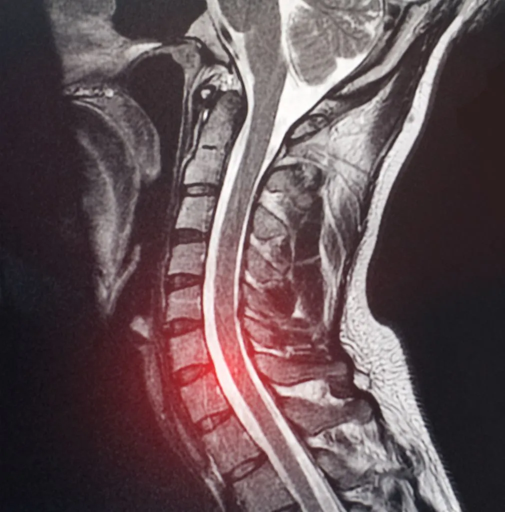
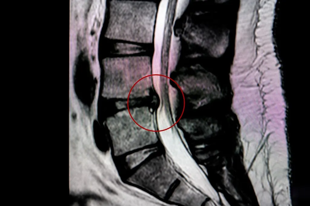
What Does A Herniated Disc Look Like On An MRI?
On an MRI, a herniated disc appears as a bulging or displaced section of the intervertebral disc material that extends beyond its normal boundaries.
In T2-weighted images, which highlight fluid content, the herniation is often visible as a dark mass against the brighter cerebrospinal fluid, indicating a disruption in the disc’s normal structure.
The severity of the herniation can vary, ranging from a minor bulge to a full extrusion where disc material has leaked out.
Understanding MRI Images: Normal Vs. Herniated Disc
A normal spinal disc appears as a well-defined, uniformly shaped structure with smooth edges and adequate spacing between adjacent vertebrae.
In T2-weighted MRI images, healthy discs have a bright central area (nucleus pulposus) and a darker, well-contained outer ring (annulus fibrosus), indicating the balance of fluid and structural integrity.
In contrast, a herniated disc shows an irregular shape with a portion of the disc extending beyond its normal boundary.
What Your Doctor Might Look For In An MRI Scan
When evaluating an MRI scan, a doctor will assess multiple factors to determine the cause of symptoms. First, they examine the alignment of the spine and the integrity of the intervertebral discs.
Any bulging, herniation, or signs of degeneration such as decreased disc height or darkened nucleus pulposus can indicate potential sources of pain.
Doctors also check for nerve involvement by looking at the neuroforamina and spinal canal. If a herniated disc is pressing on a nerve root, it may correlate with symptoms such as radiating pain, numbness, or weakness in the limbs.
What Is A Herniated Disc?
A herniated disc occurs when the soft, gel-like nucleus pulposus of an intervertebral disc pushes through a tear in the outer annulus fibrosus.
This condition deforms the normal structure of the spinal disc and can lead to inflammation (the primary cause of discogenic pain), irritation, or, in some instances, compression of nearby nerves.
Herniated discs are most commonly found in the cervical (neck) and lumbar (lower back) regions of the spine, as these areas experience the most movement and stress.
Causes Of A Herniated Disc In The Neck
Herniated discs in the cervical spine are commonly caused by disc degeneration. Over time, the discs lose water content and become less flexible, making them more prone to tears or bulging under pressure.

Based on our observations, acute injuries such as whiplash from car accidents, falls, or sports-related trauma can also lead to disc herniation.
Poor posture, repetitive strain, and improper lifting techniques contribute to excessive stress on the cervical spine, increasing the risk of disc damage.
Lifestyle factors like smoking, obesity, and lack of physical activity may further accelerate disc degeneration, making herniation more likely.
Common Symptoms Of A Herniated Disc
Symptoms of a herniated disc vary depending on its location and severity. Our findings show that herniated discs in the cervical spine can cause localized neck pain, stiffness, and discomfort that worsens with movement.
If the herniation affects nearby nerve roots, symptoms may extend into the shoulders, arms, and hands, resulting in numbness, tingling, or weakness. This condition is known as cervical radiculopathy.
In some cases, individuals may experience headaches or a reduced range of motion due to muscle tension and inflammation.
More severe cases can lead to myelopathy, where the spinal cord is affected, causing coordination difficulties, balance issues, and even bladder or bowel dysfunction.
Treatment Options After An MRI Diagnosis
Once an MRI confirms a herniated disc, treatment is recommended. Here are your options.
Non-Surgical Treatments For A Herniated Disc
Non-surgical treatments focus on reducing inflammation and managing pain while allowing the herniated disc to heal naturally. magnesium for herniated disc is one such options. Unfortunately, these claims do not hold up. If any relief is experienced, it is short-lived.
Physical therapy plays a key role in strengthening the muscles that support the spine, improving posture, and increasing flexibility.
Pain management may include anti-inflammatory medications, muscle relaxants, and corticosteroid injections to reduce swelling and discomfort.
Lifestyle modifications, such as ergonomic adjustments, weight management, and avoiding activities that strain the spine, can also aid in recovery.

Surgical Options
Surgery is considered when non-surgical treatments fail, and symptoms begin to impact daily life. Traditional procedures, such as discectomy or spinal fusion, are commonly used to remove the damaged portion of the disc and stabilize the spine.
Sadly, these procedures are invasive and may have extended recovery times.
The good news is that minimally invasive techniques reduce trauma to surrounding tissues while effectively addressing the disc herniation. Endoscopic spine surgery, like our Deuk Laser Disc Repair, allows for precise removal of the inflaming disc material with minimal disruption.
Recovery And Managing Symptoms Post-Treatment
Recovery from a herniated disc treatment varies depending on whether a patient undergoes conservative management or surgery.
Non-surgical recovery typically involves ongoing physical therapy and gradual activity modification to prevent re-injury. Patients are encouraged to maintain good posture, strengthen core muscles, and avoid heavy lifting.
For those who undergo surgery, post-operative care focuses on pain management, wound healing, and rehabilitation. Light activities are usually permitted within weeks, but full recovery can take months.
Whether you were experiencing herniated disc between shoulder blades symptoms or another herniated disc knee pain, it’s important to look after yourself so as to not cause future damage.
Conclusion
A herniated cervical disc can be diagnosed with an MRI with great success. As we’ve learned, though, an MRI alone is not always enough, and a thorough clinical evaluation helps ensure an accurate diagnosis.
This article covered how MRIs help diagnose herniated discs, the key signs to look for in scan results, and the differences between normal and herniated discs. We also explored treatment options to explore post-diagnosis.
If you’re experiencing persistent neck pain, don’t wait for it to worsen. Schedule a consultation at Deuk Spine Institute today. Book a free MRI review to get started.


