Your local surgeon recommends a 5-day hospital stay for your herniated disc surgery. The procedure requires a 3-inch incision, removal of vertebral bone, and 6-8 months of recovery before returning to work. When you ask about endoscopic options, the surgeon dismisses them as experimental techniques without proven outcomes.
I know this frustration because 90% of my patients travel to Orlando, Florida from other states and countries after local surgeons told them traditional open surgery was their only option. After performing over 2,800 endoscopic spine procedures through 4-7mm incisions with same-day discharge, I've seen how modern endoscopic care transforms outcomes for patients told they needed destructive surgery with months of recovery.
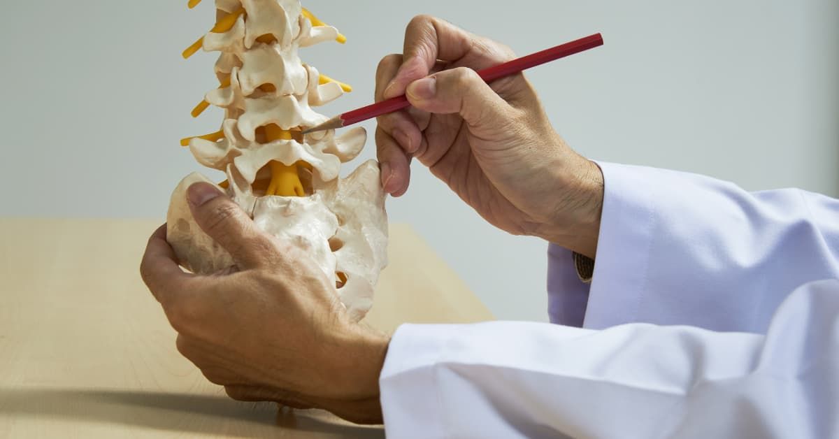
Here's what separates modern endoscopic spine surgery from traditional approaches - endoscopic surgeons use high-quality MRI to detect annular tears causing your pain, perform outpatient procedures that minimize tissue disruption, and set realistic recovery timelines focused on resolving the actual source of nerve irritation rather than removing bone and implanting hardware.
This article provides a patient-centered overview of endoscopic spine care, explaining how surgeons identify pain sources through advanced imaging, what happens during endoscopic procedures, and what realistic recovery looks like when surgery targets the actual pathology causing your chronic pain.
What Makes Endoscopic Spine Surgery Different
Traditional spine surgery evolved from techniques developed decades ago when surgeons had limited visualization tools and relied on large incisions to see the surgical field. Modern endoscopic spine surgery uses advanced camera technology and specialized instruments that allow surgeons to work through incisions measuring 4-7mm while achieving better visualization than traditional open approaches.
The fundamental differences between endoscopic and traditional spine surgery include:
- Incision size: 4-7mm endoscopic portals versus 2-4 inch traditional incisions
- Muscle disruption: Minimal with endoscopic approach versus extensive cutting and retraction with traditional surgery
- Bone preservation: Endoscopic procedures preserve vertebral structure versus bone removal in laminectomy
- Hardware avoidance: No metal implants with endoscopic techniques versus rods, screws, and cages in fusion
- Recovery location: Outpatient with same-day discharge versus 3-5 day hospital admission
- Return to activity: 4-6 weeks with endoscopic versus 6-12 months with traditional surgery
More importantly, endoscopic spine surgery targets the actual pathology causing chronic pain. Traditional procedures often address symptoms visible on imaging rather than the underlying cause. A herniated disc pressing on a nerve root represents a symptom, but the actual pain source in 85% of chronic back pain cases is the torn annular fiber with trapped herniation causing inflammation. Endoscopic techniques allow surgeons to access and treat these annular tears directly.
Advanced MRI Interpretation: Detecting Annular Tears
The foundation of successful endoscopic spine surgery begins with accurate diagnosis using high-quality MRI imaging. However, having an MRI scan doesn't mean you have an accurate diagnosis. The critical factor is whether your surgeon can identify annular tears and correlate those findings with your specific pain pattern.
Annular tears appear on MRI as high-intensity zones in the posterior annulus - the back portion of the intervertebral disc. These bright areas on T2-weighted images indicate torn annular fibers with inflammation. Standard radiological reports often miss these tears or mention them without recognizing their significance as the primary pain generator.
An endoscopic spine surgeon must demonstrate expertise in:
- High-intensity zone recognition: Identifying torn annular fibers on T2-weighted MRI sequences
- Herniation characterization: Distinguishing between loose disc material and material trapped in annular tears
- Nerve root assessment: Evaluating true compression versus irritation from disc inflammation
- Multi-level evaluation: Determining which of multiple abnormalities causes symptoms
- Pain pattern correlation: Matching imaging findings to your specific pain location and characteristics
During my training at the University of Florida - a top 3 neurosurgery program nationally - and subsequent NIH-funded fellowship, I developed expertise in identifying these subtle imaging findings that most surgeons overlook. The Deuk Spine Exam integrates high-quality MRI interpretation with detailed pain history and physical examination to achieve 99% diagnostic accuracy in identifying which specific structure causes your chronic pain.

This diagnostic precision matters because operating on the wrong level or addressing incidental findings rather than actual pain generators leads to failed surgery. Research shows that 100% of people over 45 have disc herniations visible on MRI, but only 10-15% of those herniations cause symptoms. Without expert correlation of imaging and clinical findings, surgeons perform unnecessary procedures that don't resolve your pain.
The Deuk Spine Exam: Integrating Multiple Data Points
Accurate diagnosis requires more than reviewing MRI images. The Deuk Spine Exam combines three essential elements to identify your pain source with 99% accuracy.
First, detailed pain history captures your pain pattern - where it starts, where it radiates, what makes it worse, what provides relief, and how it affects daily activities. Disc injuries at different levels create distinct pain patterns. L4-5 disc injuries typically cause low back pain with radiation into the buttocks and outer thighs. L5-S1 disc injuries create pain radiating down the back of the leg. Cervical disc injuries at C5-6 cause pain between the shoulder blades radiating into the arms.
Second, physical examination identifies which movements and positions reproduce your pain. Specific maneuvers stress different spinal structures, revealing whether discs, facet joints, or other pathology generates your symptoms. Range of motion testing, provocative maneuvers, and neurological assessment provide information that imaging alone cannot reveal.
Third, advanced MRI interpretation identifies structural pathology - annular tears, herniations, nerve compression, facet joint inflammation, and other abnormalities. The skill involves recognizing which of multiple findings actually causes your pain based on correlation with your clinical presentation.
This integrated approach achieves diagnostic accuracy that MRI interpretation alone cannot provide, ensuring that endoscopic surgery targets the actual pain source rather than incidental imaging findings.
The Endoscopic Procedure: What Actually Happens
Understanding what occurs during endoscopic spine surgery helps you evaluate whether this approach offers advantages over traditional open procedures.
Outpatient Setting and Same-Day Discharge
All endoscopic spine procedures at Deuk Spine Institute are performed in an outpatient surgical center with same-day discharge. You arrive the morning of surgery, undergo the procedure, recover for 2-3 hours while anesthesia wears off, and return home the same day. No hospital admission. No overnight stay. No extended recovery in an institutional setting.
This outpatient approach reflects the minimal tissue disruption created by endoscopic techniques. Traditional open spine surgery requires hospitalization because of extensive muscle cutting, bone removal, blood loss, and the need for intravenous pain medication. Endoscopic procedures preserve tissue, minimize bleeding, and create discomfort manageable with oral medications, allowing immediate discharge.
Surgical Approach and Incision Size
The endoscopic spine surgeon makes an incision measuring 7mm for lumbar procedures or 4mm for cervical procedures. Through this small portal, the surgeon inserts an endoscope - a narrow tube containing a high-definition camera and working channel for specialized instruments.
The endoscope provides magnified visualization of the surgical field superior to what surgeons see during traditional open procedures. The camera displays anatomical structures on high-resolution monitors, allowing precise identification of disc material, annular tears, nerve roots, and surrounding tissues.
For the Deuk Laser Disc Repair, the surgeon:
- Accesses the damaged disc through the small endoscopic portal using fluoroscopic guidance for precise positioning
- Visualizes the annular tear and trapped herniated material using the endoscopic camera
- Removes the herniated nucleus pulposus trapped in the tear causing inflammation
- Performs debridement of torn annular fibers eliminating inflamed tissue and pain nerve fibers that grew into the tear
- Uses laser energy to seal the tear promoting natural healing without fusion or artificial materials
- Confirms complete decompression of nerve roots if compression existed
The entire procedure takes 45-60 minutes. No bone removal. No muscle cutting. No hardware implantation. The surgeon directly treats the pathology causing your pain - the torn annular fiber with trapped herniation - while preserving your natural disc structure and spinal motion.
Minimizing Tissue Disruption
Traditional spine surgery creates significant collateral damage to reach the surgical target. Surgeons cut through multiple muscle layers, retract muscles away from the spine for extended periods causing ischemia and trauma, remove bone to create working space, and manipulate nerve roots to access disc material.
This tissue disruption generates the extended recovery times associated with traditional spine surgery. Muscle healing takes months. Bone regrowth requires 6-12 months. Internal scarring from extensive dissection creates chronic pain in many patients even after the original pathology is addressed.
Endoscopic techniques minimize tissue disruption by working through natural tissue planes rather than cutting through muscles. The small portal creates minimal trauma. Specialized instruments allow the surgeon to work in confined spaces without extensive retraction. Bone preservation maintains structural integrity. The result is rapid healing measured in weeks rather than months.
Recovery Timeline: Realistic Expectations
One of the most significant differences between endoscopic and traditional spine surgery involves recovery timeline and what that recovery actually means for your daily life.
Immediate Post-Operative Period (Hours 1-4)
You wake from anesthesia within 30-45 minutes after the procedure ends. Nurses monitor your vital signs, neurological function, and pain level in the recovery area. Discomfort is manageable with oral pain medication - no need for intravenous narcotics.
Within 1-2 hours, you walk with assistance from the recovery bed to the discharge area. Walking immediately after surgery demonstrates the minimal tissue disruption created by endoscopic techniques. Traditional spine surgery patients remain bedbound for 12-24 hours post-operatively.
After 2-3 hours of monitored recovery, you're discharged home with a family member or friend. You walk out of the surgical center and ride home in a car. That evening, you're in your own bed rather than a hospital room.
First Week Recovery
The first week involves managing post-operative discomfort with prescribed pain medication, limiting strenuous activities while encouraging regular walking, and avoiding positions that stress the surgical site. Most patients reduce pain medication significantly by day 3-4 as discomfort resolves.
You can shower after 48 hours with waterproof dressing over the small incision. No wound care complexity. No drain tubes. No extensive bandaging. The 4-7mm incision requires minimal attention.
Many patients return to desk work within 5-7 days post-operatively. You avoid heavy lifting, bending, and twisting during this initial healing period, but you maintain mobility and independence. No bedrest. No extended disability. No months away from work and normal life.
Weeks 2-6: Progressive Activity Increase
During weeks 2-6, you gradually increase activity levels as your body heals. Begin gentle exercises to maintain range of motion and core strength. Progressively reduce pain medication as discomfort resolves completely. Return to most normal activities including driving, light exercise, and regular work duties.
The specific pathology we treated - the torn annular fiber with trapped herniation - begins healing naturally. The inflammation that caused your chronic pain resolves. Pain nerve fibers that grew into the inflamed tissue die off. Your pain decreases progressively during this period.
Long-Term Results: 13 Months and Beyond
Published peer-reviewed research on the Deuk Laser Disc Repair demonstrates 99% elimination of pain at 13 months post-treatment. This data comes from systematic outcome tracking of patients treated for chronic back pain from disc injuries, with results published over 10 times in medical journals.
The 99% success rate reflects several factors. First, accurate diagnosis ensures we treat the actual pain source rather than incidental findings. Second, the procedure directly addresses the pathology causing pain - the annular tear with trapped herniation. Third, preserving natural disc structure and avoiding hardware eliminates complications associated with fusion including adjacent segment disease.
By 6-8 weeks post-procedure, most patients experience complete or near-complete pain elimination. Natural disc healing continues over 3-6 months. Your spine maintains normal motion and function. No stiffness from fusion. No hardware complications. No motion restrictions limiting activities you enjoy.
Why Patients Travel to Orlando, Florida for Endoscopic Care
Ninety percent of patients at Deuk Spine Institute travel from other states and countries because they cannot find endoscopic spine surgeons with equivalent expertise, outcomes, and safety records in their local areas.
Limited Endoscopic Training Among Spine Surgeons
Most spine surgeons completed residency training focused on traditional open procedures. Few received fellowship training in endoscopic techniques. The skills required for endoscopic surgery differ fundamentally from traditional approaches - working through narrow portals, operating with camera visualization, using specialized instruments in confined spaces, and identifying subtle pathology like annular tears.
Without specialized training and high case volume, surgeons cannot develop the expertise necessary for consistent excellent outcomes. A surgeon performing 10-20 endoscopic procedures annually lacks the experience to handle anatomical variations and complex cases. Someone performing 200+ endoscopic procedures yearly develops refined skills that translate to superior results.
Over 20 years, I've performed more than 2,800 endoscopic spine procedures. This volume, combined with specialized fellowship training and continuous refinement of techniques, allows me to achieve outcomes that local surgeons without endoscopic expertise cannot match.
Published Outcomes and Zero Complications
Patients travel to Orlando because they want evidence-based treatment with transparent outcomes rather than vague promises about results. The Deuk Laser Disc Repair has been published over 10 times in peer-reviewed medical journals, demonstrating 99% elimination of pain at 13 months and 95% success rates in eliminating chronic pain from disc injuries.
More importantly, over 2,800 procedures performed across 20 years have achieved zero major complications. No infections. No nerve damage. No spinal fluid leaks. No hardware failures. This safety record makes endoscopic spine surgery at Deuk Spine Institute the safest spine surgery available.
Local surgeons rarely publish their outcomes or provide specific complication data. Without systematic outcome tracking and independent peer review, patients have no way to evaluate whether a surgeon's claims of "excellent results" reflect reality or marketing.
Same-Day Outpatient Convenience
The outpatient nature of endoscopic procedures actually makes traveling for surgery more practical than you might expect. You arrive in Orlando the day before surgery, undergo the procedure the next morning, and can travel home 24-48 hours later depending on distance and comfort level.
Compare this to traditional spine surgery requiring 3-5 day hospital stays followed by weeks of restricted mobility. Traveling for traditional surgery means extended time away from home during the most difficult recovery period. With endoscopic surgery, you return home quickly while recovery is manageable, then heal in your own environment with family support.
Resolving Nerve Irritation: The Actual Goal
Traditional spine surgeons often focus on decompressing nerve roots through bone removal and disc material excision. However, nerve compression isn't the primary cause of chronic back pain in most patients. Research demonstrates that disc injuries account for 85% of chronic back pain, and the pain comes from annular tear inflammation rather than nerve compression.
Endoscopic spine surgery addresses the actual pathology:
- Removes inflammation source: Extracts the herniated material trapped in the annular tear causing inflammatory cascade
- Eliminates pain nerve fibers: Debrides torn annular tissue containing nerve fibers that grew into inflamed area
- Promotes natural healing: Seals the tear with laser energy allowing body's healing processes to repair the disc
- Preserves disc function: Maintains natural cushioning and motion rather than removing disc and fusing segments
When nerve root compression does exist alongside disc injury, the endoscopic approach addresses both pathologies simultaneously. Removing the herniation decompresses the nerve root while treating the annular tear eliminates the inflammation causing chronic pain. This comprehensive approach resolves both symptoms and underlying cause.
What to Expect During Your Consultation
If you're considering endoscopic spine surgery, understanding what happens during the consultation process helps you prepare for evaluation and decision-making.
MRI Review and Pain History
Before scheduling surgery, we perform detailed MRI review to confirm you're a candidate for endoscopic treatment. Submit your existing MRI for expert analysis. I personally review every image, identifying annular tears, herniations, nerve compression, and other pathology.
During consultation, we discuss your complete pain history - when it started, what makes it worse, what provides relief, how it affects daily activities, and what treatments you've tried. This information helps correlate MRI findings with your clinical presentation.
Physical Examination
Physical examination identifies which movements and positions reproduce your pain, tests neurological function including strength and sensation, evaluates range of motion and flexibility, and performs provocative maneuvers stressing different spinal structures.
This hands-on assessment provides information imaging cannot reveal, confirming which structure generates your pain and ensuring we target the correct pathology during surgery.
Treatment Plan Discussion
Based on diagnostic findings, we discuss whether endoscopic surgery offers the best solution for your specific condition. I explain exactly which disc or structure causes your pain, how the procedure addresses that pathology, what realistic outcomes you can expect, and what recovery timeline you should anticipate.
This consultation includes transparent discussion of success rates, potential complications, and alternatives to surgery. You receive honest assessment rather than pressure to proceed with treatment that may not be appropriate for your situation.
Making Your Decision About Endoscopic Spine Surgery
Choosing endoscopic spine surgery over traditional approaches requires evaluating several factors specific to your situation and the surgeon's expertise.
Consider endoscopic surgery when:
- MRI shows annular tears with herniation as your pain source
- Conservative treatment has failed after 8-12 weeks
- Your surgeon has fellowship training and high case volume in endoscopic techniques
- Published outcomes demonstrate excellent results and low complications
- You want to preserve spinal motion and avoid fusion
- Same-day outpatient surgery with rapid recovery appeals to you
Continue exploring options if:
- Your local surgeon dismisses endoscopic approaches without specific reasoning
- The surgeon cannot explain how they identify annular tears on MRI
- No published outcomes or complication data exists
- The surgeon performs fewer than 100 endoscopic procedures annually
- You're told traditional surgery is your "only option" without consultation with endoscopic specialist
The decision to travel for surgery depends on weighing local convenience against expertise and outcomes. If local surgeons lack endoscopic training and experience, traveling to a high-volume center with published results and zero complications offers better long-term outcomes than convenient access to traditional surgery with months of recovery and uncertain results.
Take the Next Step Toward Pain-Free Living
Endoscopic spine surgery represents modern patient-centered care that preserves your anatomy, targets the actual pain source, and allows rapid return to normal life. The approach succeeds because it addresses the fundamental pathology causing chronic pain - torn annular fibers with trapped herniation - rather than simply removing bone or fusing segments.
If you're considering spine surgery, submit your MRI for expert review to determine whether you're a candidate for endoscopic treatment. Learn whether your pain comes from disc injuries, facet joints, or other structures. Understand what realistic outcomes you can expect based on published research and systematic outcome tracking.
The decision to undergo spine surgery affects your quality of life for years or decades. Make that decision based on evidence, expertise, and outcomes rather than local convenience or vague promises about traditional approaches. Endoscopic surgery through 4-7mm incisions with same-day discharge and 4-6 week recovery offers advantages that traditional surgery with hospital stays and 6-12 month recovery cannot match.
Schedule a consultation to discuss the Deuk Laser Disc Repair® and learn how published, peer-reviewed endoscopic techniques can eliminate your chronic pain with 99% success rates and zero major complications. Your spine health deserves the most advanced, effective treatment available - not outdated approaches because that's what your local surgeon knows how to perform.