


Key Points
✓ C6-C7 is the most commonly herniated disc level in the cervical spine, responsible for 45–60% of all cervical disc herniations.
✓ A C6-C7 herniation compresses the C7 nerve root, causing burning pain down the back of the arm into the middle finger, triceps weakness, and reduced grip strength.
✓ 75–90% of patients improve within 6–12 weeks of conservative treatment — physical therapy, anti-inflammatories, and activity modification.
✓ Traditional ACDF fusion permanently removes the disc and locks C6–C7 in place; up to 25% of fusion patients require revision surgery within 10 years due to adjacent segment disease.
✓ In most herniations, only 5–10% of the disc is damaged — yet standard surgery removes the entire disc and fuses the spine to treat it.
✓ The Deuk Laser Disc Repair® removes only the damaged tissue through a 4–7 mm incision with a 99.6% success rate, zero complications, and no hospital stay.
✓ Most C6-C7 patients told they need fusion have less invasive options — a free MRI review is the fastest way to find out.
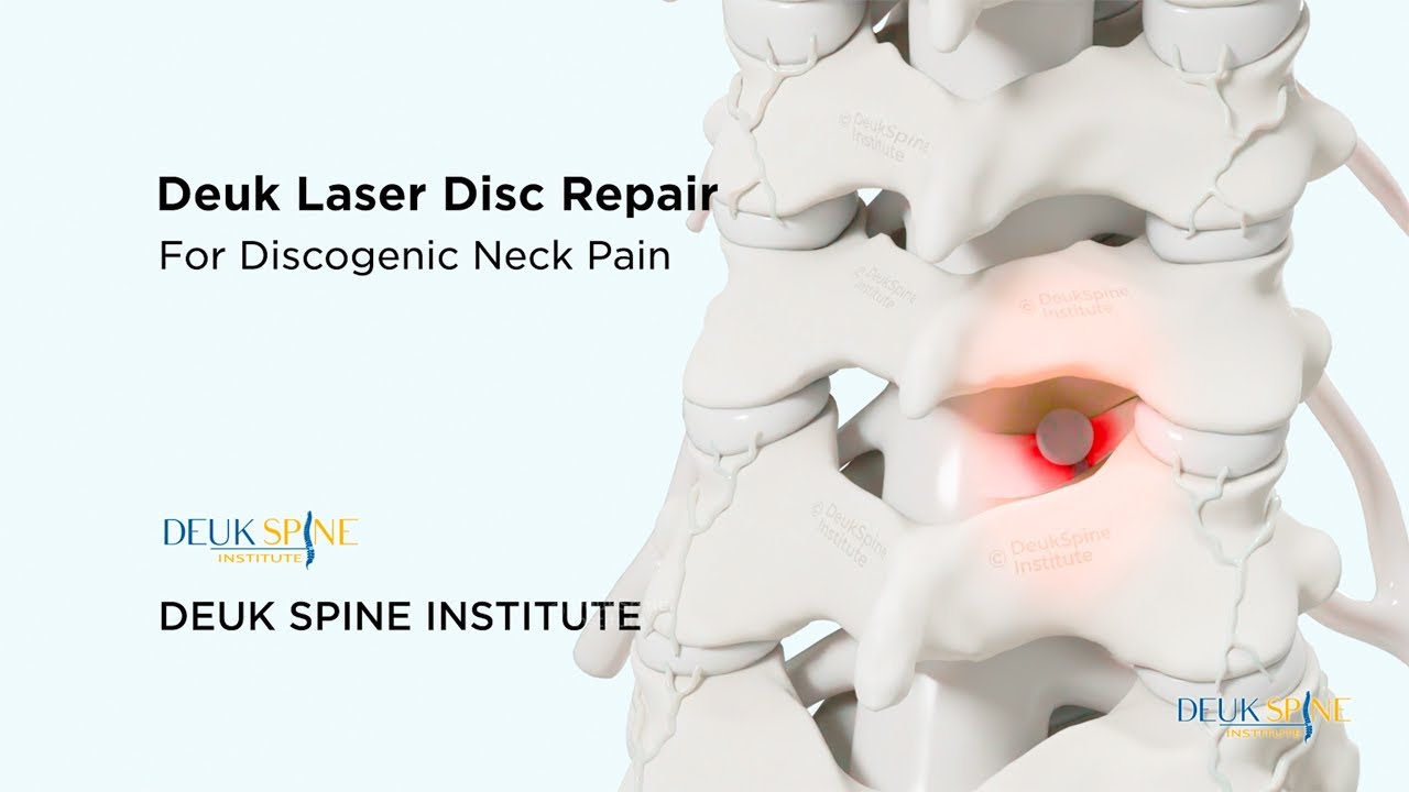
A C6-C7 disc herniation is one of the most common spinal conditions affecting the cervical spine, causing significant pain and functional limitations for millions of Americans each year. Located between the sixth and seventh vertebrae in the lower neck, this area bears substantial mechanical stress due to its role in supporting the head while allowing an extensive range of motion. When a disc herniates at this level, it can compress the C7 nerve root, leading to debilitating symptoms that radiate from the neck down through the arm and into the hand.
Understanding your diagnosis is the first step toward effective treatment. This comprehensive guide explores everything you need to know about C6-C7 disc herniation, from the underlying anatomy and causes to the latest evidence-based treatment approaches. Whether you’re experiencing symptoms for the first time or seeking alternatives after failed treatments, this guide will help you make informed decisions about your care.
Diagnosis. Answers. Relief.
Submit your MRI for a free expert review by Dr. Ara Deukmedjian, M.D. — board-certified neurosurgeon. No obligation. Real answers.
Schedule Yours Today 2,000+ procedures · Zero major complications · No cost, no obligationUnderstanding C6-C7 Anatomy and the C7 Nerve Root
The cervical spine consists of seven vertebrae, labeled C1 through C7, that extend from the base of the skull to the upper back. The C6 and C7 vertebrae sit at the lower portion of the neck, forming one of the most mobile and stress-bearing segments of the entire spine.1
Between each vertebra lies an intervertebral disc that functions as a shock absorber and allows for smooth movement. Each disc consists of two distinct components:
- Annulus fibrosus: The tough, fibrous outer ring composed of multiple layers of collagen fibers arranged in a crisscross pattern
- Nucleus pulposus: The soft, gel-like inner core that provides cushioning and distributes compressive forces
A disc herniation occurs when the annulus fibrosus develops a tear or weakness, allowing the nucleus pulposus to protrude or leak out. At the C6-C7 level, this herniated material typically compresses the C7 nerve root as it exits the spinal canal through the neural foramen.
The C7 Nerve Root Distribution
The C7 nerve root controls critical functions throughout the upper extremity:
Motor function (C7 myotome):
- Triceps muscle (extends the elbow)
- Wrist extensors and flexors
- Finger extensors
- Latissimus dorsi (large back muscle)
Sensory function (C7 dermatome):
- Posterior aspect of the upper arm
- Posterior forearm
- Back of the hand
- Middle finger (particularly characteristic)
- Sometimes portions of the index and ring fingers
Understanding this distribution pattern is crucial because the specific symptoms you experience can help pinpoint whether the C7 nerve root is the source of your pain.
Diagnosis. Answers. Relief.
Submit your MRI for a free expert review by Dr. Ara Deukmedjian, M.D. — board-certified neurosurgeon. No obligation. Real answers.
Schedule Yours Today 2,000+ procedures · Zero major complications · No cost, no obligation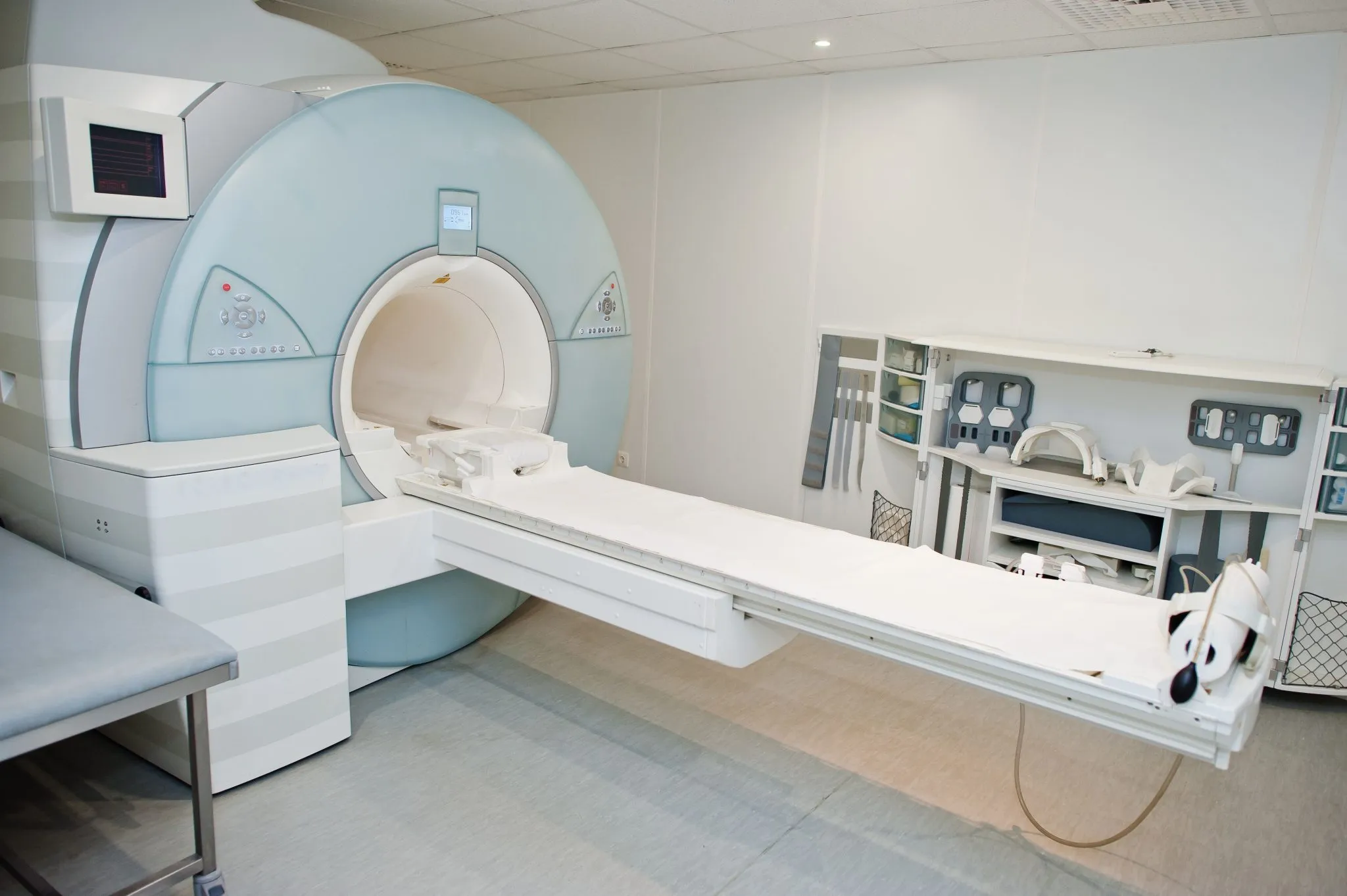
No cost · No obligation
Live Pain Free
Upload your MRI for a free expert review by Dr. Ara Deukmedjian, M.D. — board-certified neurosurgeon. Ten minutes can change your life.
C6-C7 Herniated Disc Symptoms
Compression or inflammation of the C7 nerve root produces a characteristic pattern of symptoms known as C7 radiculopathy. The specific symptoms you experience depend on the degree of nerve compression, the presence of inflammation, and individual factors.
These symptoms are part of a broader pattern – understanding herniated disc symptoms across all spinal levels can help you determine whether your pain is coming from the cervical spine or another region entirely.
Cardinal Symptoms
Radicular pain: This is perhaps the most distinctive symptom—a sharp, burning, or electric-shock-like pain that travels from the neck, through the shoulder blade, down the back of the upper arm, and into the posterior forearm. The pain typically follows the path of the C7 nerve distribution and may intensify with certain neck positions, particularly extension (looking up) and rotation toward the affected side.
Numbness and paresthesias: Many patients describe a “pins and needles” sensation or frank numbness affecting the posterior arm, back of the hand, and particularly the middle finger. This sensory disturbance may be constant or intermittent and often worsens at night.
Weakness: Muscle weakness develops when nerve compression is severe enough to affect motor function. Common manifestations include difficulty extending the elbow (weak triceps), reduced grip strength, and difficulty extending the fingers. You might notice difficulty performing tasks like opening jars, carrying grocery bags, or pushing open heavy doors.
Neck pain and stiffness: While radicular symptoms dominate the clinical picture, many patients also experience localized neck pain and reduced range of motion. The pain may be particularly severe with certain movements.
Associated symptoms: Some individuals experience additional manifestations, including:
- Shoulder blade (scapular) pain
- Headaches, particularly at the base of the skull
- Chest discomfort (which can sometimes mimic cardiac symptoms)
- Sleep disturbance due to pain intensity

Clinical Insights from Dr. Deukmedjian, MD
Over my 20-year career performing cervical spine surgery, I’ve treated thousands of patients with C6-C7 disc herniations. One pattern I’ve observed repeatedly is how these symptoms impact people’s lives far beyond the physical pain.
I remember one patient in his 40s who came to me after six months of progressively worsening symptoms. He described how the constant burning pain down his arm made it impossible to focus on his work. The numbness in his middle finger affected his typing accuracy, and the weakness in his triceps made it difficult to lift his young daughter. He’d tried physical therapy, multiple rounds of epidural injections, and various medications, but nothing provided lasting relief.
What struck me most was when he said, “I feel like I’m losing my identity. I can’t work effectively, I can’t play with my kids, and I can’t sleep through the night. This disc herniation has taken over my entire life.”
This is the reality for many patients with C6-C7 disc herniation. The symptoms don’t just cause physical discomfort—they fundamentally alter your ability to work, care for your family, exercise, and enjoy life. This is why accurate diagnosis and appropriate treatment are so crucial.
Symptom Patterns and Diagnosis
It’s important to recognize that C6-C7 disc herniation symptoms can sometimes be misdiagnosed. The radicular pain and paresthesias may be mistaken for:
- Fibromyalgia
- Carpal tunnel syndrome
- Shoulder pathology (such as rotator cuff problems)
- Peripheral nerve entrapment syndromes
- Thoracic outlet syndrome
A comprehensive evaluation, including detailed neurological examination and appropriate imaging studies, is essential for accurate diagnosis. If you’ve been given a diagnosis that doesn’t fully explain your symptoms or if treatments haven’t been effective, seeking a second opinion from a spine specialist may be warranted.
The Diagnostic Process
Accurate diagnosis of C6-C7 disc herniation requires a systematic approach that combines clinical evaluation with advanced imaging.
Clinical Examination
The diagnostic process begins with a thorough history and physical examination. Your physician should ask detailed questions about:
- The onset and duration of symptoms
- The specific distribution of pain and numbness
- Factors that worsen or alleviate symptoms
- The impact on daily activities and sleep
- Previous treatments and their effectiveness
- The presence of other medical conditions
The physical examination includes specific tests to assess nerve function:
Spurling test: The examiner extends and rotates your neck toward the symptomatic side while applying downward pressure on your head. Reproduction of your arm pain suggests cervical radiculopathy.7
Shoulder abduction relief sign: Placing your hand on top of your head may temporarily relieve symptoms by opening the neural foramen and reducing nerve root compression.
Neurological examination, includes a systematic assessment of:
- Motor strength in specific muscle groups (particularly triceps and wrist extensors)
- Sensation throughout the C7 dermatome
- Deep tendon reflexes (triceps reflex may be diminished)
- Coordination and fine motor skills
Imaging Studies
Imaging adds to the diagnostic puzzle by pointing out areas that may be responsible for generating pain. However, not all herniations cause pain and the majority of adults with show some type of abnormality in their imaging studies.
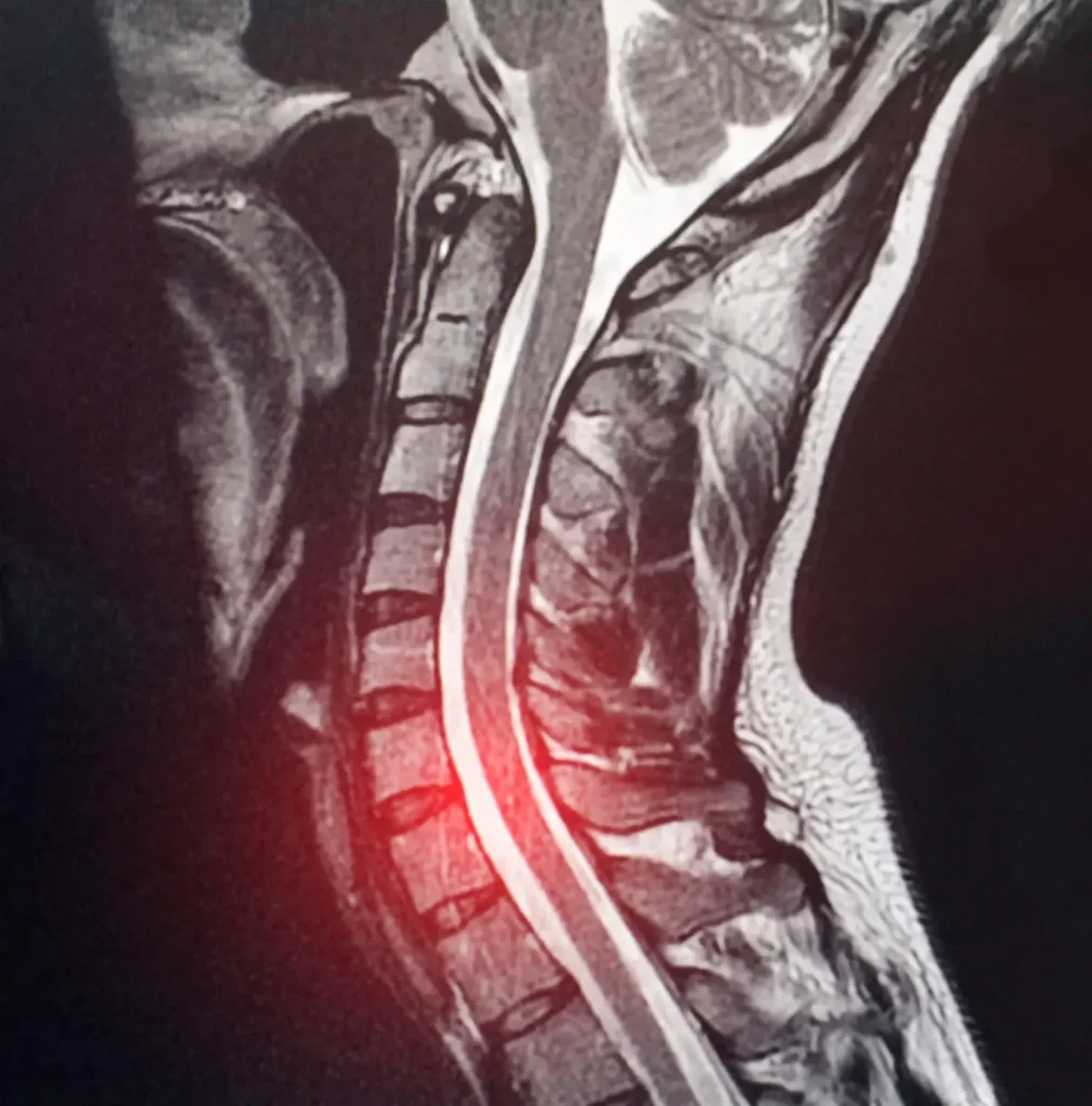
Magnetic Resonance Imaging (MRI): The gold standard for diagnosing cervical disc herniation. MRI provides detailed visualization of soft tissues, including the intervertebral discs, spinal cord, and nerve roots. It can reveal:
- The location and size of the disc herniation
- The degree of neural compression
- Spinal cord changes (if present)
- Other degenerative findings
A 2024 study emphasized that MRI is essential even when X-rays and CT scans appear normal, particularly when neurological symptoms are present (6). Don’t hesitate to request an MRI if your symptoms suggest nerve compression, but initial imaging was negative.
X-rays: While X-rays cannot visualize disc herniation directly, they provide valuable information about:
- Vertebral alignment
- Disc space height (narrowing suggests degeneration)
- Bony changes such as osteophytes (bone spurs)
- Overall cervical spine curvature
CT scan: Computed tomography offers excellent visualization of bony structures and may be useful when:
- X-rays suggest abnormalities requiring further evaluation
- MRI is contraindicated (e.g., certain metal implants)
- Evaluation of bony foraminal stenosis is needed
Electrodiagnostic studies (EMG/NCS): Electromyography and nerve conduction studies can help:
- Confirm the presence of nerve root compression
- Identify the specific nerve root affected
- Distinguish radiculopathy from peripheral nerve problems
- Assess the severity and chronicity of nerve damage
These tests are particularly valuable when clinical or imaging findings are ambiguous.
Conservative Treatment Approaches
For most patients with C6-C7 disc herniation, an initial trial of conservative (non-surgical) treatment is appropriate. Research indicates that patients can improve with conservative management, although this varies depending on herniation severity and the presence of neurological deficits.8
First-Line Therapies
Physical therapy: A structured physical therapy program is often the cornerstone of conservative treatment. A qualified physical therapist can design a program that includes:
- Cervical traction: Gentle mechanical traction may help reduce nerve root compression by temporarily increasing the space in the neural foramen. Recent studies suggest that intermittent traction combined with other modalities can provide significant pain relief.9
- Strengthening exercises: Targeted exercises to strengthen the deep neck flexors and scapular stabilizers improve spinal support and reduce mechanical stress on the affected segment.
- Postural training: Education about proper ergonomics, particularly for individuals with desk jobs, can reduce provocative stresses on the cervical spine.
- Manual therapy: Gentle mobilization techniques may improve neck mobility and reduce muscle spasm, though aggressive manipulation should be avoided in the presence of a disc herniation.
A 2024 systematic review found that manual physical therapy combined with exercise provides better outcomes than either intervention alone for cervical radiculopathy.10
Medications: Various medications can help manage symptoms during the healing process:
- Non-steroidal anti-inflammatory drugs (NSAIDs), such as ibuprofen or naproxen, reduce inflammation around the compressed nerve root. However, prolonged use can cause gastrointestinal and cardiovascular side effects.
- Neuropathic pain medications: Drugs such as gabapentin or pregabalin specifically target nerve pain and may be more effective than traditional pain relievers for radicular symptoms.
- Muscle relaxants: Short-term use can help reduce painful muscle spasms.
- Oral corticosteroids: A short course of oral steroids may be prescribed to reduce acute inflammation, though the evidence for long-term benefit is limited.
Activity modification: Making intelligent adjustments to daily activities can facilitate healing:
- Avoid activities that exacerbate symptoms, particularly overhead work or prolonged neck extension
- Take frequent breaks from static postures, especially computer work
- Use proper lifting techniques and avoid heavy lifting during the acute phase
- Modify sleeping positions to reduce neck strain
Interventional Pain Management
When conservative therapies provide insufficient relief, interventional procedures may be considered:
Cervical epidural steroid injections: These injections deliver corticosteroids directly into the epidural space surrounding the compressed nerve root. The procedure can provide significant short-term relief, allowing patients to participate more effectively in physical therapy.
Selective nerve root blocks: These diagnostic and therapeutic injections target a specific nerve root, providing both pain relief and confirmation that the suspected nerve root is the pain generator.
However, these interventions typically provide temporary relief rather than definitive solutions.
Duration of Conservative Treatment
How long should you persist with conservative treatment before considering surgery? The answer depends on several factors:
- Severity of symptoms: Mild to moderate pain without significant weakness may warrant a longer trial of conservative care
- Rate of improvement: If symptoms are gradually improving with conservative treatment, continuing this approach makes sense
- Functional impact: Severe disability or inability to work may justify earlier surgical consideration
- Presence of neurological deficits: Progressive weakness or other neurological signs may necessitate earlier intervention
Most spine specialists recommend at least 6-12 weeks of comprehensive conservative treatment before considering surgery, provided no alarming neurological signs are present.
When Surgery Becomes Necessary
While many C6-C7 disc herniations improve with conservative treatment, certain situations warrant surgical intervention.
Clear Indications for Surgery
Progressive neurological deficit: If you’re experiencing increasing weakness in your arm or hand despite conservative treatment, surgery should be considered promptly. Prolonged nerve compression can lead to permanent muscle atrophy and functional loss.
Severe, intractable pain: Pain that significantly impairs your quality of life and doesn’t respond to comprehensive conservative treatment (including physical therapy, medications, and injections) may require surgical intervention.
Cervical myelopathy: If the disc herniation is compressing the spinal cord rather than just the nerve root, symptoms may include difficulty with balance and coordination, hand clumsiness, or changes in bowel/bladder function. These signs indicate spinal cord compression and typically require surgical decompression.
Failed conservative treatment: If you’ve undergone an appropriate trial of conservative care (typically 6-12 weeks) without adequate improvement, surgery becomes a reasonable option.
The Second Opinion Advantage
After more than two decades of performing spine surgery, I’ve learned that getting a second opinion is not just advisable, it’s often essential for optimal outcomes. The spine surgery landscape includes numerous approaches with vastly different implications for your long-term health.
I regularly see patients who were told they needed multi-level fusion surgery when, in fact, a much less invasive approach could solve their problem. Conversely, I’ve also seen patients who underwent inappropriate surgery for a condition that should have been managed conservatively.
At Deuk Spine Institute, our proprietary minimally invasive techniques focus on preserving as much of the spine anatomy as possible. That way, our surgical patients can get back to their lives sooner and worry less about the condition down the road.
Upload Your MRI for Expert Review
If you’ve been diagnosed with C6-C7 disc herniation and been told you need surgery, or if conservative treatments haven’t provided relief, I encourage you to seek a second opinion. At Deuk Spine Institute, we offer complimentary MRI reviews and virtual consultations.
No cost · No obligation
Live Pain Free
Upload your MRI for a free expert review by Dr. Ara Deukmedjian, M.D. — board-certified neurosurgeon. Ten minutes can change your life.
Diagnosis. Answers. Relief.
Submit your MRI for a free expert review by Dr. Ara Deukmedjian, M.D. — board-certified neurosurgeon. No obligation. Real answers.
Schedule Yours Today 2,000+ procedures · Zero major complications · No cost, no obligation
No cost · No obligation
Live Pain Free
Upload your MRI for a free expert review by Dr. Ara Deukmedjian, M.D. — board-certified neurosurgeon. Ten minutes can change your life.


