

No cost · No obligation
Live Pain Free
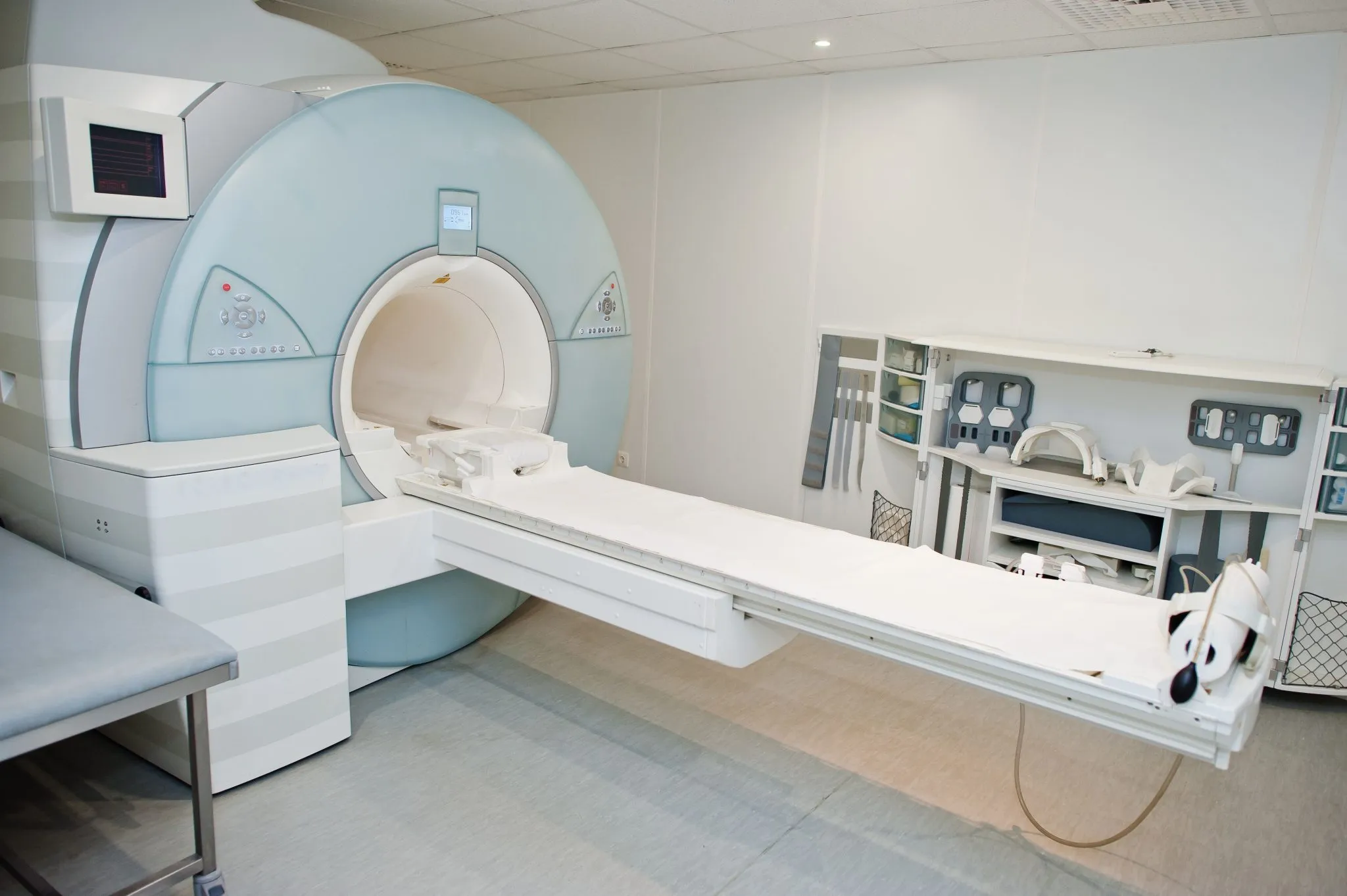
Upload your MRI for a free expert review by Dr. Ara Deukmedjian, M.D. — board-certified neurosurgeon. Ten minutes can change your life.
What Makes the L5-S1 Segment Unique (And Why It Matters to Your Recovery)
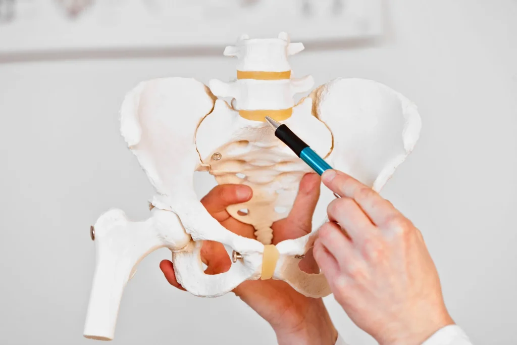
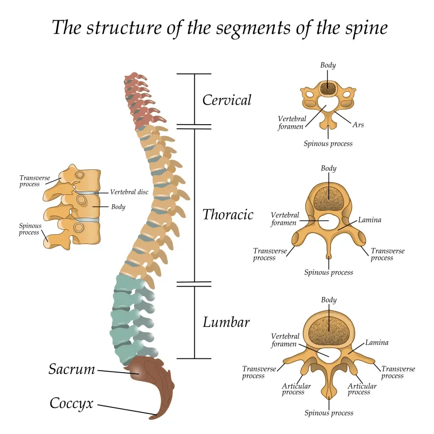
The Anatomy That Sets L5-S1 Apart
The L5-S1 segment isn’t just another spinal level—it’s a critical transition zone. Here’s why that matters:
The weight-bearing reality: Your L5-S1 disc absorbs more compressive force than any other disc in your spine. When you’re standing, walking, or lifting, this single disc bears the cumulative weight of your entire upper body. Studies show the L5-S1 experiences up to 275% more loading stress during forward bending compared to higher lumbar discs.
The curvature change: This is where your spine’s natural curve shifts from lumbar lordosis (forward curve) to sacral kyphosis (backward curve). This transition creates a shearing force that doesn’t exist at other levels—imagine a hinge point that has to bend and twist simultaneously.
The nerve root vulnerability: Unlike higher lumbar discs, where herniated material might miss the nerve root, the L5-S1 anatomy creates a “pinch point” where the S1 nerve root exits. This is why L5-S1 herniations have such a high rate of causing true sciatica.
Why Traditional “Rest and Strengthen Your Core” Advice Often Backfires
In my practice, I frequently see patients who’ve followed standard physical therapy protocols for months without improvement—or who’ve actually gotten worse. Here’s what’s usually happening:
The core strengthening trap: Many patients are told to do planks, sit-ups, or crunches to “support their spine.” But if you have a significant L5-S1 protrusion, these exercises increase intradiscal pressure at exactly the wrong spot. A 2018 study in the Journal of Biomechanics showed that standard sit-ups greatly increase L5-S1 pressure.2
The flexion problem: In 95% of cases, the L5-S1 disc herniates posteriorly (backward). Any exercise that involves forward bending—such as toe touches, certain yoga poses, or heavy deadlifts—pushes the herniated material further into the nerve. Yet many patients are given these exact exercises.
The inflammation cycle: Rest helps acute inflammation, but it doesn’t address the underlying problem: the annular tear (the actual hole in your disc). Without proper healing of this tear, every return to activity re-irritates the injury.
That annular tear and the chronic inflammation it drives are also what turn ordinary degenerative disc disease into a painful condition rather than just an incidental MRI finding, which is worth understanding if your imaging shows disc degeneration alongside your herniation.
Recognizing Your Symptoms: Beyond “My Back Hurts”
Classic L5-S1 Herniation Symptoms
If your herniation is pressing on the S1 nerve root, you won’t just experience generic “back pain.” These symptoms follow a predictable nerve root pattern – understanding the full range of herniated disc symptoms across all spinal levels can help you identify whether your pain is coming from L5-S1 or another disc entirely.
Here’s what L5-S1 compression specifically causes:
- True sciatica with a specific path: The pain follows a precise route—from your lower back or buttock, down the back of your thigh, into your calf, and often reaching your heel or the outer edge of your foot (pinky toe side). This is different from L4-L5 herniation pain, which typically affects the top of the foot and big toe.
- The “Toe Push” test: Can you stand on your tiptoes on the affected leg? S1 nerve compression specifically weakens your ability to push off with your toes when walking. You might notice you’re shuffling or can’t go up on tiptoes repeatedly on one side. This is called a positive “heel raise test” in medical terms.
- The sitting paradox: Many L5-S1 patients feel worse when sitting, especially in soft chairs or cars. Why? The slouched sitting position can increase disc pressure by 40% compared to standing, and it flexes the spine forward—exactly what pushes the herniation further back into the nerve.
- The morning stiffness pattern: You might feel relatively okay going to bed, then wake up barely able to move. This happens because discs rehydrate overnight (they can increase in height by up to 20%), and if there’s a herniation, this swelling increases nerve compression first thing in the morning.
Emergency Warning Signs: When to Seek Immediate Care
I need to be very clear about symptoms that require emergency evaluation, not a scheduled appointment:
Cauda Equina Syndrome Indicators:
- Numbness in the “saddle region” (groin, inner thighs, rectum)
- Loss of bladder or bowel control
- Sudden weakness in both legs
- Sexual dysfunction that appears suddenly
These symptoms indicate compression of multiple nerve roots and constitute a surgical emergency. If you experience any of these, go to an emergency room immediately; waiting even 24-48 hours can result in permanent nerve damage.
The L5-S1 Recovery Path: What to Actually Expect
The Three-Phase Timeline
Based on treating thousands of patients, here’s the realistic recovery timeline for different approaches:
| Recovery Phase | Timeline | Primary Goal | What Should Be Happening |
| Acute Phase | Week 1-2 | Reduce acute inflammation | Pain should decrease by 30-40%; nerve tension reducing |
| Subacute Phase | Week 3-8 | Restore basic function | Walking tolerance improving; sitting tolerance extending |
| Remodeling Phase | Month 3-6 | Prevent recurrence | Return to normal activities; disc strengthening |
| Maintenance | Month 6+ | Long-term stability | Full activity with proper body mechanics |
Important reality check: If you’re not seeing measurable improvement by week 6-8 of conservative treatment, continuing the same approach rarely leads to success. This is when specialized evaluation becomes critical.
Exercises for L5-S1: The Do’s and Critical Don’ts
The McKenzie Method uses “directional preference” exercises—movements that centralize your pain (move it away from your leg toward your spine). For posterior L5-S1 herniations, this typically means extension-based movements.
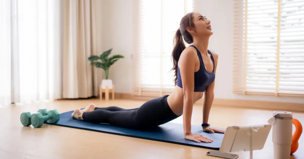
The Prone Press-Up Progression:
Phase 1 – Prone Lying (Days 1-3):
Lie face-down on a firm surface for 5-10 minutes, 4-5 times daily. This gentle extension begins shifting disc material away from the nerve.
Phase 2 – Prone on Elbows (Days 4-7):
Prop yourself on your elbows, creating a gentle arch in your lower back. Hold for 30 seconds, rest, repeat 10 times. Do this 3-4 times daily.
Phase 3 – Full Press-Ups (Week 2+):
Push up with your arms while keeping your pelvis on the floor to create a deeper arch. Hold for 1-2 seconds, lower slowly. Repeat 10 times, 3-4 times daily.
The key indicator: If these movements reduce your leg pain (even if back pain temporarily increases), you’re on the right track. If leg pain worsens, stop and consult a specialist.
Diagnosis. Answers. Relief.
Submit your MRI for a free expert review by Dr. Ara Deukmedjian, M.D. — board-certified neurosurgeon. No obligation. Real answers.
Schedule Yours TodayCritical Exercises to AVOID
Based on biomechanical studies and clinical outcomes, here are movements that consistently aggravate L5-S1 herniations:
 Seated hamstring stretches: Pulling your toes toward you while sitting places massive tension on the S1 nerve root—exactly what you’re trying to avoid. A 2019 study showed this position increases nerve tension by 60%.

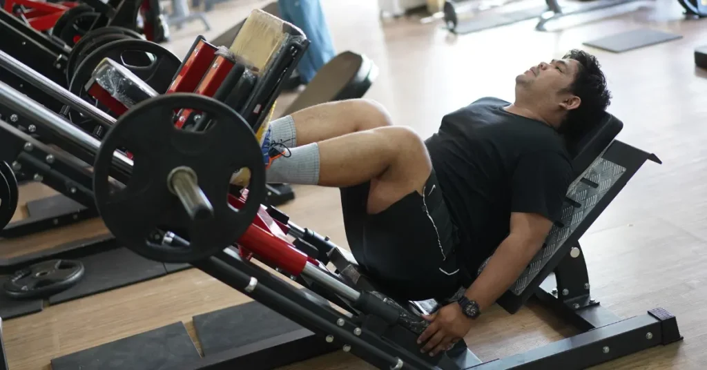
Leg press machines: This exercise combines loaded flexion (the worst position for L5-S1) with high compressive forces. I’ve seen patients re-herniate discs within days of returning to leg press exercises.
Traditional sit-ups or crunches: These create up to 3,300N of compression force at L5-S1 while flexing the spine forward—a perfect storm for worsening herniation.

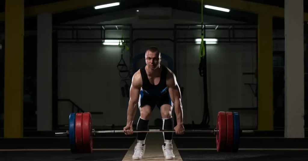
Heavy deadlifts or bent-over rows: Any exercise that loads the spine in forward flexion significantly increases the risk of herniation. Even with “perfect form,” these create stress that an injured L5-S1 cannot handle.
The Walking Prescription
Walking is one of the best activities for L5-S1 recovery, but technique matters:
- Start with 5-minute walks on flat, even surfaces
- Walk with upright posture (imagine a string pulling the top of your head toward the ceiling)
- Stop before pain increases (if you start at 5/10 pain, stop if it reaches 6/10)
- Gradually increase by 2-3 minutes weekly as tolerated
Why walking works: It promotes blood flow to the disc, maintains spinal flexibility, and activates stabilizing muscles without excessive loading.
When Conservative Treatment Isn’t Enough: Understanding Your Surgical Options
The Failed Conservative Treatment Checklist
You should consider advanced treatment if you’ve experienced:
- Persistent symptoms beyond 6-8 weeks despite appropriate therapy
- Progressive neurological deficits (increasing weakness or numbness)
- Inability to work or perform daily activities
- Failed epidural steroid injections (no relief after 2-3 properly performed injections)
- MRI showing significant nerve compression with symptoms that match
Why I Don’t Recommend Fusion for Most L5-S1 Herniations
Traditional spine surgery often leads to spinal fusion for L5-S1 problems. As someone who specializes in fusion alternatives, I need to explain why fusion should be a last resort, not a first option:
The adjacent segment disease problem: When you fuse L5-S1, you permanently eliminate motion at that level. The biomechanical stress doesn’t disappear—it transfers to the disc above (L4-L5). Studies show that 20-30% of fusion patients develop adjacent segment problems within 10 years, often requiring additional surgery.
The sacroiliac joint issue: Fusing L5-S1 also increases stress on your SI joints (where your spine meets your pelvis). This can lead to chronic SI joint pain that’s difficult to treat.
The irreversibility factor: Once you’re fused, you can’t unfuse. You’ve permanently altered your spine’s biomechanics.
The Deuk Laser Disc Repair® Difference
I developed our endoscopic approach specifically to preserve your natural disc while addressing the actual problem—the annular tear and herniation.
How It’s Different from Traditional Microdiscectomy:
| Traditional Approach | Deuk Laser Disc Repair® |
| Drills through lamina bone | Uses natural opening in spine |
| Cuts paraspinal muscles | No muscle cutting |
| Removes disc material but doesn’t heal tear | Vaporizes herniation AND seals annular tear with laser |
| Weakens spine structure | Preserves all structural integrity |
| 2-3 day hospital stay | 1-hour recovery, same-day discharge |
| 15% re-herniation rate | <1% re-herniation rate in our series |
The Procedure in Detail:
Through an incision that can be covered with a Band-Aid, I insert a specialized spinal endoscope that provides HD visualization of your disc and nerve root. Using an FDA-approved surgical laser, I:
- Carefully remove only the herniated tissue compressing the nerve
- Vaporize the damaged portions of the disc, causing inflammation
- Preserve your natural disc height and function—approximately 85-90% of your disc remains intact
The Recovery Difference:
Most of my patients leave the surgery center 1-2 hours after the procedure. They experience immediate relief from nerve compression symptoms because the pressure is gone. Over the following 6-8 weeks, the annular tear heals naturally, strengthened by the laser sealing.
There’s no hospital stay, no bone removal, no muscle damage to heal from. Patients typically return to desk work within 7-10 days and physical work within 4-6 weeks.
Physical Therapy After Diagnosis: What Actually Helps
The Proper PT Progression
Effective physical therapy for L5-S1 herniation follows a specific progression that many general PT programs miss:
Phase 1: Pain Management and Nerve Mobilization (Weeks 1-3)
- McKenzie extension exercises
- Gentle nerve gliding techniques to prevent neural adhesions
- Postural training for sitting and standing
- Activity modification education
Phase 2: Stabilization and Function (Weeks 4-8)
- Hip strengthening (glute med/max focus)
- Core stabilization (NOT crunches—think dead bugs, bird dogs)
- Gradual return to daily activities with proper mechanics
Phase 3: Return to Activity (Weeks 8-12)
- Sport-specific or work-specific training
- Advanced core stability
- Ergonomic assessment and modification
The critical component most PTs miss: Nerve-Gliding Exercises. After a herniation, the S1 nerve can develop adhesions (scar tissue) that restrict its movement. Gentle nerve mobilization techniques help prevent chronic nerve pain even after the herniation improves.
No cost · No obligation
Live Pain Free
Upload your MRI for a free expert review by Dr. Ara Deukmedjian, M.D. — board-certified neurosurgeon. Ten minutes can change your life.


