
By Dr. Ara Deukmedjian, MD
Board-Certified Neurosurgeon, Deuk Spine Institute
Medically reviewed on January 19, 2026
Medical disclaimer: This content is for educational purposes only and does not constitute medical advice. Individual results may vary. Always consult with your healthcare provider about your specific condition and treatment options.
If you’re experiencing lower back pain that radiates down your leg, there’s a significant chance you may have an L4-L5 disc herniation.1 This condition affects the intervertebral disc between the fourth and fifth lumbar vertebrae in your lower back and is one of the most common causes of sciatica and chronic lower back pain. Understanding your diagnosis, exploring all available treatment options, and making informed decisions about your care can be the difference between years of ongoing problems and complete symptom resolution.
This comprehensive guide will walk you through everything you need to know about L4-L5 disc herniation, from recognizing symptoms to understanding cutting-edge treatment approaches that preserve your spine’s natural function.
Understanding the Lumbar Spine and Its Critical Functions
The lumbar spine is the lower section of your back, located below the cervical (neck) and thoracic (mid-back) sections. This remarkable structure consists of five vertebrae numbered L1 through L5, and it serves several vital functions that we often take for granted until something goes wrong.
The Essential Roles of Your Lumbar Spine
Supporting your upper body: Your lumbar spine bears most of the weight of your upper body, acting as a foundational pillar for your entire skeletal structure.2
Distributing body weight: The lumbar spine helps distribute your body weight evenly across your pelvis, which is especially critical when you’re standing, walking, or performing physical activities.
Enabling movement and flexibility: The muscles of your lower back and the inherent flexibility of your lumbar spine allow your trunk to move in all directions—forward and backward (flexion and extension), side to side (lateral bending), and in rotational movements. The last two lumbar vertebrae, L4 and L5, facilitate most of this movement.
Protecting neural structures: Your spinal cord, encircled and protected by the bones of your spine, begins at the base of your skull and terminates at the first lumbar vertebra. Below this point, a bundle of nerves called the cauda equina (Latin for “horse’s tail”) continues down through the lumbar vertebrae.3 The vertebrae provide a protective bony enclosure for these critical nerves.
Controlling leg movement and sensation: The nerves that branch off from the cauda equina and lower spinal cord control leg sensations, muscle movement, and reflexes throughout your lower extremities.
Understanding the L4-L5 Segment
The L4-L5 spinal segment is particularly vulnerable to injury and degeneration for several reasons. This level bears tremendous mechanical stress because it sits at the junction between the mobile lumbar spine and the relatively fixed sacrum. Every time you bend, lift, twist, or even sit, your L4-L5 segment absorbs significant forces.
Additionally, the L4-L5 disc experiences the greatest range of motion in the lumbar spine, which, while beneficial for flexibility, also increases wear and tear over time. This combination of high mechanical load and extensive movement makes L4-L5 one of the most common sites for disc herniation, along with L5-S1.
Lumbar Spine Anatomy: The Five Vertebral Levels

Each vertebra in your lumbar spine has specific characteristics and potential issues:4
L1 (First Lumbar Vertebra): The topmost section of the lumbar spinal column, marking the transition from the thoracic spine.
L2 (Second Lumbar Vertebra): Contains the end of the spinal cord proper (conus medullaris). All vertebrae beyond this point house only spinal nerves, not the spinal cord itself.
L3 (Third Lumbar Vertebra): The middle vertebra of the lumbar spine, serving as a central support structure.
L4 (Fourth Lumbar Vertebra): The second-to-last section of the lumbar spinal column, bearing significant load from the upper body.
L5 (Fifth Lumbar Vertebra): The final section of the lumbar spine, articulating with the sacrum below and carrying the most weight of all lumbar vertebrae.
What Is L4-L5 Disc Herniation?
To understand disc herniation, you first need to understand disc anatomy. Intervertebral discs are specialized structures that sit between each vertebra in your spine. They consist of two distinct components:
Nucleus pulposus: The soft, gel-like inner core that provides cushioning and helps distribute compressive forces throughout the disc.
Annulus fibrosus: The tough, fibrous outer layer made of concentric rings of collagen fibers that contain the nucleus pulposus and provide structural integrity.
A disc herniation occurs when the outer annulus fibrosus develops tears or weakness, allowing the inner nucleus pulposus to push through and escape beyond the disc’s normal boundaries. When this herniated material compresses nearby nerve roots or the spinal cord, it can cause inflammation, pain, and neurological symptoms.

Types of Disc Pathology
Recent research has refined our understanding of different types of disc problems, helping physicians communicate findings more precisely:
Disc bulge: The entire disc circumference extends beyond the vertebral margins symmetrically. The annulus fibrosus remains intact with no tear. This represents early degenerative changes and may or may not cause symptoms.
Disc protrusion: The nucleus pulposus pushes against a weakened annulus, creating a focal bulge. The base of the protrusion is wider than the displaced portion, and the annulus remains unruptured but stretched.
Disc extrusion: The nucleus pulposus breaks completely through the annulus fibrosus. The displaced material remains connected to the parent disc. This typically causes more severe nerve compression and symptoms.
Disc sequestration: A fragment of disc material completely separates and becomes free-floating in the spinal canal. This represents the most severe form of disc displacement, but, paradoxically, sequestered fragments may resorb more completely over time as the immune system recognizes them as foreign material.
The Anatomy of the L4-L5 Spinal Motion Segment
Understanding the specific anatomy of the L4-L5 segment helps explain why problems at this level cause particular symptoms and why certain treatments work better than others.
Structural Components
L4 and L5 vertebrae: Each vertebra consists of a vertebral body at the front and a vertebral arch at the back. The vertebral arch includes a prominent spinous process in the middle and two transverse processes on either side. The lamina connects the spinous process to the transverse processes, while the pedicle connects the transverse process to the vertebral body. These vertebrae are joined by facet joints, which are covered with cartilage to ensure smooth movement.
The L4 and L5 vertebral bodies are taller in front than behind, contributing to the natural lumbar lordosis (inward curve) of your lower back. The upper and lower surfaces of each vertebral body are covered by bony endplates that help resist compressive loads.
L4-L5 intervertebral disc: This disc is a soft tissue joint composed of the nucleus pulposus (hydraulic gelatinous core) encased within the annulus fibrosus (firm outer collagen wall). The disc protects the spinal vertebrae and nerves from sudden impact and absorbs shock during spinal movements such as bending, twisting, and jumping.
L5 nerve root: The L5 spinal nerve roots exit the spinal canal through small bony openings called intervertebral foramina on both the right and left sides. These nerve roots combine with other nerves to form larger nerves that travel down each leg.

What Nerve Is Affected in L4-L5 Disc Herniation?
Source The L5 nerve root is most commonly affected in an L4-L5 disc herniation. This nerve root exits the spine at the level of the L4-L5 vertebrae and provides critical functions:
L5 dermatome (sensory distribution): An area of skin that receives sensations through the L5 spinal nerve, including parts of the outer leg, top of the foot, and the space between the first and second toes.
L5 myotome (motor control): A group of muscles controlled by the L5 spinal nerve, including muscles that lift the foot and toes (dorsiflexion), extend the big toe, turn the foot outward (eversion), and extend the hip.
When an L4-L5 disc herniation compresses the L5 nerve root, you may experience characteristic symptoms following these distributions, which helps physicians diagnose the specific level of nerve compression through careful clinical examination.

Causes of L4-L5 Disc Herniation
Understanding what causes disc herniation can help you make informed decisions about prevention and treatment. While some risk factors are beyond your control, others can be modified to reduce your risk of developing or worsening disc problems.
L4-L5 Treatment with Surgery
Age-related degeneration: As we age, intervertebral discs naturally lose water content through a process called disc desiccation, making them thinner, less flexible, and more prone to injury. Small tears develop in the annulus fibrosus over time, weakening its structure. This degenerative process typically begins in the third or fourth decade of life and progresses gradually.
Acute trauma and injury: Disc herniation can result from sudden impact events such as motor vehicle accidents, falls that jar the spine, sports injuries involving twisting or impact, or improper lifting technique with heavy objects. The force from these events can overwhelm the disc’s structural capacity, causing immediate herniation.5
Repetitive stress and occupational factors: Jobs requiring heavy lifting, prolonged sitting (especially with poor posture), frequent bending and twisting, or exposure to whole-body vibration (e.g., truck drivers, heavy equipment operators) can cause cumulative damage over time. Each individual stress may seem minor, but its cumulative effect over months and years can lead to a disc herniation.
Genetic predisposition: Research increasingly recognizes that some individuals inherit a tendency toward earlier disc degeneration. If multiple family members experienced lumbar disc problems at relatively young ages, genetic factors may contribute to your susceptibility.
Modifiable Risk Factors
Recent research has identified several factors you can address to potentially slow progression or reduce symptom severity:
Excess body weight: Obesity increases mechanical loads on the lumbar spine and is associated with systemic inflammation that may affect disc metabolism. Maintaining a healthy weight through diet and exercise provides multiple benefits for spine health.
Smoking: Nicotine reduces blood flow to disc tissues, impairing nutrient delivery and waste removal. Smoking also increases levels of inflammatory mediators throughout the body, and chronic coughing places repetitive stress on the spine. Studies consistently show that smokers experience more severe disc degeneration and poorer treatment outcomes.
Sedentary lifestyle: Prolonged sitting maintains sustained compression on lumbar discs, while lack of regular movement reduces nutrient flow to disc tissues, which depend on movement for nutrition. Weak core muscles provide less support for the lumbar spine, increasing disc stress.
Poor posture and ergonomics: Forward-leaning posture dramatically increases forces on lumbar discs. Work environments without proper ergonomic setup and prolonged static postures all contribute to disc problems over time.
Common Symptoms of L4-L5 Disc Herniation
The symptoms of L4-L5 disc herniation vary significantly depending on the degree of nerve compression, the presence of inflammation, and individual factors. Some people experience dramatic symptoms, while others have minimal discomfort despite significant findings on imaging studies.
Sciatica: The Hallmark Symptom
In the lumbar spine, the most common symptom associated with L4-L5 disc herniation is sciatica, also known as radicular leg pain.6 When a herniated disc at L4-L5 compresses the L5 nerve root, it typically causes:
Sharp, shooting pain that travels from the lower back or buttock down the outer side of the leg, potentially extending to the top of the foot and the area between the first and second toes. This pain often feels electric or burning in nature and can be severe enough to stop you in your tracks.
Radiating pain pattern: Unlike general lower back pain, which tends to stay localized, sciatica follows the path of the affected nerve, creating a characteristic distribution that helps physicians identify which nerve root is compressed.
Neurological Symptoms
Numbness and tingling: You may experience altered sensation in the L5 dermatome distribution, including the outer leg, top of the foot, and toes. Some patients describe this as a “pins and needles” sensation, while others report complete numbness in affected areas.
Muscle weakness: L5 nerve compression can lead to specific weakness patterns, including difficulty lifting the foot or toes (foot drop), weakness when walking on heels, reduced ability to extend the big toe, and general leg fatigue or a feeling of heaviness. These motor deficits can significantly impact your ability to walk normally and increase your risk of tripping or falling.
Altered reflexes: While reflex changes are less common with L5 radiculopathy than with other nerve root compressions, your physician may detect subtle changes during examination.
Associated Symptoms
Beyond the classic sciatica presentation, L4-L5 disc herniation can cause:
Lower back pain: Localized pain at the L4-L5 level that may be sharp, dull, or aching in character. This pain often worsens with certain movements, such as bending forward, twisting, or prolonged sitting.
Muscle spasms: Protective muscle spasms in the lower back can develop as your body attempts to stabilize the injured segment. These spasms can be painful and further limit your range of motion.
Postural changes: You may unconsciously shift your posture to avoid positions that increase pain, sometimes leading to a temporary scoliosis (side-to-side curvature) as you lean away from the affected side.
Sleep disruption: Difficulty finding comfortable sleeping positions can lead to poor sleep quality, which in turn can lower your pain threshold and worsen your overall condition.
Bowel or bladder dysfunction: In rare but serious cases where the herniated disc compresses multiple nerve roots or causes cauda equina syndrome, you may experience difficulty controlling bowel or bladder function. This is a medical emergency requiring immediate evaluation.3
Recent Research on Symptom Patterns and Natural History
Spontaneous reabsorption: A groundbreaking July 2025 review in Frontiers in Medicine examined the biological mechanisms underlying lumbar disc herniation reabsorption, in which herniated disc tissue shrinks naturally without surgical intervention. This research identified that inflammatory responses and macrophage activation play crucial roles in symptom relief during the natural healing process, providing a biological explanation for why most sciatica cases improve without surgery.7 Understanding these mechanisms helps explain why conservative treatment succeeds for most patients and why patience during the initial treatment phase is often rewarded.
AI-assisted prognosis: Emerging 2025 studies highlight the growing role of Artificial Intelligence in predicting postoperative outcomes and identifying specific symptom profiles.8 AI models are increasingly being used to determine which L4-L5 patients will benefit most from early surgical intervention versus continued conservative care, helping personalize treatment decisions and improve outcomes.
Specific L5 nerve indicators: Herniations at L4-L5 typically compress the L5 nerve root. Recent 2025 clinical guides reaffirm characteristic findings, including weakness in ankle dorsiflexion (difficulty lifting the foot upward), weakness in great toe extension, and potential gluteus medius weakness detectable through Trendelenburg gait (where the hip drops on one side when walking).9 These specific clinical signs help physicians accurately localize the level of nerve compression.
Positional factors: Recent clinical manuals emphasize that sitting increases intradiscal pressure by approximately 40% compared with standing, thereby significantly intensifying radiating pain in L4-L5 patients.10 This research validates the common patient experience that sitting is often more painful than standing or walking, and underscores the importance of limiting prolonged sitting during the acute phase.
How L4-L5 Disc Conditions Are Diagnosed
Accurate diagnosis is the foundation for successful treatment, yet many patients struggle through months or even years of unsuccessful treatments because their condition wasn’t properly assessed initially. Understanding the diagnostic process empowers you to advocate for the thorough evaluation you deserve.
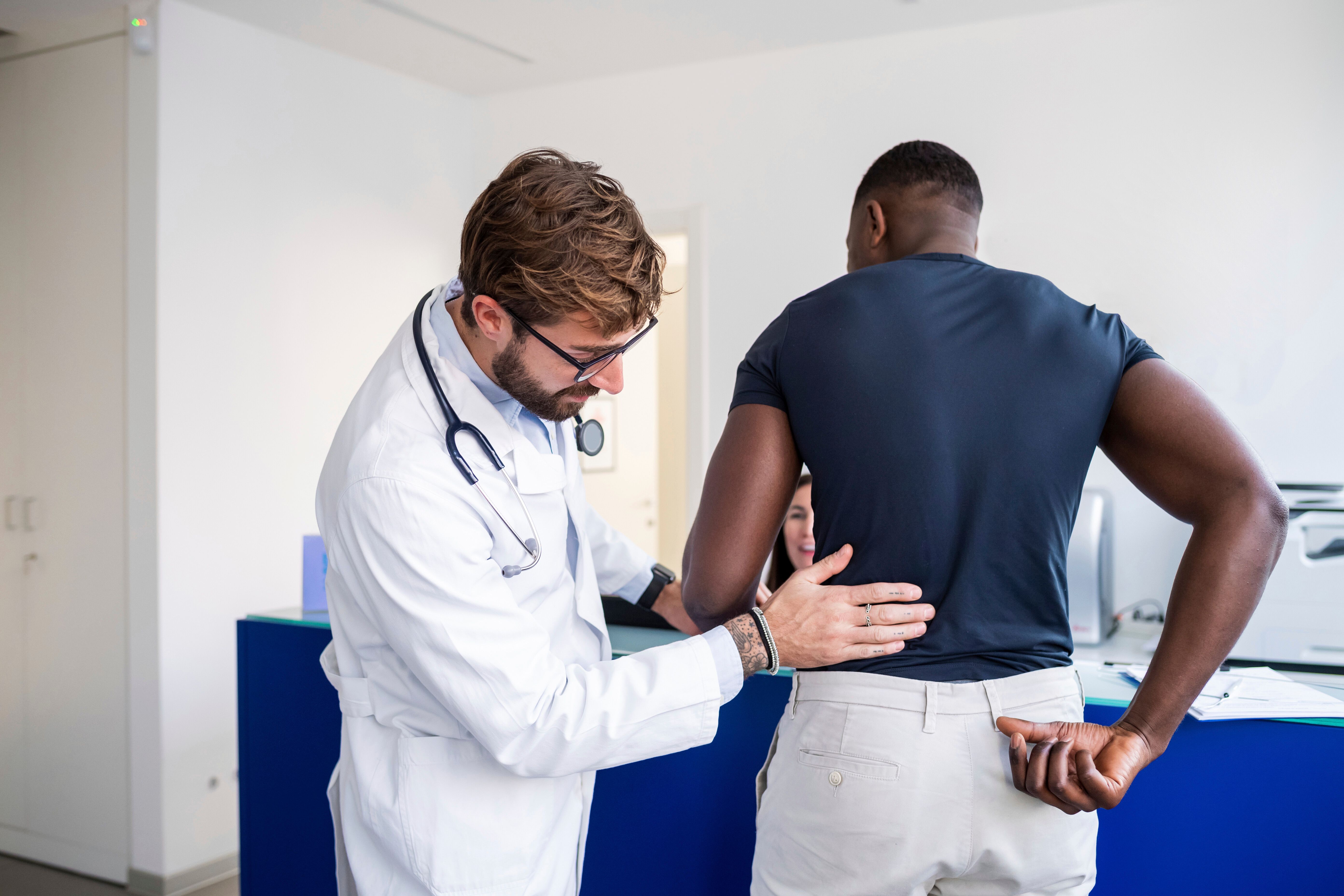
Clinical Examination
Your diagnostic journey begins with a comprehensive history and physical examination. An experienced physician should spend adequate time understanding your symptoms, including:
- When symptoms started and how they’ve progressed
- What makes symptoms better or worse
- How symptoms affect your daily activities and work
- What treatments have you tried and their effectiveness
- Whether you have other medical conditions that might influence treatment
The physical examination includes specific tests designed to assess nerve function and identify the pain source:
Straight leg raise test: You lie on your back while the examiner lifts your leg with the knee straight. Pain radiating down the leg at angles less than 60 degrees suggests nerve root compression.
Motor strength testing: The examiner evaluates specific muscle groups controlled by the L5 nerve, including ankle dorsiflexion (pulling the foot upward), great toe extension, hip abduction, and foot eversion.
Sensory examination: Testing for altered sensation in the L5 dermatome distribution along the outer leg and top of the foot.
Reflex testing: While the L5 nerve root doesn’t have a deep tendon reflex that can be easily tested, your physician may check other reflexes to rule out involvement of adjacent nerve roots.
Imaging Studies
Magnetic Resonance Imaging (MRI): MRI is the gold standard for evaluating disc herniations and nerve compression.11 It provides a detailed visualization of soft tissues, including discs, nerve roots, the spinal cord, and ligaments. MRI can reveal the size and location of disc herniations, the degree of neural foramen narrowing, whether nerve roots are compressed, and the presence of other spinal conditions.
Recent research emphasizes that an MRI should be obtained when clinical symptoms suggest nerve compression, even if initial X-rays appear normal. This is particularly important after trauma, where disc injury may be the only significant finding requiring treatment.
X-rays: While X-rays cannot visualize disc herniations directly, they provide valuable information about vertebral alignment, disc space height, bone spurs that might contribute to nerve compression, and overall lumbar spine curvature. X-rays serve as an important screening tool to rule out fractures or severe instability.
CT scans: Computed tomography offers excellent bony detail and may be useful when MRI is contraindicated, for evaluating bony foraminal stenosis, or for surgical planning when precise visualization of bony anatomy is needed.
Electrodiagnostic studies (EMG/NCS): These tests measure the electrical activity of muscles and nerves, confirming the presence and severity of nerve root compression, identifying which specific nerve root is affected, distinguishing lumbar radiculopathy from peripheral nerve problems, and assessing the chronicity and severity of nerve damage.
When Findings Don’t Match Symptoms
One of the most challenging aspects of lumbar spine diagnosis is the frequent mismatch between imaging findings and symptoms. Studies consistently show that many people without back or leg pain have disc herniations visible on MRI. Conversely, some individuals with significant symptoms have relatively minor imaging findings.
This reality underscores the importance of comprehensive clinical correlation. Your symptoms, physical examination findings, and imaging studies must all be considered together to develop an appropriate treatment plan.
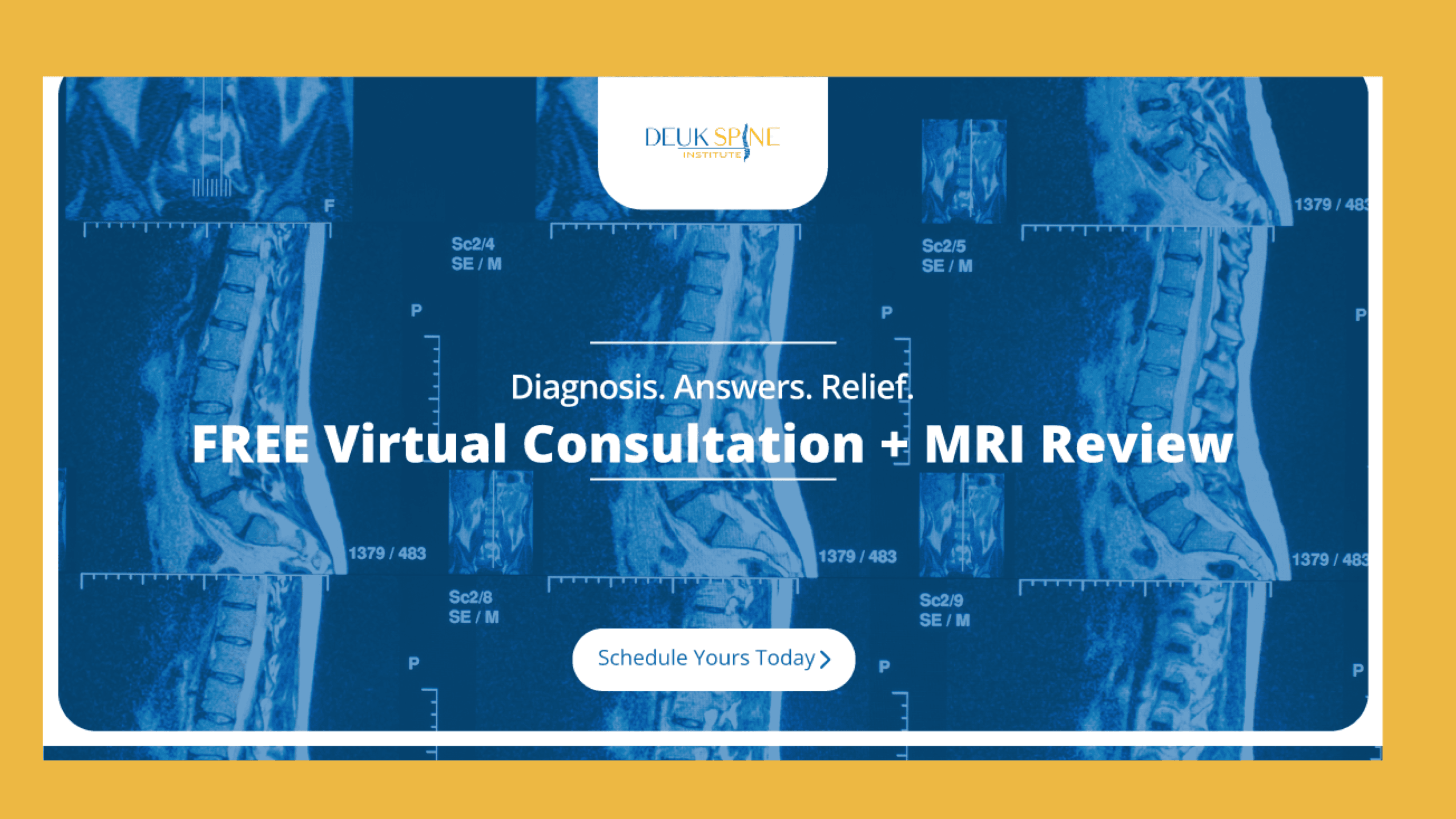
Searching for an Objective Second Opinion?
Need a new perspective? Upload your latest MRI for a free consultation and review with Dr. Ara Deukmedjian, creator of the minimally invasive Deuk Laser Disc Repair® technique.
L4-L5 Disc Herniation Treatment Options
Treatment for L4-L5 disc herniation follows a stepwise approach, starting with conservative care and progressing to surgical intervention when necessary. Understanding all available options empowers you to make informed decisions about your care.
Conservative Treatment: The First Line of Defense
For most patients with L4-L5 disc herniation, an initial trial of conservative treatment is appropriate and often successful. Research indicates that patients with lumbar disc herniations improve with comprehensive conservative care. However, the key is “comprehensive” care; not all conservative treatments are equally effective.
Physical Therapy and Exercise
Structured physical therapy forms the cornerstone of conservative treatment for many patients. A qualified therapist specializing in spine conditions should design a program tailored to your specific presentation.
Core strengthening exercises: Building strength in your abdominal and back muscles provides better support for your lumbar spine, reducing stress on the injured disc. These exercises might include planks, bridges, bird dogs, and dead bugs, all performed with proper technique to avoid worsening symptoms.
Flexibility exercises: Gentle stretching can help maintain or improve range of motion, though aggressive stretching should be avoided during the acute phase. Hamstring stretches are particularly important, as tight hamstrings can increase stress on the lumbar spine.5
McKenzie exercises: This specific approach involves repeated movements and sustained positions designed to “centralize” pain—bringing it from the leg back toward the spine, which typically indicates improvement. Some patients respond dramatically to McKenzie therapy.
Aerobic conditioning: Low-impact cardiovascular exercise, such as walking or swimming, can help reduce inflammation, improve overall fitness, and support healing without placing excessive stress on the injured disc.
Medications for Symptom Management
While medications don’t heal disc herniations, they can manage symptoms during the healing process and make physical therapy more tolerable.
Non-steroidal anti-inflammatory drugs (NSAIDs): Medications like ibuprofen or naproxen reduce inflammation around the compressed nerve root. However, prolonged use carries risks including gastrointestinal bleeding and kidney problems, so they should be used at the lowest effective dose for the shortest duration necessary.
Neuropathic pain medications: Drugs like gabapentin or pregabalin specifically target nerve pain, which often responds poorly to traditional pain relievers. Side effects may include drowsiness and dizziness, which may require dose adjustment.
Muscle relaxants: Short-term use can help break the cycle of pain and muscle spasm, though these medications typically cause drowsiness.
Oral corticosteroids: A short course of oral steroids may be prescribed for acute severe inflammation, though evidence for long-term benefit is limited, and these medications carry significant side effects with prolonged use.
Interventional Pain Management
Epidural steroid injections: These procedures deliver corticosteroids directly into the epidural space surrounding the compressed nerve root, potentially providing temporary relief by reducing inflammation. However, benefits typically last weeks to months rather than being permanent, and success rates vary considerably among patients.
Selective nerve root blocks: These injections target a specific nerve root and serve both diagnostic and therapeutic purposes. If a selective nerve root block temporarily eliminates your symptoms, it confirms that the targeted nerve root is your pain generator.
Research on conservative treatments for lumbar disc herniation demonstrates that while various approaches showed benefits, outcomes are highly variable, and many patients ultimately require more definitive intervention for lasting relief.
When Conservative Treatment Isn’t Enough
For some patients, conservative treatment doesn’t provide adequate relief. Recognizing when conservative approaches have failed and surgical intervention should be considered, is crucial for preventing permanent nerve damage and unnecessary prolonged disability.
Clear Indications for Surgical Consideration
Progressive motor weakness: If you’re experiencing increasing weakness in your leg or foot despite conservative treatment, nerve damage may be progressing. Prolonged compression can lead to permanent muscle atrophy and functional loss that won’t fully recover even after successful decompression.
Severe, intractable pain: Pain that prevents sleep, interferes with work, or limits basic daily activities represents more than an inconvenience; it’s a legitimate indication for definitive treatment when conservative approaches have been exhausted.
Failed conservative treatment: If you’ve undergone 8-12 weeks of comprehensive conservative care without meaningful improvement, continuing the same ineffective treatments indefinitely makes little sense.
Cauda equina syndrome: This medical emergency occurs when a massive disc herniation compresses multiple nerve roots, causing bowel or bladder dysfunction, saddle anesthesia (numbness in the area that would contact a saddle), and bilateral leg weakness. This condition requires urgent surgical decompression to prevent permanent neurological damage.
Surgical Treatment for L4-L5 Disc Herniation
When conservative treatment hasn’t provided adequate relief and surgery becomes necessary, understanding your options is essential. Not all surgical procedures are equal; they vary dramatically in invasiveness, recovery time, long-term outcomes, and impact on spinal function.
Traditional Surgical Approaches
Microdiscectomy: This has been the standard surgical treatment for lumbar disc herniation for decades. The procedure involves accessing the spine from the back, removing a portion of the lamina (laminotomy) to access the disc, and removing the herniated portion pressing on the nerve root.
Recovery typically requires several days in the hospital, 4-6 weeks for return to light activities, and 3-6 months for full recovery. While microdiscectomy can be effective, it involves removing bone to access the disc, which can weaken spinal stability and potentially lead to future complications.
Laminectomy: This procedure involves removing a larger portion of the lamina to decompress the spinal canal. It’s typically reserved for cases with significant spinal stenosis in addition to disc herniation.12 Recovery is generally longer and more challenging than with microdiscectomy.
Spinal Fusion: When disc problems are accompanied by spinal instability or involve multiple levels, some surgeons recommend fusion. This procedure permanently joins two or more vertebrae together using bone grafts and hardware.13
However, fusion has significant drawbacks: it eliminates motion at the fused segment, alters spinal biomechanics, increases stress on adjacent levels (potentially accelerating their degeneration), and requires extensive recovery time of 6-12 months. Recent research shows that fusion carries an increased risk of adjacent segment disease, where levels above or below the fusion develop problems requiring additional surgery.
Deuk Laser Disc Repair®: A Revolutionary Motion-Preserving Alternative
After witnessing the limitations and complications of traditional lumbar spine surgery throughout my training and career, I became convinced that a better approach was needed. This led to the development of Deuk Laser Disc Repair® (DLDR), a minimally invasive, motion-preserving procedure that addresses the pathological disc material while leaving healthy structures intact.
A Different Philosophy of Treatment
Traditional lumbar spine surgery operates on the premise of removing bone to access the disc, often removing large portions of the disc, and sometimes fusing vertebrae when instability is present. This approach has several fundamental flaws: it treats surrounding healthy tissue as collateral damage, permanently alters spinal biomechanics during fusion, requires significant tissue dissection, leading to prolonged recovery, and carries substantial risk of complications.
DLDR® takes an entirely different approach:
- Precisely removes only the damaged portion of the disc causing symptoms
- Leaves the healthy disc intact to preserve function
- Maintains natural spinal motion without fusion or hardware
- Minimizes tissue trauma through advanced endoscopic techniques
How Deuk Laser Disc Repair® Works
DLDR® is performed through a tiny 4-7mm incision, so small it can be covered with a simple band-aid. Through this minimally invasive opening, a specialized endoscope provides high-definition visualization of the surgical field.
Using advanced laser technology, only the damaged disc material compressing the nerve root is targeted and removed, typically just 5-10% of the total disc. The laser’s precision allows work around delicate neural structures without the collateral damage inherent in traditional surgery.
Because most of the disc remains intact and functional, there’s no need for fusion, bone grafts, or metal hardware. The natural disc continues to provide cushioning and allow normal motion. The procedure typically takes approximately one hour to perform.
The Science Behind Precision
The key to DLDR®'s success lies in its precision. Traditional surgery requires large incisions, muscle stripping, and significant bone removal to access the problem disc. These approaches are inherently traumatic to surrounding tissues, explaining the prolonged recovery and potential complications.
DLDR® causes minimal tissue disruption through several mechanisms:
- The endoscope accesses the disc through natural tissue planes
- The laser removes only pathological disc material without damaging healthy tissue
- No bone removal is required, preserving structural integrity
- Surrounding muscles, nerves, and blood vessels remain undisturbed
This precision directly translates into the patient experience. Because tissues aren’t unnecessarily traumatized, patients typically experience minimal postoperative pain, walk out of recovery within an hour, and return to work within days rather than months.
A 2025 study examining endoscopic discectomy techniques found that minimally invasive approaches provided excellent clinical outcomes while preserving spinal stability and reducing surgical complications compared to traditional open procedures.14
Recovery Timeline and Outcomes
One of the most dramatic differences between DLDR® and traditional surgery is the recovery experience:
Day of surgery: Most patients notice significant pain relief as soon as they wake from twilight anesthesia. They leave the recovery room within an hour and go home the same day with no hospital stay required.
First week: The tiny incision heals within days. Most patients require only over-the-counter pain medication, if anything. Many return to desk work within 3-5 days.
2-4 weeks: Complete healing occurs. Patients can resume most normal activities with minor restrictions depending on their condition.
Long-term: Because no fusion was performed, no adjacent segment disease develops. Patients maintain a full range of motion and can expect normal function for decades.
Benefits Beyond Pain Relief
The advantages of DLDR® extend far beyond simply relieving symptoms:
Preservation of natural anatomy: Because we’re not removing entire discs or fusing vertebrae, normal anatomy and biomechanics remain intact. This preservation of natural function is perhaps the most significant long-term benefit.
No hardware complications: Without metal screws or plates, there are no hardware-related complications, no metal detectors to worry about, and no hardware removal surgery needed.
Minimal scarring: The 4-7mm incision heals with virtually no visible scar.
No narcotics needed: The procedure is so precise that postoperative narcotic pain medication is typically unnecessary, increasingly important given the opioid crisis.
Proven outcomes: DLDR® has been published in peer-reviewed medical literature documenting its safety and effectiveness. This isn’t experimental; it’s an established, proven procedure with a 99% success rate of eliminating pain and zero complications in over 2,000 patients treated.
See How it Works
Learn how DLDR® is minimally invasive, motion preserving technique.
Patient Perspectives: Real Recovery Stories
Understanding treatment options through research and medical explanations is important, but hearing from real patients who have walked this path provides an invaluable perspective. Here are experiences from patients who faced lumbar disc problems and chose minimally invasive treatment.
Ten Years of Back Pain Resolved in Hours
A patient from Michigan came to our facility after suffering for 10 years with debilitating lower back pain from disc herniations at multiple levels. She had exhausted conservative treatments, including physical therapy, medications, and injections, without meaningful relief.
After reviewing her MRI, we performed DLDR® along with treatment for her piriformis syndrome and SI joint dysfunction—addressing all sources of her pain in a comprehensive fashion. She described experiencing relief she hadn’t felt in a decade, with her pain eliminated within 12 hours of the procedure.
Her story illustrates an important point: comprehensive evaluation can identify multiple pain generators that must be addressed for complete relief. She traveled from Michigan specifically because traditional surgeons in her area had recommended fusion, which she wanted to avoid.15
Retired Firefighter Finds Relief After 25 Years
A 30-year firefighter veteran came to us after battling chronic back pain for 25 years, an occupational hazard of repeatedly carrying heavy equipment and patients. Conservative treatments provided only temporary relief, and he was facing retirement from a career he loved due to his worsening condition.
Using DLDR®, we addressed his L4-L5 disc herniation while preserving his spinal motion and stability. His recovery was remarkably quick, and he was able to return to his active lifestyle without the limitations that had plagued him for years.16
His experience demonstrates that even long-standing disc problems can be successfully treated with motion-preserving techniques and that years of pain don’t necessarily mean permanent disability.
Indiana Patient Overcomes Two Disc Herniations
Another patient from Indiana had been unable to work for years due to severe pain from disc herniations at both L1-L2 and L5-S1. Multiple conservative treatments had failed, and she was told she might need extensive fusion surgery.
After reviewing her case, we determined she was a candidate for DLDR® at both levels. By addressing both problem discs in a single minimally invasive procedure, we were able to eliminate her pain while preserving motion throughout her lumbar spine. She returned to work and normal activities within weeks.17
These patient stories reinforce the central message: taking time to thoroughly understand your condition, seeking second opinions when appropriate, and exploring all available treatment options can make the difference between years of ongoing problems and complete symptom resolution.
Living with L4-L5 Disc Herniation: Practical Strategies
While pursuing definitive treatment for your L4-L5 disc herniation, implementing practical strategies can help manage symptoms and potentially prevent worsening. These recommendations are based on biomechanical principles and clinical experience with thousands of patients.
Sleep Position and Pillow Selection
Poor sleeping positions can significantly aggravate lumbar disc problems. Proper sleep ergonomics is essential for symptom management.
Back sleeping with knee support (optimal): Lying on your back with a pillow under your knees reduces stress on the lumbar spine by maintaining a neutral curve. This position evenly distributes weight and minimizes disc pressure.
Side sleeping with a pillow between your knees (acceptable): If you prefer side sleeping, place a pillow between your knees to keep your spine aligned and reduce rotational stress on your lower back.
Stomach sleeping (avoid completely): This position forces your lower back into extension, placing maximum stress on lumbar discs and facet joints. It’s the worst possible position for disc problems.
Ergonomics and Posture
Modern life, particularly desk work and prolonged sitting, places significant stress on the lumbar spine. Proper ergonomics can substantially reduce this burden.
Workstation setup: Use a chair with proper lumbar support to maintain your spine’s natural curve. Position your feet flat on the floor or on a footrest. Keep your keyboard and mouse at elbow height to avoid slouching. Stand and move for 2-3 minutes every 30 minutes of sitting.
Lifting technique: Bend at the knees, not the waist. Keep objects close to your body. Avoid twisting while lifting. Use leg muscles rather than back muscles to perform the lift. Ask for help with heavy items—there’s no prize for injuring yourself.
Driving: Adjust your seat so your knees are level with your hips. Use lumbar support, either built into your seat or via an aftermarket cushion. Take breaks every hour on long drives to stand and stretch.
Exercise and Physical Activity
Appropriate exercise can be beneficial, but choosing the right activities is crucial.
Recommended activities: Walking provides low-impact cardiovascular exercise that doesn’t stress the back. Water aerobics and swimming offer excellent exercise with minimal spinal load. Stationary cycling (with proper posture) provides cardiovascular benefits. Gentle yoga focusing on core strength and flexibility can be helpful.
Activities to avoid: Running and high-impact sports create jarring forces on the spine. Heavy weightlifting, especially deadlifts and squats, should be avoided during the acute phase. Contact sports carry the risk of additional injury. Golf and tennis involve twisting motions that can aggravate disc problems.
What to Avoid
Certain activities and positions can significantly worsen L4-L5 disc herniation symptoms:
Prolonged sitting: Sitting increases disc pressure by approximately 40% compared to standing. Take frequent breaks, use proper lumbar support, and consider a standing desk option.
Forward bending: Activities requiring prolonged forward flexion (gardening, mopping floors, prolonged computer work with poor posture) should be minimized or modified.
Heavy lifting: Avoid lifting anything over 10-15 pounds during the acute phase. When you must lift, use proper technique.
Sudden movements: Quick, jerking movements can exacerbate symptoms or cause additional disc injury. Move deliberately and avoid sudden twisting.
Stress Management and Sleep Quality
Chronic pain creates stress, and stress can amplify pain perception, creating a vicious cycle. Implementing stress management strategies can help break this pattern:
- Mindfulness meditation and relaxation techniques
- Cognitive behavioral therapy for chronic pain
- Maintaining social connections despite limitations
- Adequate sleep (though pain often disrupts sleep, creating another cycle to break)
Key Takeaways
L4-L5 disc herniation is a common condition that can significantly impact quality of life, but understanding your diagnosis and exploring all treatment options empowers you to make informed decisions about your care.
Understanding the condition: The L4-L5 segment bears tremendous mechanical stress and is one of the most common sites for disc herniation. When the disc herniates and compresses the L5 nerve root, characteristic symptoms develop, including sciatica (leg pain), numbness and tingling in the outer leg and top of foot, weakness in foot and toe movements (potentially leading to foot drop), and lower back pain that worsens with certain positions.
Importance of accurate diagnosis: Not all disc herniations cause symptoms, and not all leg pain originates from disc problems. Comprehensive clinical correlation between your symptoms, physical examination findings, and imaging studies is essential. MRI is the gold standard for visualizing disc herniations and nerve compression.
Conservative treatment success rates: Approximately 75-90% of patients improve with comprehensive conservative treatment, including physical therapy, appropriate medications, activity modifications, and sometimes epidural injections. However, those who don’t respond within 8-12 weeks or who develop progressive weakness should consider surgical intervention.
Recent research insights: A 2024 systematic review found that while conservative treatments show benefits for lumbar disc herniation, outcomes vary significantly, and many patients ultimately require definitive intervention for lasting relief. Another 2024 study emphasized that patients with severe radicular pain often have better surgical outcomes than those with predominantly axial back pain, highlighting the importance of accurate diagnosis in predicting treatment success.
Surgical options range widely: Traditional approaches like microdiscectomy and fusion permanently alter spinal mechanics, carry risks of adjacent segment disease, and require lengthy recovery periods. Modern minimally invasive techniques, particularly Deuk Laser Disc Repair®, offer superior alternatives with precise removal of only damaged disc material, preservation of healthy disc structure and natural motion, a proven 99% success rate of eliminating pain with zero surgical complications in over 2,000 patients, same-day outpatient procedure with recovery measured in days, and no need for fusion, hardware, or long-term activity restrictions.
Patient advocacy matters: Seeking second opinions and understanding all available options is not a sign of distrust but an act of informed self-advocacy. Many patients discover treatment options they didn’t know existed when consulting specialists with different training and expertise.
Don’t accept unnecessary suffering: While avoiding surgery when possible is often reasonable, unnecessarily delaying surgery when genuinely needed carries risks, including irreversible nerve damage, chronic pain sensitization, and prolonged disability affecting work and quality of life.
Frequently Asked Questions About L4-L5 Disc Herniation
Q: What are the most common symptoms of L4-L5 disc herniation?
A: The hallmark symptom of L4-L5 disc herniation is sciatica, sharp, shooting pain that radiates from your lower back or buttock down the outer leg, potentially extending to the top of your foot. This occurs because the herniated disc typically compresses the L5 nerve root.
Additional symptoms include numbness and tingling in the outer leg, top of foot, and between the first and second toes; weakness in lifting your foot or toes (dorsiflexion), which can lead to foot drop; difficulty walking on your heels; localized lower back pain that worsens with bending, sitting, or twisting; and muscle spasms in the lower back.
In rare but serious cases, a massive disc herniation can cause cauda equina syndrome, characterized by bowel or bladder dysfunction, saddle anesthesia (numbness in the groin area), and bilateral leg weakness. This is a medical emergency requiring immediate treatment.
It’s important to note that imaging findings don’t always correlate with symptoms; you can have a disc herniation visible on MRI without pain, or experience significant symptoms with relatively mild imaging findings.
Q: How is an L4-L5 disc herniation diagnosed?
A: Diagnosis involves a combination of clinical examination and imaging studies.
Clinical examination: Your physician will take a detailed history of your symptoms and perform specific tests including the straight leg raise test (lifting your leg while you lie on your back—pain radiating down the leg suggests nerve compression), motor strength testing of specific muscle groups controlled by the L5 nerve, sensory examination to check for numbness or altered sensation, and reflex testing.
Imaging studies: MRI is the gold standard for diagnosing disc herniation, as it provides detailed visualization of discs, nerve roots, and soft tissues.1 X-rays can show disc space narrowing and bone spurs, but cannot visualize the disc herniation itself. CT scans provide excellent bony detail and may be used when an MRI is contraindicated. Electrodiagnostic studies (EMG/NCS) can confirm nerve compression and identify which specific nerve root is affected.
The key is comprehensive clinical correlation—your symptoms, examination findings, and imaging must all align to ensure accurate diagnosis and appropriate treatment planning.
Q: Can an L4-L5 disc herniation heal on its own?
A: Yes, many L4-L5 disc herniations can improve significantly without surgical intervention. Research indicates that approximately 75-90% of patients experience substantial improvement with appropriate conservative treatment.
The natural healing process involves several mechanisms: acute inflammation around the nerve root subsides over time, reducing pain even if some compression persists; the immune system can gradually resorb some herniated disc material, particularly with disc extrusions where material has escaped beyond the disc boundaries; the nervous system can adapt to chronic compression through neuroplastic mechanisms; and the disc may gradually desiccate and retract somewhat from neural structures.
However, “healing” doesn’t mean the disc returns to its pre-injury state. Once the annulus fibrosus develops tears, permanent structural changes typically persist. While symptoms may resolve, underlying disc degeneration remains.
Key factors influencing whether conservative treatment will succeed include the size and type of disc pathology, the degree of nerve compression, your age and overall health, and adherence to treatment recommendations.
If symptoms persist beyond 6-12 weeks of comprehensive conservative treatment, or if progressive weakness develops, surgical evaluation is warranted.
Q: How long does recovery take after L4-L5 disc herniation surgery?
A: Recovery time varies dramatically depending on which surgical procedure is performed.
Traditional microdiscectomy: Hospital stay of 1-2 days is typical. Return to light activities takes 4-6 weeks. Full recovery requires 3-6 months. Physical therapy is usually needed for several months. Some activity restrictions may be permanent.
Spinal fusion: Hospital stay of 2-4 days. Bone fusion takes 3-6 months to solidify. Return to work takes 3-6 months. Full recovery can take up to a year. Permanent loss of motion at the fused segment. Risk of adjacent segment disease requiring future surgery.
Deuk Laser Disc Repair® (minimally invasive): Same-day outpatient procedure with no hospital stay. Most patients experience immediate relief from leg pain. Return to desk work within 3-5 days. Complete healing within 2-3 weeks. No prolonged activity restrictions. Full range of motion preserved. No risk of adjacent segment disease.
The dramatic difference stems from the fundamental approach; traditional surgeries involve significant tissue trauma and often permanent alteration of spinal anatomy, while minimally invasive endoscopic approaches preserve healthy tissue and maintain normal biomechanics.
Q: What’s the difference between conservative and surgical treatment?
A: Conservative (non-surgical) treatment aims to reduce inflammation, manage symptoms, and allow natural healing to occur. It includes physical therapy and exercise to strengthen supporting muscles, medications to manage pain and inflammation, activity modifications and ergonomic improvements, epidural steroid injections for temporary relief, and typically requires 8-12 weeks to assess effectiveness.
Conservative treatment is appropriate as the first-line approach for most patients. However, it doesn’t directly remove the herniated disc material—it relies on natural healing processes.
Surgical treatment directly addresses the structural problem by removing the herniated disc material and decompressing the nerve. Options range from traditional microdiscectomy (removing bone to access and remove herniated disc material) and spinal fusion (permanently joining vertebrae when instability is present) to advanced minimally invasive procedures like Deuk Laser Disc Repair® (precise removal of only damaged disc material while preserving healthy structures and natural motion).
Surgery should be considered when conservative treatment has failed after an appropriate trial, progressive muscle weakness is developing, severe pain significantly impairs quality of life despite conservative care, or cauda equina syndrome is present (medical emergency).
The key is matching treatment to your specific situation based on symptom severity, functional impact, and response to conservative approaches.
Q: Will I need to avoid certain activities permanently?
A: This depends on your treatment approach and individual situation.
With conservative treatment success: If symptoms resolve with conservative care, you can typically return to most activities with appropriate precautions. You may need to permanently modify activities that place excessive stress on the lumbar spine, such as heavy lifting, high-impact sports, or prolonged sitting without breaks. Maintaining core strength, proper body mechanics, and a healthy weight becomes a lifelong commitment.
After traditional fusion surgery: Fusion eliminates motion at the treated segment, and certain activities may need permanent modification, including high-impact activities, sports requiring extensive spinal flexibility, heavy physical labor, and activities with repetitive twisting. You’ll also face an increased risk of adjacent segment disease as other levels compensate for the fused segment.
After Deuk Laser Disc Repair®: Because DLDR® preserves natural spinal motion and doesn’t fuse vertebrae, most patients can return to full activities within weeks with no long-term restrictions. The procedure maintains normal biomechanics, reducing the risk of future problems at adjacent levels. Patients typically return to work, sports, travel, and all normal activities without the limitations imposed by fusion.
The bottom line: motion-preserving surgical techniques offer the best opportunity for returning to unrestricted activity, while fusion surgery typically requires permanent activity modifications.
Q: Should I get a second opinion for my L4-L5 disc herniation?
A: Absolutely. Getting a second opinion is highly advisable for anyone diagnosed with a lumbar disc herniation, particularly if surgery has been recommended.
Here’s why: treatment approaches vary dramatically among surgeons, with some primarily performing fusion procedures and others offering motion-preserving alternatives. The stakes are high—spinal surgery can profoundly impact your quality of life for years or decades. Many patients discover treatment options they didn’t know existed when consulting specialists with different training and expertise. You deserve to understand all available options before committing to any intervention.
A meaningful second opinion comes from a provider with different capabilities from your first consultation. If you saw a fusion surgeon, seek an opinion from a motion-preservation specialist. If you have consulted a pain management physician, consider seeing a spine surgeon to determine whether definitive treatment is appropriate.
At Deuk Spine Institute, we offer a complimentary MRI review and virtual consultation. You can upload your imaging studies through our secure portal to receive an expert assessment with no cost or obligation. Many patients who thought fusion was their only option discovered they were candidates for motion-preserving procedures that offered superior outcomes with dramatically faster recovery.
Q: What are the risks of delaying necessary surgery?
A: While conservative treatment is appropriate as a first-line approach, unnecessarily delaying surgery when it’s genuinely needed carries several significant risks:
Irreversible nerve damage: Prolonged nerve compression can cause permanent damage to the nerve fibers. If you’re experiencing progressive weakness, delaying surgery may result in muscle atrophy and functional loss that won’t fully recover even after successful decompression. Nerve tissue has limited regenerative capacity, and there’s often a “window of opportunity” for optimal recovery.
Chronic pain sensitization: Extended periods of severe pain can cause changes in your nervous system called central sensitization, where the nervous system becomes hyperreactive to pain signals. This can make pain more difficult to treat, even after the underlying structural problem is addressed.
Disability and quality of life impact: Months or years of severe symptoms can lead to job loss and financial stress, social isolation and depression, loss of physical fitness and muscle conditioning, and development of unhealthy pain coping mechanisms, including excessive medication use.
Muscle atrophy: Muscles that aren’t used due to pain or nerve damage begin to waste away. Once significant atrophy has occurred, full strength may not return even after successful nerve decompression.
Cauda equina syndrome: If you develop bowel or bladder dysfunction, saddle anesthesia, or bilateral leg weakness, this is a surgical emergency. Delaying treatment for cauda equina syndrome can result in permanent loss of bowel/bladder control and permanent leg weakness.
The key is finding the right balance—giving conservative treatment an adequate trial (typically 8-12 weeks) while remaining vigilant for signs that surgical intervention should not be delayed further. An experienced spine specialist can help you determine when you’ve reached that point.
Q: What is the success rate of L4-L5 disc herniation treatment?
A: Success rates vary depending on the treatment approach and how “success” is defined.
Conservative treatment: Studies show that approximately 75-90% of patients with lumbar disc herniation experience significant improvement with comprehensive conservative care. However, improvement doesn’t always mean complete symptom resolution, and some patients have recurrent episodes.
Traditional microdiscectomy: Success rates of 85-95% for leg pain relief are commonly reported. However, success for back pain is lower (60-70%). Recurrent herniation occurs in approximately 5-15% of patients. Patient satisfaction varies widely depending on expectations and activity level.
Spinal fusion: While fusion can be effective for specific indications, it carries a 10-15% risk of adjacent segment disease requiring reoperation. Fusion doesn’t always eliminate pain, particularly if the disc itself was the main pain generator rather than instability.
Deuk Laser Disc Repair®: Our findings show a 99% success rate in eliminating disc-related pain with zero surgical complications in over 2,00 patients treated. Patient satisfaction is exceptionally high due to rapid recovery and preservation of natural motion.
It’s important to understand that “success” means different things to different people. For some, any reduction in pain represents success. For others, only complete elimination of symptoms and return to all activities qualifies as success. Discuss your specific goals and expectations with your treatment provider to ensure you’re aligned on what constitutes a successful outcome.
Sources
1: https://deukspine.com/blog/l5-s1-disc-herniation-guide
2: https://www.aans.org/en/Patients/Neurosurgical-Conditions-and-Treatments/Lumbar-Spinal-Stenosis
3: https://www.aans.org/en/Patients/Neurosurgical-Conditions-and-Treatments/Cauda-Equina-Syndrome
4: https://mayfieldclinic.com/pe-anatspine.htm
5: https://www.youtube.com/watch?v=mHyTJ4DCzuo
6: https://deukspine.com/blog/sciatica-surgery-overview
7: https://www.frontiersin.org/journals/medicine/articles/10.3389/fmed.2025.1633762/full
8: https://pmc.ncbi.nlm.nih.gov/articles/PMC12180165
9: https://www.ncbi.nlm.nih.gov/books/NBK554393/
10: https://www.ncbi.nlm.nih.gov/books/NBK560878/
11: https://deukspine.com/blog/herniated-disc-mri
12: https://deukspine.com/blog/lumbar-laminectomy-surgery-for-back-pain
13: https://deukspine.com/blog/true-cost-of-spinal-fusion
14: https://jmisst.org/journal/view.php?viewtype=pubreader&number=349
16: https://deukspine.com/patient-testimonials/retired-firefighter-ends-25-years-back-pain-dldr
17: https://deukspine.com/patient-testimonials/indiana-patient-two-disc-herniations-laser-surgery-relief